. 2013 Mar 15;288(11):7506-7518.
doi: 10.1074/jbc.M112.439935. Epub 2013 Jan 22.

Autophagy protects the retina from light-induced degeneration

Affiliations

Autophagy protects the retina from light-induced degeneration

Yu Chen et al. J Biol Chem. .

Abstract

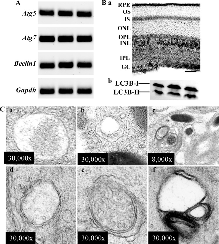
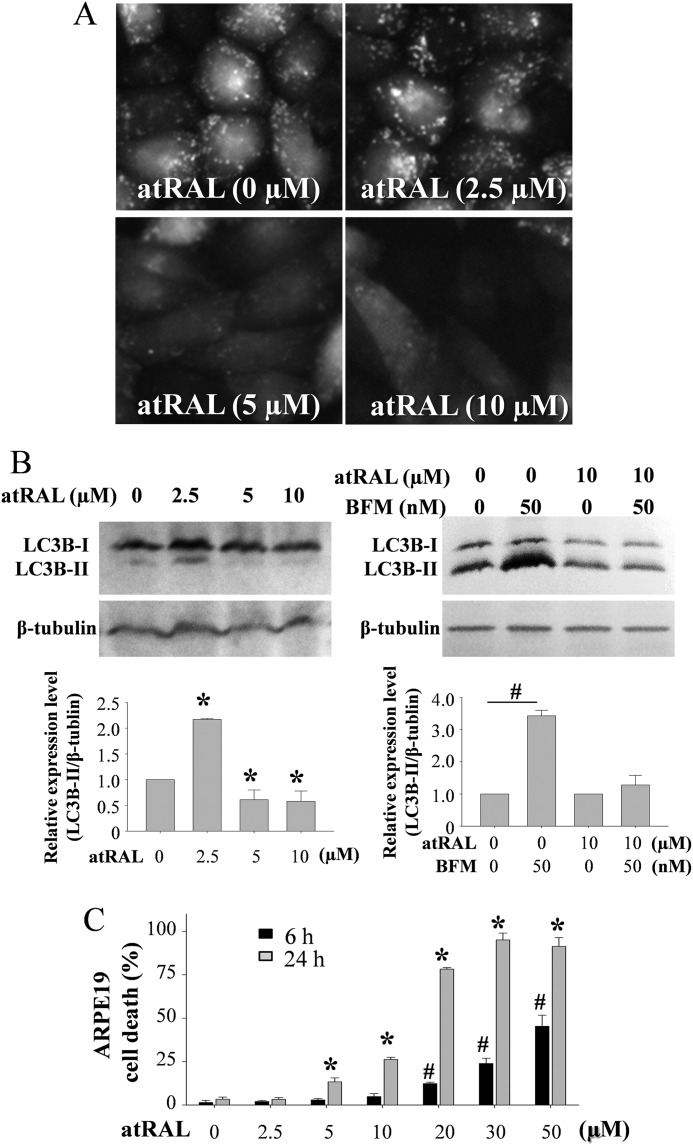
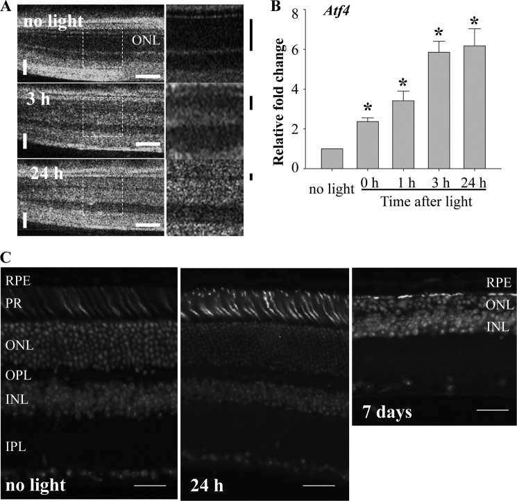
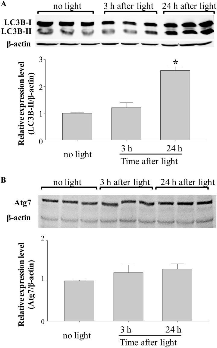
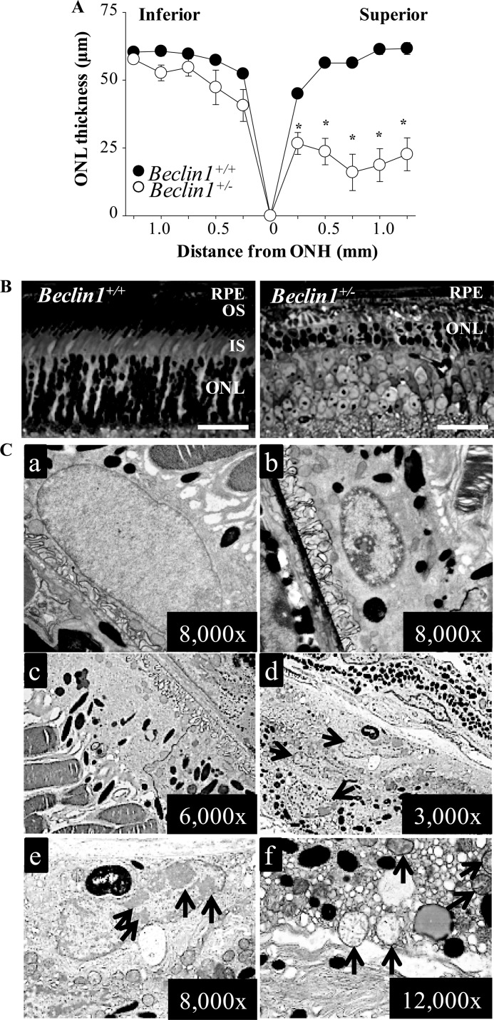
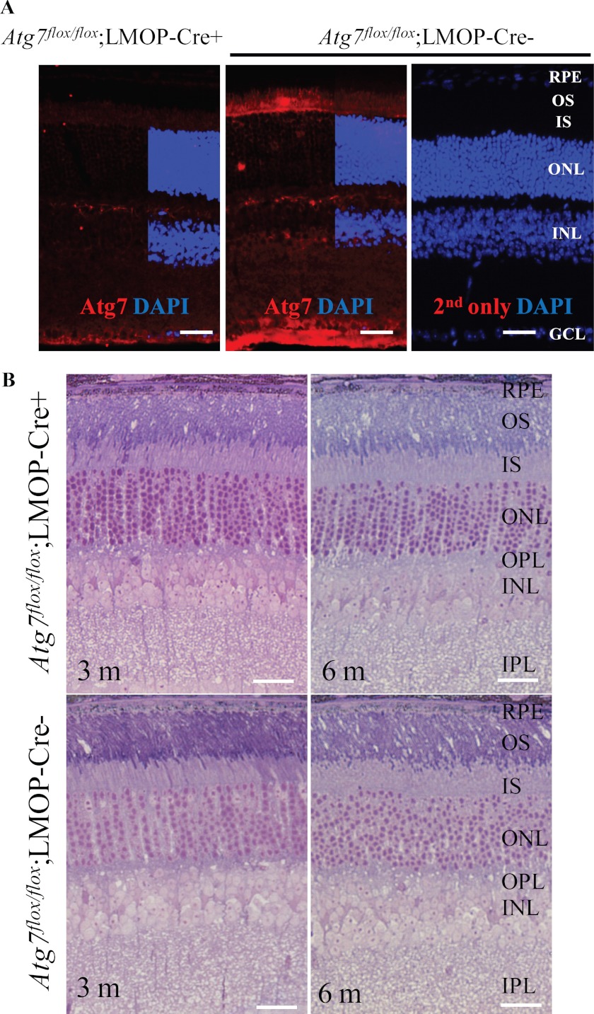
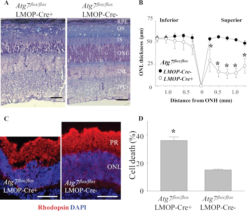
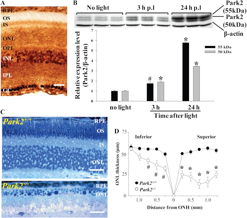
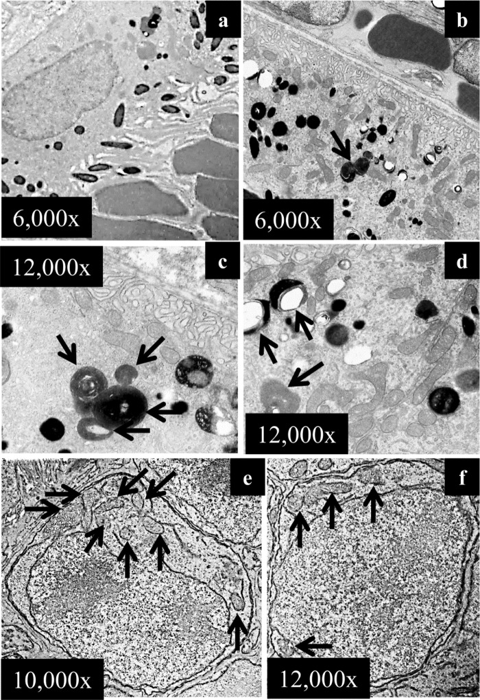
Autophagy is a conserved feature of lysosome-mediated intracellular degradation. Dysregulated autophagy is implicated as a contributor in neurodegenerative diseases; however, the role of autophagy in retinal degeneration remains largely unknown. Here, we report that the photo-activated visual chromophore, all-trans-retinal, modulated autophagosome formation in ARPE19 retinal cells. Increased formation of autophagosomes in these cells was observed when incubated with 2.5 μM all-trans-retinal, a condition that did not cause cell death after 24 h in culture. However, autophagosome formation was decreased at concentrations, which caused cell death. Increased expression of activating transcription factor 4 (Atf4), which indicates the activation of oxidative stress, was recorded in response to light illumination in retinas of Abca4(-/-)Rdh8(-/-) mice, which showed delayed clearance of all-trans-retinal after light exposure. Expression of autophagosome marker LC3B-II and mitochondria-specific autophagy, mitophagy, regulator Park2, were significantly increased in the retinas of Abca4(-/-)Rdh8(-/-) mice after light exposure, suggesting involvement of autophagy and mitophagy in the pathogenesis of light-induced retinal degeneration. Deletion of essential genes required for autophagy, including Beclin1 systemically or Atg7 in only rod photoreceptors resulted in increased susceptibility to light-induced retinal damage. Increased photoreceptor cell death was observed when retinas lacking the rod photoreceptor-specific Atg7 gene were coincubated with 20 μM all-trans-retinal. Park2(-/-) mice also displayed light-induced retinal degeneration. Ultra-structural analyses showed mitochondrial and endoplasmic reticulum impairment in retinas of these model animals after light exposure. Taken together, these observations provide novel evidence implicating an important role of autophagy and mitophagy in protecting the retina from all-trans-retinal- and light-induced degeneration.


Figures

FIGURE 1.
FIGURE 1.
Autophagy in the retina of wild-type mice. A, the expression of genes essential for autophagy was readily detected in 6-week-old WT mouse retinas. The mRNA expression of Atg5, Atg7, Beclin1, and Gapdh was detected in isolated mouse retinas by RT-PCR done in triplicate. Ba, the expression and immunolocalization of LC3B was revealed by immunohistochemistry using the retina of albino C57BL mice, indicating the expression of LC3B in RPE, photoreceptor inner segment (IS), outer plexiform layer (OPL), inner nuclear layer (INL), inner plexiform layer (IPL), and ganglion cell (GC). The bar indicates 20 μm. b, the protein expression of LC3B was detected in mouse retina. LC3B was resolved into two bands in SDS-PAGE, the cytoplasmic form LC3B-I and the autophagosome-associated form of LC3B-II. C, autophagic events were frequently encountered in the mouse retina under electromicroscopic examination. Autophagosomes containing portions of cytoplasm (a), a small amount of cytoplasm and dilated endoplasmic reticulum (ER) (b), membranous structure and a melanosome (c) were found in RPE. Autophagosomes enclosing cytoplasm (d), a damaged mitochondrion and small portion of cytoplasm (e), and partially membranous structure (f) were observed in mouse photoreceptor inner segment.
FIGURE 2.
FIGURE 2.
All-trans-retinal modulates autophagosome formation. A, LC3B-GFP was stably expressed in ARPE19 cells, and cells were incubated with 0, 2.5, 5, and 10 μm atRAL for 24 h at 37 °C. GFP signals were visualized under a fluorescent microscope (Leica DMI 6000 System). B, LC3B expression in ARPE19 cells after coincubation with atRAL for 24 h was examined by Western blotting. Increased expression of LC3B-II was detected ARPE19 cells incubated with 2.5 μm atRAL, whereas decreased expression of LC3B-II was noted under coincubation with 5 and 10 μm atRAL (left panel). *, compared with cells without coincubation with atRAL, p < 0.01. Balifomycin A1 (BFM) at 50 nm, an inhibitor for lysosome/autolysosome degradation, was admixed to ARPE19 cells cultured with 0 or 10 μm atRAL (right panel). Balifomycin A1 caused increased expression of LC3B-II in ARPE19 cells, whereas this increase was not observed when cells were coincubated with 10 μm atRAL. #, compared with cells without addition of balifomycin A1, p < 0.01. C, cell death rate was calculated by measuring lactate dehydrogenase (LDH) production (LDH-Cytotoxicty Assay kit, BioVision, Milpitas, CA). ARPE19 cells were coincubated with atRAL at indicated concentrations for 6 h or 24 h. # and *, compared with vehicle-treated cells for 6 h (#) or 24 h (*), p < 0.01.
FIGURE 3.
FIGURE 3.
Early changes in light-induced retinal damage in Abca4−/−Rdh8−/− mice. A, Abca4−/−Rdh8−/− mice at 6 weeks of age were exposed to white light at the intensity of 10,000 lux for 30 min, and SD-OCT imaging was carried out to monitor the retinal morphology at 3 and 24 h after light exposure (left). Horizontal or vertical bars in white indicate 200 or 50 μm, respectively. Magnified images of the areas with dash lines are presented (right). Black bars indicated outside show the thickness of dark area related to ONL observed in OCT imaging. Thickness of the dark area was decreased after light exposure. B, expression of Atf4 was examined by quantitative real-time-PCR and exhibited significant and rapid up-regulation in response to light exposure of 10,000 lux for 30 min. *, compared with no light, p < 0.01. C, cryosections of Abca4−/−Rdh8−/− mice after light exposure at 10,000 lux for 30 min are presented. Cone photoreceptors and nuclei were stained with peanut agglutinin and DAPI, respectively. Although decreased ONL thickness was displayed 7 days after light, such change was not detected 24 h after light. Bars indicate 20 μm.
FIGURE 4.
FIGURE 4.
Increased expression of autophagic marker LC3B-II in the retina is associated with light-induced retinopathy in Abca4−/−Rdh8−/− mice. A, expression of LC3B was examined by SDS-PAGE followed by Western blotting with triplicate samples. LC3B-II expression was significantly up-regulated by light 24 h after illumination. *, compared with no light, p < 0.01. B, expression of Atg7 was examined by SDS-PAGE followed by Western blotting with triplicate samples. Shown are retinas of 6-week-old Abca4−/−Rdh8−/− mice after exposure to white light at the intensity of 10,000 lux for 30 min.
FIGURE 5.
FIGURE 5.
Increased susceptibility to light-induced retinal damage as a result of Beclin1 deficiency. Beclin1+/− mice and Beclin1+/+ littermate controls at 6 weeks of age were exposed to white light at 5,000 lux for 2 h. A, the ONL thickness was measured by SD-OCT and plotted (*, p < 0.01). Light-induced photoreceptor degeneration was prominent in the superior retina in Beclin1+/− mice compared with intact retinal morphology displayed by Beclin1+/+ mice. ONH, optic nerve head. B, plastic sections were prepared from eye cups collected from light-exposed Beclin1+/− mice and Beclin1+/+ littermate controls, which was followed by histological examination after toluidine blue staining. Severely disrupted photoreceptor structure was marked by degenerated photoreceptor outer segment and inner segment and significantly reduced thickness of ONL in Beclin1+/− eyes compared with the intact photoreceptor structure displayed by Beclin1+/+ eyes. Bars indicate 20 μm. IS, inner segment. OS, outer segment. C, EM was carried out to examine the RPE structure in Beclin1+/− mice and Beclin1+/+ littermate control 24 h and 10 days after light exposure, respectively. a, EM images from the RPE of Beclin1+/+ mice 24 h after light exposure show normal looking nucleus. B, EM images from the RPE of Beclin1+/− mice 24 h after light exposure show changes in nucleus including chromatin condensation and vacuolation. c, shown are EM images from the RPE of Beclin1+/+ mice 10 days after light exposure. d–f, EM images from the RPE of Beclin1+/− mice 10 days after light exposure reveal chromatin condensation and accumulation of large amounts of damaged mitochondria and dilated EM causing cytoplasmic vacuolation. Arrows in d, RPE hyperplasia indicated by presence of multiple nuclei; arrow in e, nucleus showing chromatin condensation; Arrows in f, damaged mitochondria.
FIGURE 6.
FIGURE 6.
Retinal morphology is maintained in mice with rod photoreceptor-specific Atg7 deletion under room lighting conditions. A, Atg7 expression was examined in Atg7flox/flox;LMOP-Cre+ and littermate Atg7flox/flox;LMOP-Cre− mice at 6 weeks of age. Immunohistochemistry was conducted with anti-Atg7 Ab (red) and DAPI (blue). Atg7 signal was much weaker in the inner segment of Atg7flox/flox;LMOP-Cre+ mice than that of Atg7flox/flox;LMOP-Cre− mice. INL, inner nuclear layer; IS, inner segment; OS, outer segment. Bars indicate 20 μm. B, the steady-state retinal morphology under room lighting conditions in Atg7flox/flox;LMOP-Cre+ mice was examined by histological analysis with epon-embedment together with their wild-type counterparts (Atg7flox/flox;LMOP-Cre−) at the ages of 3 and 6 months. No overt morphological changes were observed in Atg7flox/flox;LMOP-Cre+ mice compared with that from Atg7flox/flox;LMOP-Cre- littermate controls. Bars indicate 20 μm.
FIGURE 7.
FIGURE 7.
Increased photoreceptor death caused by light exposure and all-trans-retinal in retinas of rod photoreceptor-specific Atg7 deletion. Atg7flox/flox;LMOP-Cre+ mice and Atg7flox/flox;LMOP-Cre− littermate controls at 5–6 weeks of age were exposed to white light at 5,000 lux for 2 h. Mice were kept in the dark, and retinal morphology was examined 7 days after light exposure. A, plastic sections were prepared from eye cups collected from light-exposed Atg7flox/flox;LMOP-Cre+ mice and Atg7flox/flox;LMOP-Cre− littermate controls, which was followed by histological examination after toluidine blue staining. B, the ONL thickness was measured by SD-OCT and plotted (*, p < 0.01). Light-induced photoreceptor degeneration was prominent in the superior retina in Atg7flox/flox;LMOP-Cre+ mice compared with intact retinal morphology displayed by Atg7flox/flox;LMOP-Cre− littermate mice. C, retinas were collected from Atg7flox/flox;LMOP-Cre+ and Atg7flox/flox;LMOP-Cre− mice, and these retinas were cultured with or without 20 μm atRAL for 6 h at 37 °C. Immunohistochemistry of these retinas were conducted with anti-rhodopsin Ab (red) and DAPI (blue). Bars indicate 20 μm. PR, photoreceptor layer. D, amounts of lactate dehydrogenase (LDH) were measured by lactate dehydrogenase-cytotoxicity assay kit (BioVision) with supernatant of ex vivo retinal culture with 20 μm atRAL for 6 h at 37 °C, and cell death rate was calculated. *, compared with no light, p < 0.01. Rod photoreceptor-specific deletion of Atg7 increased the susceptibility to light-induced and atRAL-induced retinal degeneration.
FIGURE 8.
FIGURE 8.
Enhanced expression of Park2 is associated with light-induced retinopathy and Park2 deficiency results in light-induced retinopathy. A, Park2 expression in the retina of albino WT mice at 6 weeks of age was examined by immunohistochemistry, revealing that Park2 is ubiquitously expressed in the retina. Bars indicate 20 μm. B, Abca4−/−Rdh8−/− mice at 6 weeks of age were exposed to white light at 10,000 lux for 30 min. Park2 expression in the retina was then examined by SDS-PAGE followed by Western blotting with triplicate samples. Up-regulated Park2 expression was observed in response to light exposure. Compared with no light. *, p < 0.01; #, p < 0.05. Is, inner segment; OPL, outer plexiform layer; INL, inner nuclear layer; GC, ganglion cell; OS, outer segment. C, Park2−/− mice together with Park2+/+ littermate controls were exposed to intermittent white light for 40 min given by 20 min each with a 2-h dark adaption between each illumination. Histological examination on plastic sections after toluidine blue staining revealed the photoreceptor damage in Park2−/− mice compared with the intact photoreceptor structure in Park2+/+ mice. Bars indicate 20 μm. D, the ONL thickness was measured by SD-OCT and plotted (*, p < 0.01; #, p < 0.05).
FIGURE 9.
FIGURE 9.
Impaired phagosomal digestion in RPE and ectopic mitochondria in photoreceptors in light-exposed Park2-deficient mice. Park2−/− mice together with Park2+/+ littermate controls at 6 weeks of age were exposed to intermittent light at 10,000 lux for 20 min per exposure, two exposures with a 2-h interval of dark adaptation. The ultra-structure of RPE and photoreceptors were examined 24 h after light exposure. No overt abnormality in RPE ultra-structure was displayed by Park2+/+ mice (a). In Park2−/− RPE cells, undigested or partially digested phagosomes were readily detected (arrows in b–d). A large amount of mitochondria was also present in Park2+/+ RPE cells (d). The ectopic mitochondria were also frequently detected in Park2−/− photoreceptors (e and f).

Similar articles

Cited by

References

    1. Yang Z., Klionsky D. J. (2010) Eaten alive. A history of macroautophagy. Nat. Cell Biol. 12, 814–822 - PMC - PubMed
    1. Pickford F., Masliah E., Britschgi M., Lucin K., Narasimhan R., Jaeger P. A., Small S., Spencer B., Rockenstein E., Levine B., Wyss-Coray T. (2008) The autophagy-related protein beclin 1 shows reduced expression in early Alzheimer disease and regulates amyloid β accumulation in mice. J. Clin. Invest. 118, 2190–2199 - PMC - PubMed
    1. Hara T., Nakamura K., Matsui M., Yamamoto A., Nakahara Y., Suzuki-Migishima R., Yokoyama M., Mishima K., Saito I., Okano H., Mizushima N. (2006) Suppression of basal autophagy in neural cells causes neurodegenerative disease in mice. Nature 441, 885–889 - PubMed
    1. Komatsu M., Waguri S., Ueno T., Iwata J., Murata S., Tanida I., Ezaki J., Mizushima N., Ohsumi Y., Uchiyama Y., Kominami E., Tanaka K., Chiba T. (2005) Impairment of starvation-induced and constitutive autophagy in Atg7-deficient mice. J. Cell Biol. 169, 425–434 - PMC - PubMed
    1. Mizushima N., Komatsu M. (2011) Autophagy. Renovation of cells and tissues. Cell 147, 728–741 - PubMed

Publication types

MeSH terms

Substances