

3'-Deoxy-3'-18F-fluorothymidine PET predicts response to (V600E)BRAF-targeted therapy in preclinical models of colorectal cancer
- PMID: 23341544
- PMCID: PMC3633462
- DOI: 10.2967/jnumed.112.108456
3'-Deoxy-3'-18F-fluorothymidine PET predicts response to (V600E)BRAF-targeted therapy in preclinical models of colorectal cancer
Abstract
Selective inhibition of oncogenic targets and associated signaling pathways forms the basis of personalized cancer medicine. The clinical success of (V600E)BRAF inhibition in melanoma, coupled with the emergence of acquired resistance, underscores the importance of rigorously validating quantitative biomarkers of treatment response in this and similar settings. Because constitutive activation of BRAF leads to proliferation in tumors, we explored 3'-deoxy-3'-(18)F-fluorothymidine ((18)F-FLT) PET to noninvasively quantify changes in tumor proliferation that are associated with pharmacologic inhibition of (V600E)BRAF downstream effectors and that precede changes in tumor volume.
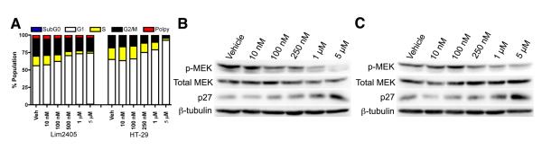
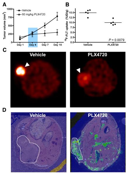
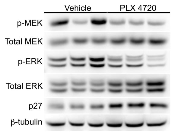
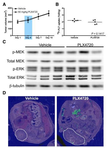
Methods: Human colorectal cancer (CRC) cell lines expressing (V600E)BRAF were used to explore relationships between upregulation of p27 and phosphorylation of BRAF downstream effectors on small-molecule (V600E)BRAF inhibitor exposure. Athymic nude mice bearing (V600E)BRAF-expressing human CRC cell line xenografts were treated with a small-molecule (V600E)BRAF inhibitor (or vehicle) daily for 10 d. Predictive (18)F-FLT PET was conducted before changes in tumor volume occurred. Correlations were evaluated among PET, inhibition of phosphorylated MEK (p-MEK) and phosphorylated-ERK (p-ERK) by Western blot, tumor proliferation by histology, and small-molecule exposure by matrix-assisted laser desorption/ionization (MALDI) imaging mass spectrometry (IMS).
Results: Treatment of CRC cell lines with PLX4720 reduced proliferation associated with target inhibition and upregulation of p27. In vivo, PLX4720 treatment reduced (18)F-FLT uptake, but not (18)F-FDG uptake, in Lim2405 xenografts before quantifiable differences in xenograft volume. Reduced (18)F-FLT PET reflected a modest, yet significant, reduction of Ki67 immunoreactivity, inhibition of p-MEK and p-ERK, and elevated tumor cell p27 protein levels. Both (18)F-FLT PET and (18)F-FDG PET accurately reflected a lack of response in HT-29 xenografts, which MALDI imaging mass spectrometry suggested may have stemmed from limited PLX4720 exposure.
Conclusion: We used preclinical models of CRC to demonstrate (18)F-FLT PET as a sensitive predictor of response to (V600E)BRAF inhibitors. Because (18)F-FLT PET predicted reduced proliferation associated with attenuation of BRAF downstream effectors, yet (18)F-FDG PET did not, these data suggest that (18)F-FLT PET may represent an alternative to (18)F-FDG PET for quantifying clinical responses to BRAF inhibitors.
Figures

Similar articles
-
Non-Invasive Glutamine PET Reflects Pharmacological Inhibition of BRAFV600E In Vivo.Mol Imaging Biol. 2017 Jun;19(3):421-428. doi: 10.1007/s11307-016-1008-z. Mol Imaging Biol. 2017. PMID: 27770401 Free PMC article.
-
Therapy response monitoring of the early effects of a new BRAF inhibitor on melanoma xenograft in mice: evaluation of (18) F-FDG-PET and (18) F-FLT-PET.Contrast Media Mol Imaging. 2015 May-Jun;10(3):203-10. doi: 10.1002/cmmi.1619. Epub 2014 Sep 9. Contrast Media Mol Imaging. 2015. PMID: 25204436
-
3'-deoxy-3'-[18F]fluorothymidine positron emission tomography is a sensitive method for imaging the response of BRAF-dependent tumors to MEK inhibition.Cancer Res. 2007 Dec 1;67(23):11463-9. doi: 10.1158/0008-5472.CAN-07-2976. Cancer Res. 2007. PMID: 18056475 Free PMC article.
-
A comprehensive overview of the molecular features and therapeutic targets in BRAFV600E-mutant colorectal cancer.Clin Transl Med. 2024 Jul;14(7):e1764. doi: 10.1002/ctm2.1764. Clin Transl Med. 2024. PMID: 39073010 Free PMC article.
-
BRAF Targeting Across Solid Tumors: Molecular Aspects and Clinical Applications.Int J Mol Sci. 2025 Apr 16;26(8):3757. doi: 10.3390/ijms26083757. Int J Mol Sci. 2025. PMID: 40332392 Free PMC article. Review.
Cited by
-
Multimodal Imaging of Orthotopic Mouse Model of Endometrial Carcinoma.PLoS One. 2015 Aug 7;10(8):e0135220. doi: 10.1371/journal.pone.0135220. eCollection 2015. PLoS One. 2015. PMID: 26252891 Free PMC article.
-
Non-Invasive Glutamine PET Reflects Pharmacological Inhibition of BRAFV600E In Vivo.Mol Imaging Biol. 2017 Jun;19(3):421-428. doi: 10.1007/s11307-016-1008-z. Mol Imaging Biol. 2017. PMID: 27770401 Free PMC article.
-
18F-FLT Positron Emission Tomography (PET) is a Pharmacodynamic Marker for EWS-FLI1 Activity and Ewing Sarcoma.Sci Rep. 2016 Sep 27;6:33926. doi: 10.1038/srep33926. Sci Rep. 2016. PMID: 27671553 Free PMC article.
-
Molecular imaging for early prediction of response to Sorafenib treatment in sarcoma.Am J Nucl Med Mol Imaging. 2013 Dec 15;4(1):70-9. eCollection 2013. Am J Nucl Med Mol Imaging. 2013. PMID: 24380047 Free PMC article.
-
Monitoring of anti-cancer treatment with (18)F-FDG and (18)F-FLT PET: a comprehensive review of pre-clinical studies.Am J Nucl Med Mol Imaging. 2015 Oct 12;5(5):431-56. eCollection 2015. Am J Nucl Med Mol Imaging. 2015. PMID: 26550536 Free PMC article. Review.
References
-
- Davies H, Bignell GR, Cox C, Stephens P, Edkins S, Clegg S, et al. Mutations of the BRAF gene in human cancer. Nature. 2002;417(6892):949–54. - PubMed
-
- Satyamoorthy K, Li G, Gerrero MR, Brose MS, Volpe P, Weber BL, et al. Constitutive mitogen-activated protein kinase activation in melanoma is mediated by both BRAF mutations and autocrine growth factor stimulation. Cancer Res. 2003;63(4):756–9. - PubMed
Publication types
MeSH terms
Substances
Grants and funding
- P50 CA128323/CA/NCI NIH HHS/United States
- P50 CA095103/CA/NCI NIH HHS/United States
- R01 CA046413/CA/NCI NIH HHS/United States
- R01 CA140628/CA/NCI NIH HHS/United States
- R01 CA46413/CA/NCI NIH HHS/United States
- S10 RR017858/RR/NCRR NIH HHS/United States
- S10 RR17858/RR/NCRR NIH HHS/United States
- U24 CA126588/CA/NCI NIH HHS/United States
- RC1 CA145138/CA/NCI NIH HHS/United States
- RC1 145138/RC/CCR NIH HHS/United States
- R25 CA136440/CA/NCI NIH HHS/United States
- P50 951903/PHS HHS/United States
- P30 DK058404/DK/NIDDK NIH HHS/United States
- K25 CA127349/CA/NCI NIH HHS/United States
LinkOut - more resources
Full Text Sources
Other Literature Sources
Medical
Research Materials
Miscellaneous