. 2013 Feb 5;110(6):2360-5.
doi: 10.1073/pnas.1221077110. Epub 2013 Jan 22.

Transcription factor Nurr1 maintains fiber integrity and nuclear-encoded mitochondrial gene expression in dopamine neurons

Affiliations

Transcription factor Nurr1 maintains fiber integrity and nuclear-encoded mitochondrial gene expression in dopamine neurons

Banafsheh Kadkhodaei et al. Proc Natl Acad Sci U S A. .

Abstract

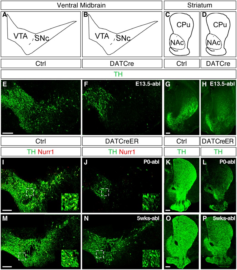
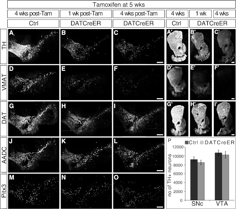
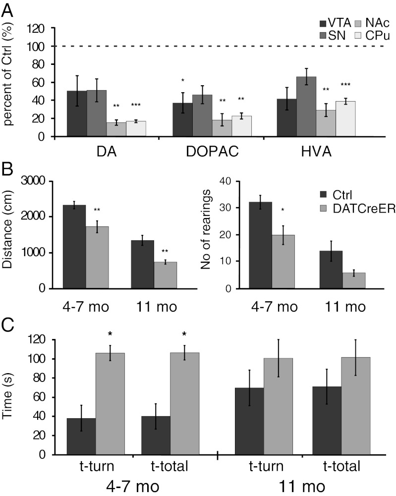
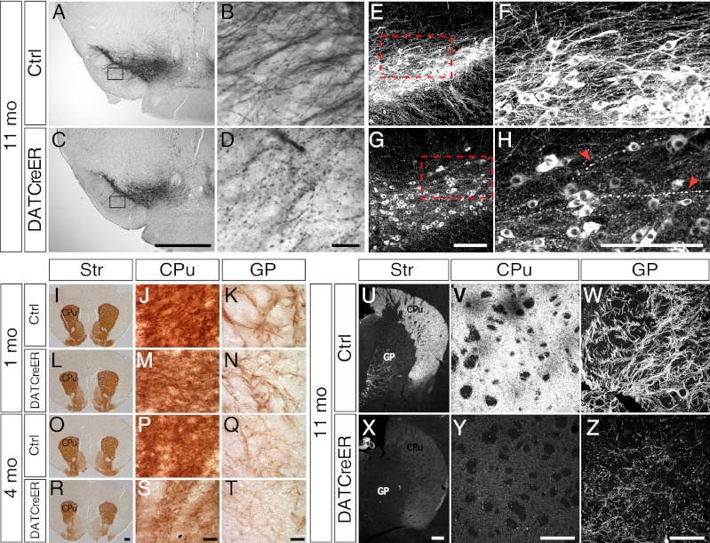
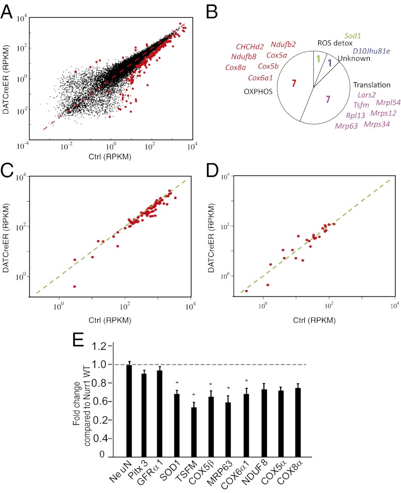
Developmental transcription factors important in early neuron specification and differentiation often remain expressed in the adult brain. However, how these transcription factors function to mantain appropriate neuronal identities in adult neurons and how transcription factor dysregulation may contribute to disease remain largely unknown. The transcription factor Nurr1 has been associated with Parkinson's disease and is essential for the development of ventral midbrain dopamine (DA) neurons. We used conditional Nurr1 gene-targeted mice in which Nurr1 is ablated selectively in mature DA neurons by treatment with tamoxifen. We show that Nurr1 ablation results in a progressive pathology associated with reduced striatal DA, impaired motor behaviors, and dystrophic axons and dendrites. We used laser-microdissected DA neurons for RNA extraction and next-generation mRNA sequencing to identify Nurr1-regulated genes. This analysis revealed that Nurr1 functions mainly in transcriptional activation to regulate a battery of genes expressed in DA neurons. Importantly, nuclear-encoded mitochondrial genes were identified as the major functional category of Nurr1-regulated target genes. These studies indicate that Nurr1 has a key function in sustaining high respiratory function in these cells, and that Nurr1 ablation in mice recapitulates early features of Parkinson's disease.


Conflict of interest statement

The authors declare no conflict of interest.

Figures

Fig. 1.
Fig. 1.
Nurr1 ablation in developing and adult DA neurons. (A–D) Cross-sections at the level of VMB showing the VTA and SNc and at the level of striatum showing the CPu and NAc. (E–P) Immunostaining of TH (green) and Nurr1 (red) at the level of the VMB and in the striatum of cNurr1Ctrl (Ctrl), cNurr1DatCre (DatCre), and cNurr1DatCreER (DatCreER) mice. Nurr1 was ablated by DAT-Cre crosses (cNurr1DatCre mice) at approximately E13.5 and analyzed at P0 (E–H), or treated with tamoxifen at P0 (I–L) or at 5 wk after birth (M–P). In I–P, mice were killed at 1 wk after tamoxifen treatment. Nurr1 immunoreactivity was almost completely lost in TH-positive cells in the cNurr1DatCre and cNurr1DatCreER mice (compare the high-magnification insets in I, J, M, and N).
Fig. 2.
Fig. 2.
Expression analysis of DA neuron markers and cell counting in Nurr1-ablated mice. Nurr1 was ablated at age 5 wk by tamoxifen treatment. Analyses were performed in cNurr1Ctrl (Ctrl) and cNurr1DatCreER (DatCreER) mice at either 1 wk or 4 wk after tamoxifen treatment. (A–O) Marker expression at the level of the VMB. (A′–I) Marker expression at the level of the striatum. (P) Stereology of TH-positive neurons in the SNc and VTA in cNurr1Ctrl (Ctrl) and cNurr1DatCreER (DatCreER) mice at 2 mo after tamoxifen treatment.
Fig. 3.
Fig. 3.
Analysis of catecholamines and motor behavior in Nurr1-ablated mice. (A) Striatal levels of DA, 3,4-Dihydroxyphenylacetic acid (DOPAC), and homovanillic acid (HVA) in cNurr1Ctrl (Ctrl) and cNurr1DatCreER (DatCreER) mice at 11 mo after tamoxifen treatment in 5-wk-old mice. Separate analyses were performed on tissue extracts from the VTA, SNc, NAc, and CPu, as indicated. (B and C) Open-field measures of horizontal (distance in cm over a 5-min measurement) and vertical (number of rearings during a 5-min measurement) locomotion (B) and vertical pole test of posture control (C) of cNurr1Ctrl (Ctrl) and cNurr1DatCreER (DatCreER) mice at 4–7 mo and 11 mo after tamoxifen treatment in 5-wk-old mice. Here t-turn is the time required to orient downward, and t-total is the total time taken to turn and descend the pole. Note that overall performance was also poor in controls at 11 mo.
Fig. 4.
Fig. 4.
Fiber integrity in Nurr1-ablated mice. (A–D) TH immunostaining by horseradish peroxidase/diaminobenzidine (DAB) staining in either low magnification (4×; A and C) or high magnification (40×; B and D) showing dendrites extending into the substantia nigra reticulata in cNurr1Ctrl (Ctrl) and cNurr1DatCreER (DatCreER) mice at 12 mo after tamoxifen treatment in 5-wk-old mice. Boxed regions in A and C are shown in B and D. (E–H) TH immunofluorescence in either low magnification (20×) or high magnification (35×) showing fibers extending dorsorostrally from the substantia nigra cell bodies in the VMB. Arrowheads in H indicate abnormal fibers. (I–T) DAT immunostaining (DAB) of the striatum in cNurr1Ctrl (Ctrl) and cNurr1DatCreER (DatCreER) mice at 1 mo and 4 mo after tamoxifen treatment in 5-wk-old mice. Images of the striatum (Str) in I, L, O, and R are at low magnification (2×). Images at high magnification (100×) (J, K, M, N, P, Q, S, and T) show fibers in either the CPu or in the GP, as indicated. (U–Z) TH immunofluorescence of the striatum in cNurr1Ctrl (Ctrl) and cNurr1DatCreER (DatCreER) mice at 11 mo after tamoxifen treatment in 5-wk-old mice. Images at low magnification (U and X) and high magnification (V–Z) show the Str, CPu, or GP, as indicated.
Fig. 5.
Fig. 5.
RNA sequencing of control and Nurr1-ablated DA neurons. (A) Scatter diagram showing all detected genes. Differentially expressed genes are highlighted by red dots. The x-axis shows mRNA sequencing RPKM values from control cells; the y-axis, mRNA sequencing RPKM values from Nurr1-ablated cells. (B) Circle diagram showing functional categories and gene names of significantly regulated nuclear-encoded mitochondrial genes. The majority of these genes encode either proteins involved in oxidative phosphorylation or mitochondrial translation. One gene (Sod1) is implicated in radical oxygen species (ROS) detoxification. (C) Scatter diagram highlighting genes encoding proteins involved in oxidative phosphorylation. The x-axis shows mRNA sequencing RPKM values from control cells; the y-axis, mRNA sequencing RPKM values from Nurr1-ablated cells. (D) Scatter diagram highlighting genes encoding proteins involved in fatty acid metabolism. The x-axis shows mRNA sequencing RPKM values from control cells; the y-axis, mRNA sequencing RPKM values from Nurr1-ablated cells. (E) qPCR analysis in Nurr1 ablated and control mice 8–10 wk after tamoxifen treatment. Data are expressed as mean ± SEM (n = 5 per group). *P < 0.05 compared with Nurr1WT/WT group.

Similar articles

Cited by

References

    1. Graf T, Enver T. Forcing cells to change lineages. Nature. 2009;462(7273):587–594. - PubMed
    1. Holmberg J, Perlmann T. Maintaining differentiated cellular identity. Nat Rev Genet. 2012;13(6):429–439. - PubMed
    1. Hobert O. Regulation of terminal differentiation programs in the nervous system. Annu Rev Cell Dev Biol. 2011;27:681–696. - PubMed
    1. Cheng H-C, Ulane CM, Burke RE. Clinical progression in Parkinson disease and the neurobiology of axons. Ann Neurol. 2010;67(6):715–725. - PMC - PubMed
    1. Wang Z, et al. Structure and function of Nurr1 identifies a class of ligand-independent nuclear receptors. Nature. 2003;423(6939):555–560. - PubMed

Publication types

MeSH terms

Substances

Associated data