. 2013 Feb 5;110(6):2366-70.
doi: 10.1073/pnas.1221891110. Epub 2013 Jan 22.

Aberrant splicing of HTT generates the pathogenic exon 1 protein in Huntington disease

Affiliations

Aberrant splicing of HTT generates the pathogenic exon 1 protein in Huntington disease

Kirupa Sathasivam et al. Proc Natl Acad Sci U S A. .

Abstract

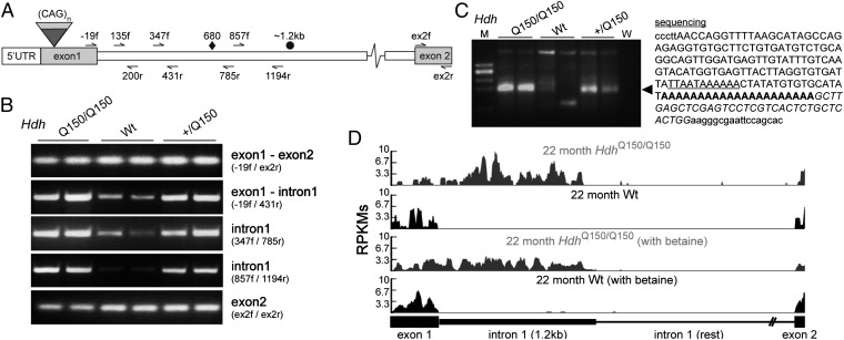
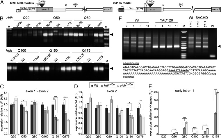
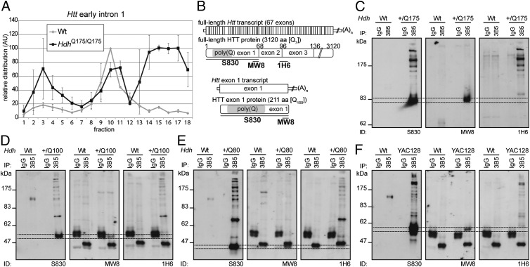
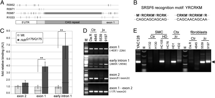
Huntington disease (HD) is a devastating, late-onset, inherited neurodegenerative disorder that manifests with personality changes, movement disorders, and cognitive decline. It is caused by a CAG repeat expansion in exon 1 of the HTT gene that translates to a polyglutamine tract in the huntingtin protein (HTT). The formation of HTT fragments has been implicated as an essential step in the molecular pathogenesis of HD and several proteases that cleave HTT have been identified. However, the importance of smaller N-terminal fragments has been highlighted by their presence in HD postmortem brains and by the fact that nuclear inclusions are only detected by antibodies to the N terminus of HTT. Despite an intense research effort, the precise length of these fragments and the mechanism by which they are generated remains unknown. Here we show that CAG repeat length-dependent aberrant splicing of exon 1 HTT results in a short polyadenylated mRNA that is translated into an exon 1 HTT protein. Given that mutant exon 1 HTT proteins have consistently been shown to be highly pathogenic in HD mouse models, the aberrant splicing of HTT mRNA provides a mechanistic basis for the molecular pathogenesis of HD. RNA-targeted therapeutic strategies designed to lower the levels of HTT are under development. Many of these approaches would not prevent the production of exon 1 HTT and should be reviewed in light of our findings.


Conflict of interest statement

The authors declare no conflict of interest.

Figures

Fig. 1.
Fig. 1.
Aberrant splicing of Htt exon 1 to exon 2 results in a short polyadenylated mRNA in the HdhQ150 knock-in mouse model. (A) Schematic representation of the mouse Htt gene and primers used for the RT-PCR analysis. ♦, cryptic polyadenylation signal; ●, end of overrepresented intronic sequences. (B) RT-PCR analysis of exon 1, the exon 1–intron 1 boundary, intron 1, and exon 2. (C) 3′RACE product was generated from HdhQ150/Q150 and Hdh+/Q150 brain RNA (◄), but not from WT controls, and contained a polyA tail located ∼700 bp into intron 1. The cryptic polyadenylation signal is underlined, the polyA tail is shown in bold, the primer sequence is in italics, and vector sequence is in lowercase. (D) RNA-Seq reads from cortex of 22 mo HdhQ150/Q150 and WT mice mapping to the Htt exon 1–exon 2 region. M, low-molecular-weight marker (New England Biolabs); W, water.
Fig. 2.
Fig. 2.
Aberrant splicing is CAG repeat length–dependent and occurs in the context of both mouse Htt and human HTT. (A) Schematic representations of chimeric human/mouse Htt genes in the HdhQ20, HdhQ80, and zQ175 lines. The 5′UTR and first 28 bp of exon 1 are always of mouse origin, whereas the remaining exon 1 sequence is human. The HdhQ20 and HdhQ80 lines contain 268 bp of human intron 1, 124 bp of mouse intron 1 is deleted, and there is a loxP site 5′ to the ATG. Line zQ175 contains 10 bp of human intron 1 with 94 bp of mouse intron 1 deleted and an intact neo-cassette 1.3-kb 5′ to the ATG. X, XmnI; E, EcoRV; K, KpnI; ♦, cryptic polyadenylation signal. (B) 3′RACE indicates the presence of the identical polyadenylated transcript (◄) in all lines except HdhQ20. (C and D) The relative levels of the spliced exon 1–exon 2 and exon 2 transcripts are shown relative to WT. (E) The expression level of early intron 1 transcripts is shown relative to the geometrical mean of three housekeeping (HK) genes (Atp5b, Eif4a3, Sdha). Primers for quantitative RT-PCR are specified in Table S3. Exon 1–exon 2, 19f-ex2r; exon 2, ex2f-ex2r; early intron 1, 135f–200r. n = 8/genotype; *P < 0.05; **P < 0.01; ***P < 0.001. Data are mean ± SEM. (F) A 3′RACE product was generated from YAC128 and BACHD brain RNA (◄), but not from WT controls, and contained a polyA tail, ∼7.3 kb into intron 1. The cryptic polyadenylation signal is underlined, the polyA tail is in bold, the primer sequence (UAPdT18) is shown in italics, and the vector sequence is in lowercase letters. M is HaeIII-digested ΦX174 (B) and low-molecular-weight markers (D) (New England Biolabs).
Fig. 3.
Fig. 3.
The aberrantly spliced Htt transcript is translated and produces an exon 1 HTT protein. (A) Polysome gradients showing the relative distribution of early Htt intron 1 (135f/200r) transcripts (n = 2). Data are mean ± SEM. (B) Schematic shows the position of the HTT antibody epitopes. (CF) HTT proteins were immunoprecipitated with 3B5H10-coupled magnetic beads from WT and (C) zQ175, (D) HdhQ100, (E) HdhQ80, and (F) YAC128 brain lysates, and Western immunoblots were immunoprobed with S830, MW8, and 1H6 antibodies. Dotted lines indicate the gel migration of the exon 1 HTT proteins, which are retarded by the polyglutamine tract and do not migrate as would be predicted by their molecular weight. To resolve exon HTT proteins with different polyQ tracts, the gel depicted in (C) was run for a longer time than those in (DF). On the HdhQ80 blots, exon 1 HTT comigrates with a nonspecific band (*). 3B5 = 3B5H10.
Fig. 4.
Fig. 4.
The splicing factor SRSF6 binds to expanded CAG repeats in Htt transcripts and the aberrantly spliced transcript is present in HD patient tissues. (A) RegRNA predicts a cluster of R0987 regulatory motifs (CTGN) in the expanded CAG repeat of Htt. Using ESEfinder 3.0, this motif was mapped to the binding site of SRSF6. (B) The RNA recognition motif of SRSF6 is YRCRKM, closely resembling a CAG or CAGCAA repeat. Nucleotide abbreviations: Y, T or C; R, A or G; K, G or T; M, A or C. (C) RNA-IP of SRSF6 from zQ175 brain resulted in the coprecipitation of higher levels of early intronic (135f-200r) and exon 1 (5′UTR 1f-5′UTR 1r) transcripts compared with WT. In contrast, the coprecipitated levels of zQ175 and WT exon 2 transcripts were not significantly different. n = 6/genotype. Horizontal bar, IgG control immunoprecipitation. Data are mean ± SEM; **P < 0.01. (D) RT-PCR of human samples. Human fibroblast lines can be found in Table S2. (E) 3′RACE of human samples. ◄, the expected RACE product size of about 260 bp; Ctr, control subject; Ctx, cortex; HD, adult onset HD; ju, juvenile-onset HD; M, low-molecular-weight markers (New England Biolabs); SMC, sensory motor cortex; tg, transgenic; W, water.

Comment in

References

    1. Bates GP, Harper PS, Jones AL, eds (2002) Huntington's Disease (Oxford University Press, Oxford), 3rd Ed.
    1. Ross CA, Tabrizi SJ. Huntington’s disease: From molecular pathogenesis to clinical treatment. Lancet Neurol. 2011;10(1):83–98. - PubMed
    1. Wellington CL, et al. Caspase cleavage of mutant huntingtin precedes neurodegeneration in Huntington’s disease. J Neurosci. 2002;22(18):7862–7872. - PMC - PubMed
    1. Kim YJ, et al. Caspase 3-cleaved N-terminal fragments of wild-type and mutant huntingtin are present in normal and Huntington’s disease brains, associate with membranes, and undergo calpain-dependent proteolysis. Proc Natl Acad Sci USA. 2001;98(22):12784–12789. - PMC - PubMed
    1. Gafni J, et al. Inhibition of calpain cleavage of huntingtin reduces toxicity: Accumulation of calpain/caspase fragments in the nucleus. J Biol Chem. 2004;279(19):20211–20220. - PubMed

Publication types

MeSH terms