. 2013 Feb 5;110(6):2276-81.
doi: 10.1073/pnas.1218295110. Epub 2013 Jan 22.

Fas expression by tumor stroma is required for cancer eradication

Affiliations

Fas expression by tumor stroma is required for cancer eradication

Joanna J Listopad et al. Proc Natl Acad Sci U S A. .

Abstract

The contribution of molecules such as perforin, IFN-γ (IFNγ), and particularly Fas ligand (FasL) by transferred CD8(+) effector T (T(E)) cells to rejection of large, established tumors is incompletely understood. Efficient attack against large tumors carrying a surrogate tumor antigen (mimicking a "passenger" mutation) by T(E) cells requires action of IFNγ on tumor stroma cells to avoid selection of antigen-loss variants. Because "cancer-driving" antigens (CDAs) are rarely counterselected, IFNγ may be expected to be dispensable in elimination of cancers by targeting a CDA. Here, initial regression of large, established tumors required neither IFNγ, FasL, nor perforin by transferred CD8(+) T(E) cells targeting Simian Virus (SV) 40 large T as CDA. However, cytotoxic T(E) cells lacking IFNγ or FasL could not prevent relapse despite retention of the rejection antigen by the cancer cells. Complete tumor rejection required IFNγ-regulated Fas by the tumor stroma. Therefore, T(E) cells lacking IFNγ or FasL cannot prevent progression of antigenic cancer because the tumor stroma escapes destruction if its Fas expression is down-regulated.


Conflict of interest statement

The authors declare no conflict of interest.

Figures

Fig. 1.
Fig. 1.
Failure of cytotoxic IFNγ−/−, but not Pfp−/− TE cells, to prevent tumor relapse. (A) Histological analysis of established tumors was performed 70 d after s.c. injection of 106 16.113 cells into Rag1−/− mice (Upper, HE and Lower, Tag staining). One representative example out of seven analyzed tumors is shown. ATT of established tumors was performed by i.v. injection of 106 purified CD8+ TE cells either from immunized WT (B and C), Pfp−/− (B), or IFNγ−/− (C) mice. Tumor volume ± SD is shown over time. Arrows indicate ATT. Tables indicate the number of mice rejecting tumors per total number of mice in the experiment. Data are representative of three independent experiments (in parentheses). (D) Immunostaining of CD8+ T cells of 16.113 tumor sections from Rag1−/− mice after ATT with WT TE cells (Upper, day 7) or in tumors relapsing after IFNγ−/− TE therapy (Lower, days 80–100 after ATT). Tumor tissues were counterstained with hematoxylin. One representative example out of four is shown. (E) Summary of in vivo cytotoxicity for pIV 80–100 d after ATT of 16.113 tumors treated with WT, IFNγ−/−, or Pfp−/− CD8+ T cells. Data from four to five mice are shown. Horizontal bars indicate median values. Naive (B6N) and immunized C57BL/6 (B6I) mice served as controls. ATT of established 9.27 tumors was performed as before with purified CD8+ TE cells from immunized WT (F and G), Pfp−/− (F), or IFNγ−/− (G) mice.
Fig. 2.
Fig. 2.
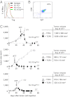
Relapsed tumors of mice treated with IFNγ−/− TE cells retained Tag as rejection antigen. Three different 16.113 tumors, relapsing after ATT with IFNγ−/− CD8+ TE, were reisolated (designated 16.113-pIFNγ-1 to 16.113-pIFNγ-3). (A) Intracellular Tag expression of 16.113 cells of tumors relapsing in mice treated with IFNγ−/− CD8+ TE cells (16.113-pIFNγ), parental 16.113 cells, and 16.113 cells passaged in a Rag1−/− mouse (16.113-p). (B) Purity of TCR-I TE cells following CD8+ cell enrichment was 97%, measured by CD8/Vβ7 staining and flow cytometry. One representative out of two experiments is shown. (C) Three different 16.113-pIFNγ cell lines were injected (106 cells) s.c. into Rag1−/− mice and established tumors (days 16–35) were treated with 106 TCR-I TE cells. Arrows show start of ATT and data represent mean tumor volume ± SD of indicated mice.
Fig. 3.
Fig. 3.
IFNγR expression by host cells is required to prevent tumor relapse. (A) Rag1−/−/IFNγR−/− and Rag1−/− mice bearing 60-d-old large, established 16.113 tumors were treated with 106 purified WT CD8+ TE cells or left untreated and tumor growth was monitored. The arrow indicates start of ATT. The table indicates the number of mice with rejected tumors per total number of mice in the experiment. Data are representative of three independent experiments (in parentheses). Data represent mean tumor volume ± SD of indicated mice and individual mice for TE cell-treated Rag1−/−/IFNγR−/− mice. (B) pIV-specific in vivo cytotoxic activity of WT CD8+ TE cells in Rag1−/−/IFNγR−/− mice 140–170 d after ATT. Horizontal bars indicate median values. Naive C57BL/6 (B6N) and tumor-bearing Rag1−/−/IFNγR−/− mice without T-cell therapy were used as negative controls and immunized C57BL/6 (B6I) as positive controls.
Fig. 4.
Fig. 4.
Loss of MHC I expression by tumor stroma does not contribute to tumor relapse. (A) Sections from relapsed tumors after IFNγ−/− CD8+ TE cell therapy (days 80–100) and from untreated tumors were stained for Tag, H-2Kb, and Fas expression, as visualized by immunofluorescence (red) and counterstaining with DAPI (blue). Five different tumors were analyzed. (B) Luciferase-expressing 16.113-pFluc cells (H-2b) (106) were injected s.c. into SCID mice (H-2d) and 25 d later (tumor size 391 ± 125 mm3) 106 purified TCR-I CD8+ TE cells were transferred. The arrow indicates start of ATT. Data represent mean tumor volume ± SD of indicated mice. One mouse died after ATT and was excluded from the experiment. (C) Bioluminescence signals of TE cell-treated 16.113-pFluc tumors of three out of six mice, as in B, were measured over time. (D) A representative image of mice shown in C (1-s exposure time) is shown.
Fig. 5.
Fig. 5.
FasL expression by TE cells is essential to prevent relapse of tumors that retained the rejection antigen. (A) When 16.113 tumors were established in Rag1−/− mice (73 d after s.c. injection), purified WT or FasL−/− CD8+ TE cells were transferred. Arrow indicates start of ATT. Table indicates number of mice with rejected tumors per total number of mice in the experiment. Data are representative of three independent experiments (in parentheses). Experiments shown in Figs. 5A and 1C were conducted in parallel; therefore, the same controls (−TE, + WT TE) are shown. (B) Relapsed tumors from FasL−/− TE cells (80–100 d after ATT) were analyzed for Tag and β-actin expression by Western blot. (C) Reisolated 16.113-pFasL1-3 cells were reinjected into Rag1−/− mice and established tumors were treated with TCR-I CD8+ TE cells. Arrows indicate start of ATT. Data represent mean tumor volume ± SD of indicated mice.
Fig. 6.
Fig. 6.
Fas expression on stroma cells is essential to prevent tumor relapse. (A) 16.113 and (B) 9.27 cells were injected into Rag1−/−/Fas−/− and Rag1−/− mice. Established tumors were treated with 106 purified WT CD8+ TE cells. The table indicates the number of mice with rejected tumors per total number of mice in the experiment. Data are representative of three independent experiments (in parentheses). Data represent mean ± SD of indicated mice and individual mice for TE cell-treated Rag1−/−/Fas−/− mice.

References

    1. Dudley ME, et al. Adoptive cell transfer therapy following non-myeloablative but lymphodepleting chemotherapy for the treatment of patients with refractory metastatic melanoma. J Clin Oncol. 2005;23(10):2346–2357. - PMC - PubMed
    1. Zippelius A, et al. Effector function of human tumor-specific CD8 T cells in melanoma lesions: A state of local functional tolerance. Cancer Res. 2004;64(8):2865–2873. - PubMed
    1. Baitsch L, et al. Exhaustion of tumor-specific CD8⁺ T cells in metastases from melanoma patients. J Clin Invest. 2011;121(6):2350–2360. - PMC - PubMed
    1. Wen FT, Thisted RA, Rowley DA, Schreiber H. A systemic analysis of experimental immunotherapies on tumors differing in size and duration of growth. Oncoimmunology. 2012;1:172–178. - PMC - PubMed
    1. Schreiber K, Rowley DA, Riethmüller G, Schreiber H. Cancer immunotherapy and preclinical studies: Why we are not wasting our time with animal experiments. Hematol Oncol Clin North Am. 2006;20(3):567–584. - PubMed

Publication types

MeSH terms

LinkOut - more resources