. 2013 Feb 5;110(6):E517-25.
doi: 10.1073/pnas.1218933110. Epub 2013 Jan 22.

Human retinal gene therapy for Leber congenital amaurosis shows advancing retinal degeneration despite enduring visual improvement

Affiliations

Human retinal gene therapy for Leber congenital amaurosis shows advancing retinal degeneration despite enduring visual improvement

Artur V Cideciyan et al. Proc Natl Acad Sci U S A. .

Abstract

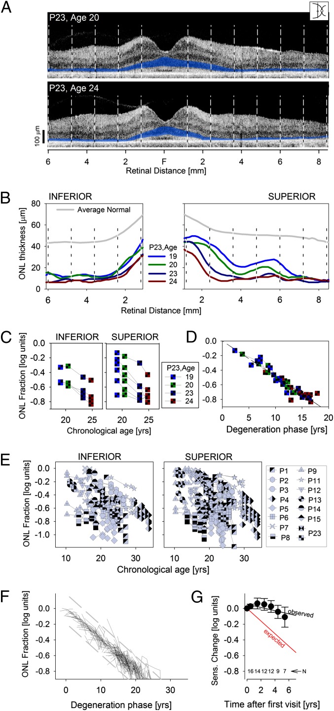
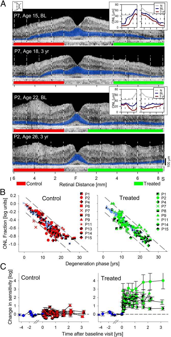
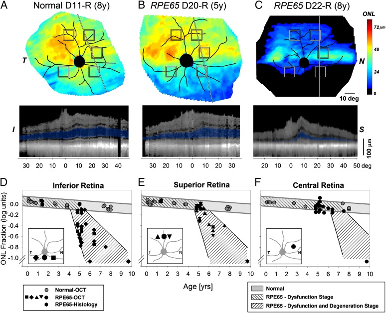
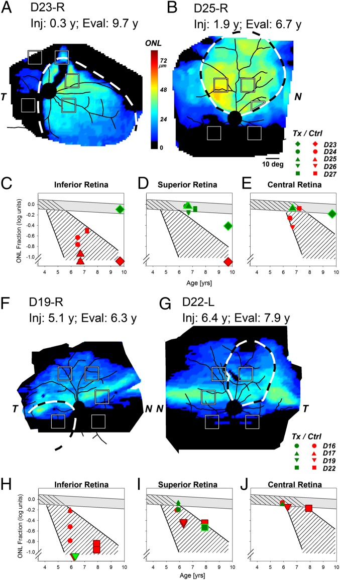
Leber congenital amaurosis (LCA) associated with retinal pigment epithelium-specific protein 65 kDa (RPE65) mutations is a severe hereditary blindness resulting from both dysfunction and degeneration of photoreceptors. Clinical trials with gene augmentation therapy have shown partial reversal of the dysfunction, but the effects on the degeneration are not known. We evaluated the consequences of gene therapy on retinal degeneration in patients with RPE65-LCA and its canine model. In untreated RPE65-LCA patients, there was dysfunction and degeneration of photoreceptors, even at the earliest ages. Examined serially over years, the outer photoreceptor nuclear layer showed progressive thinning. Treated RPE65-LCA showed substantial visual improvement in the short term and no detectable decline from this new level over the long term. However, retinal degeneration continued to progress unabated. In RPE65-mutant dogs, the first one-quarter of their lifespan showed only dysfunction, and there was normal outer photoreceptor nuclear layer thickness retina-wide. Dogs treated during the earlier dysfunction-only stage showed improved visual function and dramatic protection of treated photoreceptors from degeneration when measured 5-11 y later. Dogs treated later during the combined dysfunction and degeneration stage also showed visual function improvement, but photoreceptor loss continued unabated, the same as in human RPE65-LCA. The results suggest that, in RPE65 disease treatment, protection from visual function deterioration cannot be assumed to imply protection from degeneration. The effects of gene augmentation therapy are complex and suggest a need for a combinatorial strategy in RPE65-LCA to not only improve function in the short term but also slow retinal degeneration in the long term.


Conflict of interest statement

Conflict of interest statement: W.W.H. and the University of Florida have a financial interest in the use of adeno-associated virus therapies and own equity in a company (AGTC Inc.) that might, in the future, commercialize some aspects of this work. The remaining authors declare no conflict of interest. University of Pennsylvania, University of Florida, and Cornell University hold a patent on the described gene therapy technology (United States Patent 20070077228, “Method for Treating or Retarding the Development of Blindness”).

Figures

Fig. 1.
Fig. 1.
Natural history of disease in untreated human RPE65-LCA. (A) Cross-sectional OCT retinal scans in P23 performed along the vertical meridian (Inset) at ages 20 and 24 y. Vertical dashed lines delimit the 10 extrafoveal regions quantified. ONL is highlighted in blue for visibility. F, fovea. (B) ONL thickness of P23 as a function of eccentricity at ages 19, 20, 23, and 24 y shows progressive thinning at most retinal locations. (C) Logarithm of the ONL fraction against age supports an underlying exponential decay function describing progression. (D) Data from C replotted after horizontal shifts accounting for differences in the onset of degeneration at each retinal region. (E) ONL thickness in untreated RPE65-LCA eyes shows an extremely wide spectrum of results with a weak tendency of thinning as a function of chronological age. (F) Data from E replotted after horizontal shifts accounting for differences in age of onset. Results show a predictable natural history underlying the progressive degeneration in most patients. Dashed lines delimit the range of deviation (±2 SD) of the data from the model. (G) Average visual sensitivities measured at the same retinal locations in the same patients are stable over time and do not conform to the expected slope (red) estimated from the ONL degeneration. Error bars are SE. Numbers of eyes contributing to each data point are shown.
Fig. 2.
Fig. 2.
Photoreceptor degeneration in human RPE65-LCA eyes that have had gene therapy. (A) Cross-sectional OCT scans in two patients, P7 and P2, at baseline (BL) and 3 y after treatment. Green lines demarcate the region superior to the fovea that received a subretinal injection and that visual function measurements showed significant improvements in sensitivity. Red lines demarcate the untreated control retinal regions. ONL is highlighted in blue for visibility. (Insets) Thickness of the ONL at BL and posttreatment time points compared with mean normal (N) shows degeneration occurring at most loci. (B) ONL measures in control (Left) and treated (Right) retinal regions plotted on the natural history of RPE65-LCA disease defined in Fig.1. The progression in control and treated regions is, for the most part, indistinguishable from the natural history. (C) Visual sensitivities measured at control (Left) and treated (Right) retinal locations in the same patients. Control locations show stable sensitivities before and after surgery. Treated locations show a dramatic improvement after the surgery that is retained over time. Pretreatment measures are shown with purple symbols in B and C.
Fig. 3.
Fig. 3.
Natural history of retinal degeneration in Rpe65-mutant dogs shows regional effects. (A–C) Photoreceptor (ONL) thickness topography in a normal dog compared with two Rpe65-mutant dogs at different ages. ONL thickness topography is mapped to a pseudocolor scale. I, inferior; N, nasal; S, superior retina; T, temporal. Reconstituted OCT scans along a superior–inferior meridian (line) are shown in Lower with the ONL layer highlighted in blue. Retinal distances are specified relative to the point closest to the optic nerve. Note that the OCTs shown are reconstituted from ultra-wide angle maps covering more than 80° × 80° (∼24 × 24 mm) sampled at 1°. (D–F) ONL thickness quantified as a function of age at five retinal locations (shown as squares in A–C): two inferior loci (D), two superior loci (E), and a nasal central visual streak locus (F). Gray symbols represent normal animals; black symbols represent RPE65-mutant dogs. Gray lines approximate the expected range of normal results. Hashed regions within gray lines show the dysfunction-only stage, and hashed regions within black lines show the dysfunction and degeneration stage of RPE65 disease. Insets are 60° × 60° and show the five retinal locations sampled with OCT and two locations sampled with histology. Overlapping symbols have been moved horizontally to aid in their visibility.
Fig. 4.
Fig. 4.
Gene therapy applied before and after the onset of retinal degeneration in Rpe65-mutant dogs. (A and B) Photoreceptor (ONL) thickness topography in two dogs (D23 and D25) treated before the onset of the degeneration and evaluated ∼5–9 y later. There is much better retention of ONL thickness within the treatment region (dashed lines) compared with outside the treatment region. (C–E) ONL thickness quantified as a function of age at five retinal locations in five dogs treated before the initiation of degeneration. Red symbols correspond to retinal locations outside the treatment region, and green symbols correspond to locations within the treatment region. (F and G) ONL thickness topography in two dogs (D19 and D22) treated after the onset of degeneration. There is no evidence for thicker ONL within the treatment regions compared with outside the treatment regions. (H–J) ONL thickness quantified as a function of age at five retinal locations in treated eyes. Both untreated control (red symbols) and treated (green symbols) regions are not substantially different compared with the natural history of disease. Larger symbols in C–E correspond to data shown in A and B, and larger symbols in H–J correspond to data shown in F and G.
Fig. 5.
Fig. 5.
Histologic evaluation of the RPE65-mutant dog D23 treated at 0.3 y before the onset of retinal degeneration shows remarkable rescue of photoreceptors from degeneration when assessed over a decade later. (A) Schematic representation of the en face image showing the treatment area (dashed lines), on which is superimposed the ONL rows (mean of three values in area sampled) and disease staging (a, advanced atrophy with gliosis and loss of retinal layer organization; m, moderate photoreceptor loss with 1/3 to 1/2 of ONL remaining; n, normal) assessed at 11.2 y. (B–D) Representative photomicrographs taken from areas identified in A. In the treatment area, there is normal retinal preservation (B), although the RPE shows vacuolated inclusions typical of the disease (arrowheads). At the edge of the treatment border (C), the photoreceptor layer becomes markedly attenuated and is absent (D) outside of the treatment region. (B1, C1, and D1) Double immunolabeling with RPE65 (red) and rod opsin (green) taken from regions corresponding to B–D. Labeling in RPE is present inside the treatment region (B), with some cells intensely labeled and others (vertical arrows) showing weaker immunolabeling. In the transition zone (C1), RPE65 labeling is absent, and opsin labeling reflects rod OS shortening and disorientation; outside the treatment area (D1), the photoreceptor layer is absent, and the inner nuclear layer (INL) is disorganized. (E and F) Control sections from WT dog (E) and RPE65-mutant dog in early stages of degeneration (F) show, respectively, the presence (red) or absence of specific RPE65 immunolabeling in the RPE. GCL, ganglion cell layer; NFL, nerve fiber layer; OPL, outer plexiform layer; OS/IS, outer and inner segment layer; INL, inner nuclear layer; IPL, inner plexiform layer. (Calibration: 20 µm.)

Comment in

Similar articles

Cited by

References

    1. Bramall AN, Wright AF, Jacobson SG, McInnes RR. The genomic, biochemical, and cellular responses of the retina in inherited photoreceptor degenerations and prospects for the treatment of these disorders. Annu Rev Neurosci. 2010;33:441–472. - PubMed
    1. Palczewski K. Chemistry and biology of vision. J Biol Chem. 2012;287(3):1612–1619. - PMC - PubMed
    1. Cideciyan AV. Leber congenital amaurosis due to RPE65 mutations and its treatment with gene therapy. Prog Retin Eye Res. 2010;29(5):398–427. - PMC - PubMed
    1. Redmond TM, et al. Rpe65 is necessary for production of 11-cis-vitamin A in the retinal visual cycle. Nat Genet. 1998;20(4):344–351. - PubMed
    1. Aguirre GD, et al. Congenital stationary night blindness in the dog: Common mutation in the RPE65 gene indicates founder effect. Mol Vis. 1998;4:23. - PubMed

Publication types

MeSH terms

Substances