. 2013 Jan;9(1):e1003132.
doi: 10.1371/journal.ppat.1003132. Epub 2013 Jan 17.

Atomic model of rabbit hemorrhagic disease virus by cryo-electron microscopy and crystallography

Affiliations

Atomic model of rabbit hemorrhagic disease virus by cryo-electron microscopy and crystallography

Xue Wang et al. PLoS Pathog. 2013 Jan.

Abstract

Rabbit hemorrhagic disease, first described in China in 1984, causes hemorrhagic necrosis of the liver. Its etiological agent, rabbit hemorrhagic disease virus (RHDV), belongs to the Lagovirus genus in the family Caliciviridae. The detailed molecular structure of any lagovirus capsid has yet to be determined. Here, we report a cryo-electron microscopic (cryoEM) reconstruction of wild-type RHDV at 6.5 Å resolution and the crystal structures of the shell (S) and protruding (P) domains of its major capsid protein, VP60, each at 2.0 Å resolution. From these data we built a complete atomic model of the RHDV capsid. VP60 has a conserved S domain and a specific P2 sub-domain that differs from those found in other caliciviruses. As seen in the shell portion of the RHDV cryoEM map, which was resolved to ~5.5 Å, the N-terminal arm domain of VP60 folds back onto its cognate S domain. Sequence alignments of VP60 from six groups of RHDV isolates revealed seven regions of high variation that could be mapped onto the surface of the P2 sub-domain and suggested three putative pockets might be responsible for binding to histo-blood group antigens. A flexible loop in one of these regions was shown to interact with rabbit tissue cells and contains an important epitope for anti-RHDV antibody production. Our study provides a reliable, pseudo-atomic model of a Lagovirus and suggests a new candidate for an efficient vaccine that can be used to protect rabbits from RHDV infection.


Conflict of interest statement

The authors have declared that no competing interests exist.

Figures

Figure 1
Figure 1. Electron microscopy and 3D image reconstruction of RHDV.
(A) Micrograph of purified, negatively stained RHDV (bar = 100 nm). (B) CryoEM micrograph of purified RHDV (bar = 40 nm). The black arrows and stars point to RHDV particles with differing amounts of internal density (also see Figure S1). (C) Reconstructed cryoEM map of the RHDV virion, color-coded by radius. Icosahedral 2-, 3- and 5-fold axes are indicated by black symbols and AB and CC capsomers are identified. On the right, the closest half of the density map has been removed to reveal internal features in the RHDV density map. The contour threshold of the cryoEM map here was set to 3.3σ above the mean. (D) Same as (C) but with the capsomer density removed to show just the RHDV inner shell. Insert: A region of the density map is shown as a transparent grey isosurface into which is fitted the backbone structures of the S domains from three copies of the RHDV major capsid protein, VP60 (A in red, B in yellow and C in blue). Magenta arrows point to a few representative density features that represent residue side chains.
Figure 2
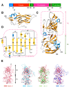
Figure 2. Crystal structures of the S and P domains of RHDV major capsid protein VP60.
(A) Domain organization of VP60. (B) Ribbon representation of the crystal structure of the VP60 S domain showing the classic viral, BIDG-CHEF β-barrel motif. Secondary structures are colored blue for helices, gold for β-strands, and grey for loops and are labeled sequentially (see also Figure S2). (C) Ribbon representation of the crystal structure of the VP60 P domain. P1 (green) and P2 (pink) sub-domains are indicated and colored according to their secondary structure elements and labeled sequentially (see also Figure S4). (D) Topology diagram of the VP60 P2 sub-domain. Labels and residue numbers correspond to those shown in panel (C). (E) Crystal structure of the VP60 P domain of RHDV (red) is superimposed with the crystal structures of the VP60 P domains in rNV (green), SMSV (cyan), and FCV (blue). The L1 loop of the RHDV VP60 P domain is labeled.
Figure 3
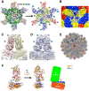
Figure 3. Atomic model of RHDV capsid.
(A) Modeling the NTA domain. The crystal structure of the VP60 S domain was fitted to and refined against the cryoEM map of RHDV. The S domains from quasi-equivalent VP60 monomers A, B and C, are colored red, yellow, and blue, respectively. Dashed arrows point to the N-terminal, T67 residues resolved in the crystal structures. A/B and C/C dimers are highlighted by dashed orange and pink polygons, respectively. Difference densities between the fitted models and cryoEM map, shown in dark-green around a 3-fold axis, were used to trace and build the NTA segments of VP60. The final pseudo-atomic model of the RHDV inner shell, including the NTA segments (green) around the 3-fold axis, is shown in ribbon form on the right. (B) CryoEM density reveals that the NTA segments of the B and C monomers extend to the proximal 3-fold axis and interact with each other. Densities corresponding to the A, B, and C monomers are colored red, yellow, and blue, respectively and those for the NTA domains are colored grey and are surrounded by a dashed cyan hexagon. (C, D, E) The match between the final model and the cryoEM map for the A/B capsomer in (C), for the C/C capsomer in (D) and for the whole virus in (E). The color scheme is that used in (A). In (C) and (D), the L1 loop at the top surface of capsomers A/B and C/C is labeled. In (E), the whole pseudo-atomic model of RHDV is shown in ribbon representation and fitted into the cryoEM map and viewed along a 5-fold axis. Black triangles and an ellipse mark the positions of two 3-fold axes and a single 2-fold axis, respectively. (F) Atomic models of three quasi-equivalent RHDV VP60 monomers, A (red), B (yellow), and C (blue). The three models, represented in ribbon form, are superimposed with the P domains aligned. Conformational differences among the three subunits are shown in schematic form at the right, which highlights alternative orientations that the S domains adopt relative to P.
Figure 4
Figure 4. Structural comparison of histo-blood group antigens (HBGAs) binding sites in RHDV and NV VP60s.
(A) RHDV C/C capsomer (blue and green) is superimposed with the crystal structure of the norovirus strain (VA207, GII.9) (PDB code: 3PUN) P dimer (gold and magenta). A close-up, top-down view of the HBGAs binding sites in VA207 is shown at the right and compared with the RHDV P2 sub-domains. Ley represents the HBGAs Lewis y. (B, C) Surface representations of the dimeric P domains in NV VA207 (B) and RHDV (C) are shown in equivalent top views and colored as in (A, right panel). The portions of RHDV that correspond to the Ley binding sites in NV VA207 are indicated with orange dashed circles in (C).
Figure 5
Figure 5. Sequence alignment of VP60s of representative RHDV isolates and location of variation regions on the capsomer surface.
(A) Multiple sequence alignments of VP60s among six genetic groups of RHDV isolates according to Nystrom, K. et.al. . The alignment is just shown for the P2 sub-domain region for residues from 301 to 480. The GeneBank accession numbers for those isolates shown are JF438967 (G1), Z24757 (AST/89), Z49271 (RHDV-AST89), FR823355 (G2), AF231353 (NZ), FR823354 (G3), AJ535092 (95-05FR), AJ535094 (G4), Y15424 (Frankfurt), AJ006019 (Rainham), AM085133 (G5), Y15427 (Wriezen), AY926883 (Ireland12), AY928269 (Ireland19), AJ969628 (G6), DQ069280 (whn/China/01/2005), AF453761 (China/Harbin/TP), DQ205345 (JX/CHA/97), AJ303106 (00-ReuFR), AY269825 (NJ/China/1985), DQ530363 (China-Yingling(YL)), and DQ069281 (whn/China/02/2005). The seven variation regions (V1–V7) that distinguish these isolates are highlighted in yellow. Sequence alignments were performed and plotted using Chimera . (B) Locations of variation regions on the RHDV capsomer surface. The degrees of sequence conservation based on the multiple sequence alignments in (A) are mapped onto the surface of the RHDV capsomer (shown in top view on the left). The most highly conserved regions (between 80 and 100% conservation) are shown in colors varying from white to cyan and then green, whereas less conserved regions (from 80 down to 43%) are represented in shades from white to violet. The seven variation regions in each of the two P2 sub-domains are indicated by dashed black ellipses and are labeled in gold in one monomer and blue in the other. The right panel shows a view of the variation sites from a different angle and three putative binding sites for HBGAs are highlighted with dashed red circles and labeled C1, C2 and C3.
Figure 6
Figure 6. Variation region V1 of RHDV VP60.
(A) Binding assay of three types of rabbit tissue cells (hepatocytes, splenocytes, and RK13 cells) for peptides derived from loop L1. (B) Western blot assay against RHDV for the sera containing anti-NJ85-KLH and anti-NJ85Δ-KLH antibodies. The black arrow indicates the band of the major capsid protein VP60 (see also Figure S9). (C) Virus neutralization assays for the sera raised against the NJ85 and NJ85Δ peptides. Rabbits were challenged by an RHDV sample that was mixed with the respective sera and monitored for the number of animals that survived as a function of time. (D) Efficiency of KLH-conjugated peptides (NJ85-KLH and NJ85Δ-KLH) in developing vaccines. Rabbits immunized with NJ85-KLH, NJ85Δ-KLH and KLH (control), were subjected to challenge against the HYD strain of RHDV. Each experimental group consisted of five rabbits and survival was monitored every day.

Similar articles

Cited by

References

    1. Xu ZJ, Chen WX (1989) Viral haemorrhagic disease in rabbits:a review. Vet Res Commun 13: 205–212. - PubMed
    1. Liu SJ, Xue HP, Pu BQ, Qian NH (1984) A new viral disease in rabbits. AnimHusbVet Med 16: 253–255.
    1. Nowotny N, Bascunana CR, Ballagi-Pordany A, Gavier-Widen D, Uhlen M, et al. (1997) Phylogenetic analysis of rabbit haemorrhagic disease and European brown hare syndrome viruses by comparison of sequences from the capsid protein gene. Arch Virol 142: 657–673. - PubMed
    1. Abrantes J, van der Loo W, Le Pendu J, Esteves PJ (2012) Rabbit haemorrhagic disease (RHD) and rabbit haemorrhagic disease virus (RHDV): a review. Vet Res 43: 12. - PMC - PubMed
    1. Meyers G, Wirblich C, Thiel HJ (1991) Genomic and subgenomic RNAs of rabbit hemorrhagic disease virus are both protein-linked and packaged into particles. Virology 184: 677–686. - PMC - PubMed

Publication types

Substances