. 2013;9(1):e1003193.
doi: 10.1371/journal.pgen.1003193. Epub 2013 Jan 17.

Secondary metabolism and development is mediated by LlmF control of VeA subcellular localization in Aspergillus nidulans

Affiliations

Secondary metabolism and development is mediated by LlmF control of VeA subcellular localization in Aspergillus nidulans

Jonathan M Palmer et al. PLoS Genet. 2013.

Abstract

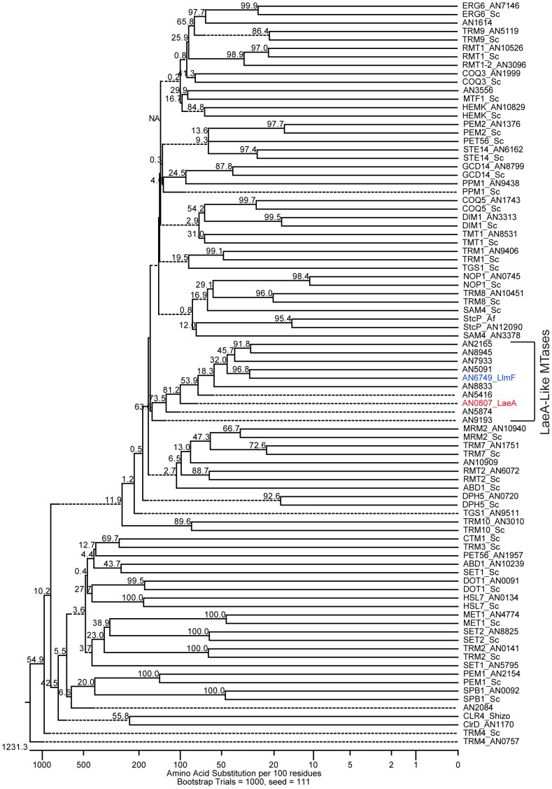
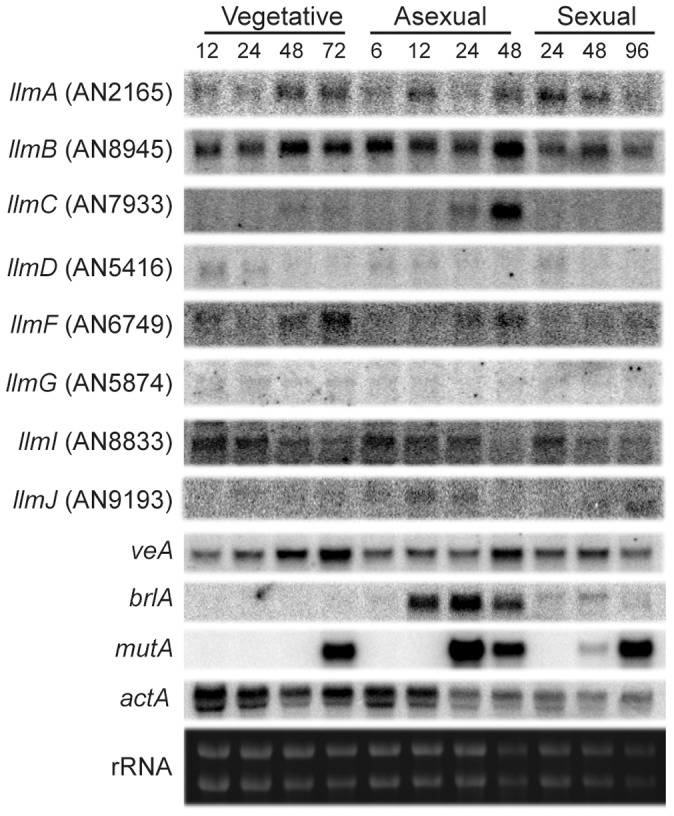
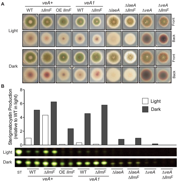
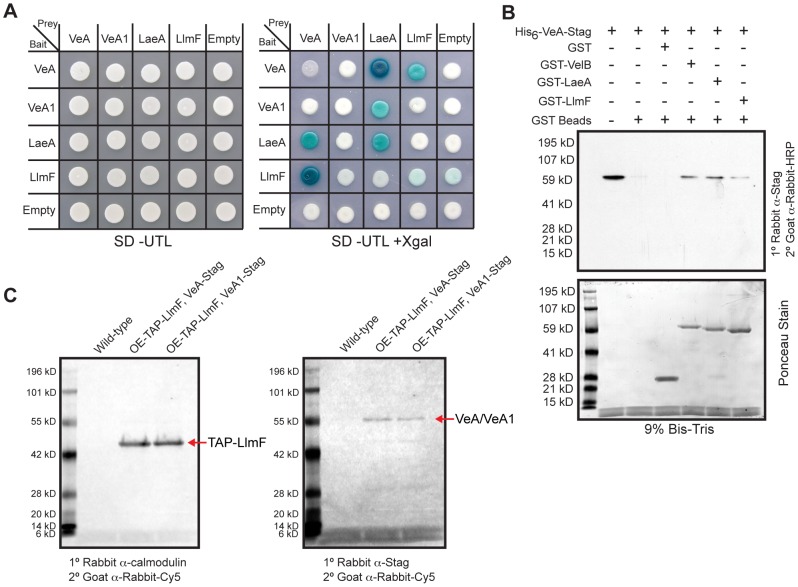
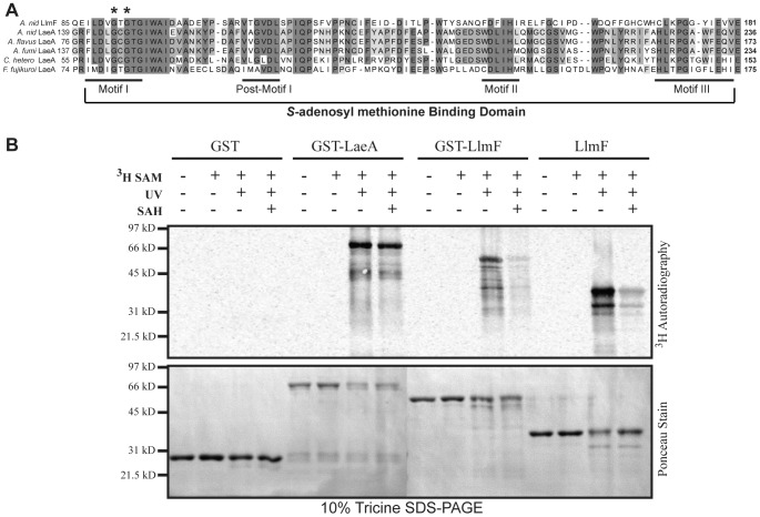
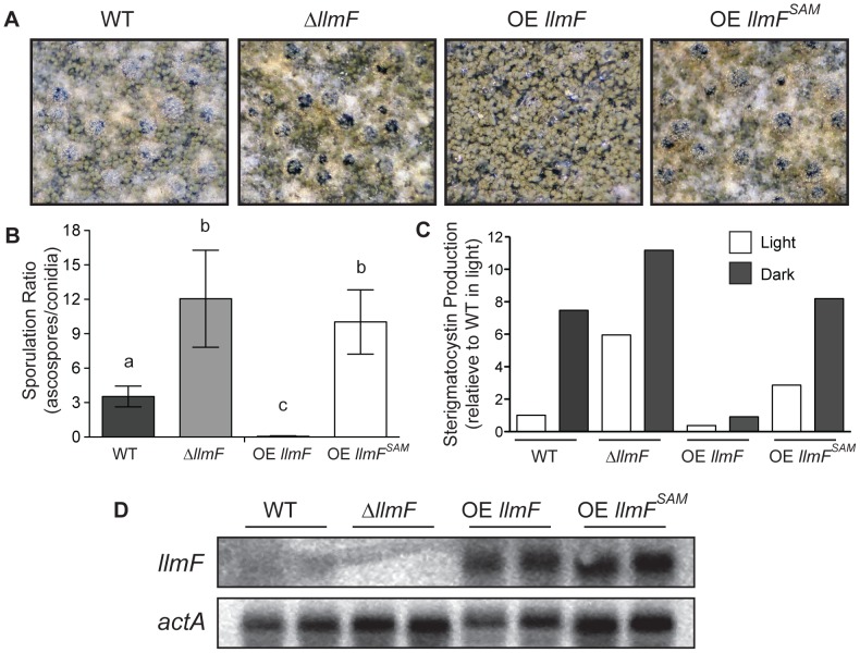
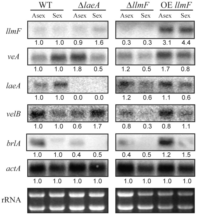
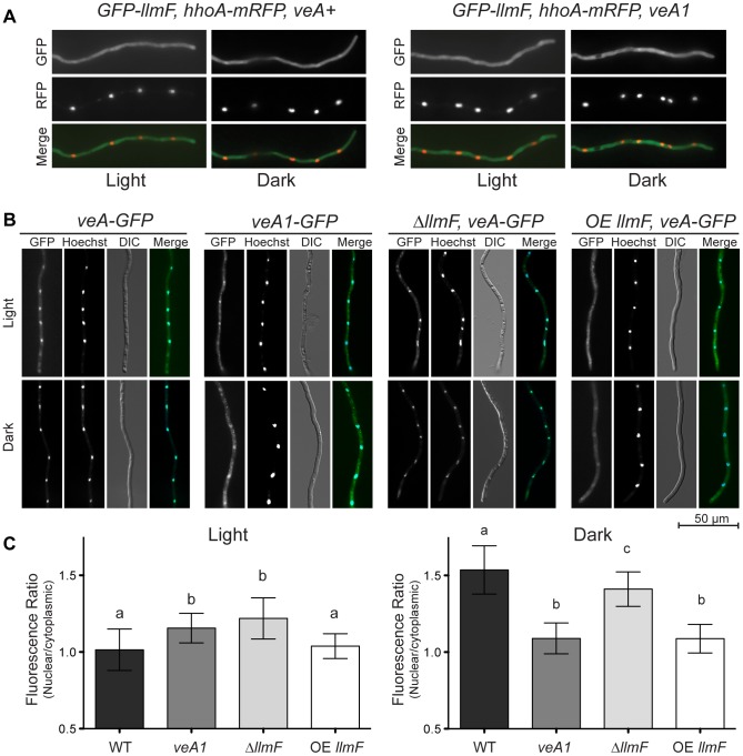
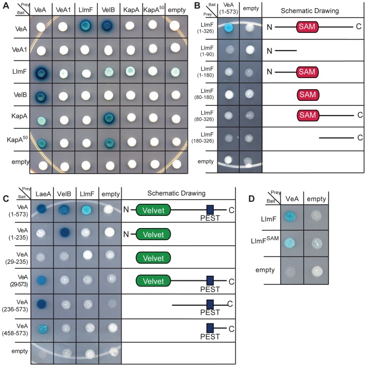
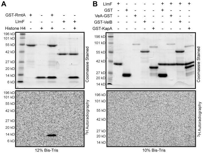
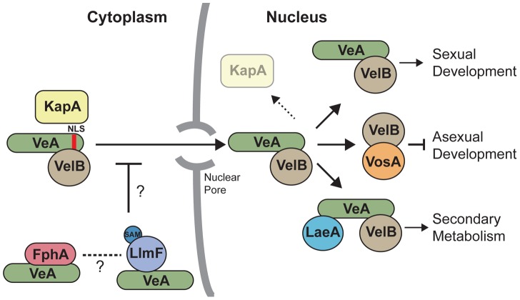
Secondary metabolism and development are linked in Aspergillus through the conserved regulatory velvet complex composed of VeA, VelB, and LaeA. The founding member of the velvet complex, VeA, shuttles between the cytoplasm and nucleus in response to alterations in light. Here we describe a new interaction partner of VeA identified through a reverse genetics screen looking for LaeA-like methyltransferases in Aspergillus nidulans. One of the putative LaeA-like methyltransferases identified, LlmF, is a negative regulator of sterigmatocystin production and sexual development. LlmF interacts directly with VeA and the repressive function of LlmF is mediated by influencing the localization of VeA, as over-expression of llmF decreases the nuclear to cytoplasmic ratio of VeA while deletion of llmF results in an increased nuclear accumulation of VeA. We show that the methyltransferase domain of LlmF is required for function; however, LlmF does not directly methylate VeA in vitro. This study identifies a new interaction partner for VeA and highlights the importance of cellular compartmentalization of VeA for regulation of development and secondary metabolism.


Conflict of interest statement

The authors have declared that no competing interests exist.

Figures

Figure 1
Figure 1. Reverse genetics identified LaeA-like methyltransferases in A. nidulans.
Putative methyltransferases were identified from the genome annotation of A. nidulans at the Broad Institute, based on the presence of a predictive S-adenosyl methionine (SAM) binding domain. A ClustalW multiple sequence alignment was preformed on 88 amino acid sequences that corresponded to 50 identified from the A. nidulans genome, 37 previously characterized from Saccharomyces cerevisiae and Schizosaccharomyces pombe, and one from A. flavus (StcP). Bootstrapping analysis was done using the neighbor joining method with 1000 trials and a seed of 111 (MegAlign, DNASTAR), the numbers at each node indicate bootstrap values, and dashed lines represent collapsed nodes. This analysis identified nine uncharacterized loci that are homologous to LaeA. These nine loci have been named LaeA-like methyltransferases. AN5091 (LlmE) has previously been published and therefore is not included in this manuscript .
Figure 2
Figure 2. LaeA-like methyltransferases expression over different development stages.
Expression profiling of all of the LaeA-like methyltransferases via northern blot indicates that some are expressed at very low levels (llmD, llmG, and llmJ), some are expressed constantly through development (llmA, llmB, and llmI), and llmC is expressed only during late asexual development. Controls for developmental time points were as follows: brlA was used as a control for asexual induction, mutA for sexual development, and actin (actA) as a loading control. Expression of llmF increases during both late vegetative and asexual development, however it does not increase during sexual development.
Figure 3
Figure 3. LlmF is a negative regulator of sterigmatocystin production.
(A) Colony growth phenotypes are shown from strains grown on glucose minimal media (GMM) for 3 days in either light or dark conditions. (B) Overlay inoculated cultures were grown in either the light or the dark and 3 cores were taken from each plate after 4 days of growth at 37°C. The cores were homogenized in water and extracted with an equal volume of ethyl acetate. Thin layer chromatography using toluene: ethyl acetate: acetic acid (8∶1∶1) as a solvent of organic extracts followed by spraying with 15% aluminum chloride allows for the analysis of sterigmatocystin. Quantification of sterigmatocystin was achieved using ImageJ software.
Figure 4
Figure 4. LlmF interacts with VeA in the yeast-two-hybrid, in vitro GST pull-down, and in vivo co-purification.
(A) A directed yeast-two-hybrid approach measured protein-protein interactions and indicated LlmF interacts with VeA, but not the truncated VeA1. Yeast cells harboring the indicated bait and prey plasmids were grown in liquid shaking culture to a density of approximately 2×10⁷ cells/ml and 10 µl was spotted on synthetic dropout media (SD) containing the appropriate supplements (uracil (U), tryptophan (T), leucine (L), and/or X-gal). A positive interaction results in the activation of the lacZ reporter, which turns the media blue in the presence of X-gal. (B) Recombinant GST, GST-LlmF, GST-LaeA, and GST-VelB were incubated with recombinant His6-VeA-S-tag and subsequently purified by glutathione sepharose 6B to look for co-purification of VeA with any of the GST labeled proteins. An immunoblot using anti-S-tag antibody detected the presence of the His6-VeA-S-tag protein and Ponceau stain of the membrane served as an indication of the amount of GST fusion proteins in each lane. GST tagged LlmF, VelB, and LaeA were capable of pulling down His6-VeA-S-tag, while GST alone did not. (C) Crude protein extracts were prepared from one-liter liquid shaking culture of each strain and subjected to the TAP protein purification protocol. The resulting eluate was electrophoresed on a 10% Bis-Tris SDS-PAGE gel and transferred to a nitrocellulose membrane where an anti-calmodulin antibody confirmed TAP-LlmF and an anti-S-tag antibody was used to detect VeA-S-tag and VeA1-S-tag. Strains used are: WT =  RJMP103.5, OE-TAP-llmF veA-S-tag = RJMP249.1, and OE-TAP-llmF veA1-S-tag = RJMP250.2.
Figure 5
Figure 5. Recombinant LlmF binds the methyl donor molecule S-adenosyl methionine.
(A) Multiple sequence alignment of LlmF with previously characterized LaeA proteins from A. nidulans, A. flavus, A. fumigatus, Cochliobolus heterostrophus, and Fusarium fujikuroi identifies that LlmF harbors a conserved S-adenosyl methionine (SAM) binding domain , . The SAM binding domain can be further broken down into four motifs that correspond to β-strands in the binding pocket: motif I, post-motif I, motif II, and motif III. LlmF and LaeA proteins contain these conserved motifs. Asterisks indicate the conserved glycine residues in motif I that were previously mutated in LaeA to render the protein inactive . (B) An ultraviolet (UV) crosslinking assay was used to test the SAM binding site prediction and show that both LaeA and LlmF are capable of binding SAM. 3H-SAM was incubated with recombinantly purified GST, GST-LaeA, GST-LlmF and LlmF for 30 minutes at room temperature. To crosslink 3H-SAM into the binding site of the enzyme, some samples were incubated on ice while being exposed to UV light for 30 minutes and then all samples were subsequently electrophoresed on a 10% Tricine SDS-PAGE gel, transferred to nitrocellulose membrane, and then exposed to a tritium phosphor screen. Active site crosslinking of SAM was confirmed by incubation with the active site competitive inhibitor, S-adenosyl-homocysteine (SAH) at a concentration of 1 µM.
Figure 6
Figure 6. The methyltransferase domain of LlmF is required for negative regulation of sexual development and sterigmatocystin.
(A) Cultures grown for five days under sexual developmental induction were imaged under a dissecting microscope. Wild type and ΔllmF strains produce abundant cleistothecia and few conidia, while OE llmF strains produce abundant conidia and few cleistothecia under these conditions. Additionally, the OE llmFSAM mutant displays a ΔllmF phenotype indicating the requirement of the SAM binding domain. (B) Quantification of spores produced under sexual developmental conditions (materials and methods) supports the macroscopic images, as the ΔllmF strain produces an increased ratio of ascospores to conidia (sexual to asexual) and the OE llmF strain produces a decrease in this ratio. These data demonstrate that the OE llmFSAM mutant has a sporulation ratio similar to that of the ΔllmF strain. Lowercase letters refer to statistical significance that measured with a student T-test of significance using Prism (Graphpad). (C) Analysis of sterigmatocystin was done in light and dark conditions as described in the materials and methods. Relative quantification of sterigmatocystin was achieved using ImageJ software and the bar graph indicates the average of two replicates. Notably, OE llmFSAM does not repress the production of sterigmatocystin that is displayed in the OE llmF strain. (D) Strains were confirmed by northern analysis of llmF and actA transcripts from RNA extracted from sexual developmental induction.
Figure 7
Figure 7. Expression of the velvet complex members is not increased in ΔllmF strains.
Mycelia was grown in liquid shaking culture for 20 hours in minimal media and then subsequently transferred to solid minimal media plates. Asexual development was induced by incubation in constant light for 24 hours while sexual development was induced by incubation in constant darkness for 48 hours. Total RNA was extracted from these conditions and expression was analyzed with a northern blot. Actin (actA) was included as a loading control. Numbers represent relative expression levels that have been normalized to actin from each condition and then normalized to wild type expression for each growth condition (asexual or sexual). Quantification was done using ImageJ software. Strains are as follows: WT = RJMP144.6, ΔlaeA = RJMP153.7, ΔllmF = RJMP144.9, and OE llmF = RJMP143.5.
Figure 8
Figure 8. LlmF localizes to the cytoplasm and controls the subcellular localization of VeA.
(A) The localization of LlmF was determined by constructing an N terminal GFP-LlmF fusion protein driven by the gpdA promoter. Germlings were grown on coverslips submerged in liquid minimal media overnight at 30°C. GFP fluorescence appears throughout the hyphae independent of light or the veA allele, as GFP-LlmF localization does not change in a veA+ versus veA1 background. Nuclei are shown with histone H1 labeled with mRFP (hhoA-mRFP). (B) VeA localization is determined by illumination. In the light VeA is found in the cytoplasm and the nucleus, while in the dark VeA concentrates in the nucleus. The truncated VeA1 protein is blind to light, as its localization does not change depending on illumination. In an OE llmF background, VeA does not accumulate in the nucleus when grown in the dark, while ΔllmF strains show similar localization patterns as wild type. (C) Quantification of VeA-GFP and VeA1-GFP fluorescence was done by measuring 50 nuclei and 50 cytoplasmic regions using Zeiss AxioVision Software 4.7 and statistical significance was calculated separately for light and dark conditions using an ANOVA and represented on the graph using lower case letters (a, b, and c). Error bars indicate standard deviation.
Figure 9
Figure 9. LlmF does not interact with VelB or KapA and only full-length LlmF interacts with VeA.
(A) A directed yeast-two-hybrid analysis indicates that LlmF is not able to interact with VelB, full length KapA, or KapA50, which is missing the first 79 amino acids containing the importin β interaction domain. A positive interaction is indicated by a blue change in color via the lacZ reporter when grown on media containing X-gal. (B) Five truncation mutants of LlmF were constructed and tested against full length VeA in the yeast-two-hybrid assay. (C) Similarly, five different VeA truncations were created based on the locations of the predicted domain structure; the velvet domain is located from amino acid position 29–235 and the PEST domain is located from 458–573. The VeA truncations were tested in the yeast-two-hybrid assay against full length LaeA, VelB, and LlmF. (D) VeA is capable of interacting with LlmFSAM, which contains a mutation in motif I of the SAM binding site of LlmF.
Figure 10
Figure 10. LlmF does not methylate the velvet complex in vitro.
(A) An in vitro methylation assay was conducted by incubation of the recombinant proteins with 3H-SAM for 1 hour at 30°C. The reactions were stopped by addition of SDS sample buffer and electrophoresed on Bis-Tris SDS-PAGE gels. The gels were treated with fluorography enhancing solution (En3Hance, Perkin Elmer), dried, and exposed to a tritium phosphor screen for 2 weeks. GST-RmtA and human recombinant histone H4 (NEB) served as a positive control. Methylation of histone H4 by GST-RmtA was visualized via 3H autoradiography. (B) Using the same in vitro methylation assay, members of the velvet complex (VeA, VelB, and KapA) were tested as methylation substrates for LlmF. Under these conditions, LlmF was unable to methylate any of the proteins tested.
Figure 11
Figure 11. A model illustrates that LlmF controls VeA subcellular localization through methylation of an unknown substrate.
Cytoplasmic VeA-VelB dimer is recognized by the importin α (KapA) and subsequently imported through the nuclear pore complex. After nuclear import, KapA dissociates where the VeA-VelB dimer functions to activate sexual development, VelB forms a dimer with VosA to repress asexual development, and the LaeA-VeA-VelB heterotrimeric complex forms that activates secondary metabolism. The data presented here depicts a role for a LlmF-VeA transient complex repressing the nuclear import of VeA primarily through the putative methylation activity of LlmF. The red light sensing phytochrome, FphA, also controls the subcellular localization of VeA , however it is unknown if FphA and LlmF share a pathway or independently regulate VeA subcellular localization.

Similar articles

Cited by

References

    1. Brown DW, Yu JH, Kelkar HS, Fernandes M, Nesbitt TC, et al. (1996) Twenty-five coregulated transcripts define a sterigmatocystin gene cluster in Aspergillus nidulans . Proc Natl Acad Sci U S A 93: 1418–1422. - PMC - PubMed
    1. Bok JW, Keller NP (2004) LaeA, a regulator of secondary metabolism in Aspergillus spp. Eukaryot Cell 3: 527–535. - PMC - PubMed
    1. Bayram O, Krappmann S, Ni M, Bok JW, Helmstaedt K, et al. (2008) VelB/VeA/LaeA complex coordinates light signal with fungal development and secondary metabolism. Science 320: 1504–1506. - PubMed
    1. Sarikaya Bayram O, Bayram O, Valerius O, Park HS, Irniger S, et al. (2010) LaeA control of velvet family regulatory proteins for light-dependent development and fungal cell-type specificity. PLoS Genet 6: e1001226 doi:10.1371/journal.pgen.1001226. - DOI - PMC - PubMed
    1. Bayram O, Braus GH (2012) Coordination of secondary metabolism and development in fungi: the velvet family of regulatory proteins. FEMS Microbiol Rev 36: 1–24. - PubMed

Publication types

MeSH terms

LinkOut - more resources