. 2012:6:216-31.
doi: 10.2174/1874357901206010216. Epub 2012 Dec 28.

A Decade of Global mRNA and miRNA Profiling of HPV-Positive Cell Lines and Clinical Specimens

Affiliations

A Decade of Global mRNA and miRNA Profiling of HPV-Positive Cell Lines and Clinical Specimens

Bogumil Kaczkowski et al. Open Virol J. 2012.

Abstract

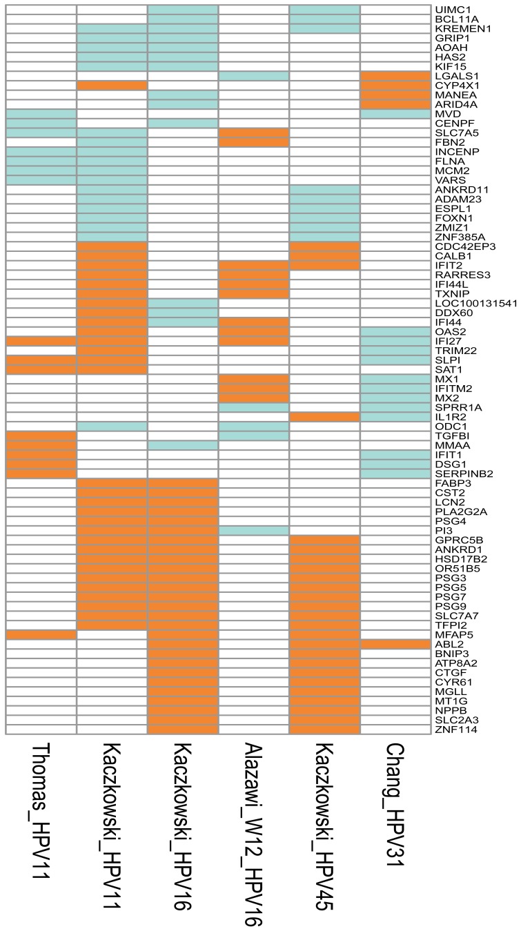
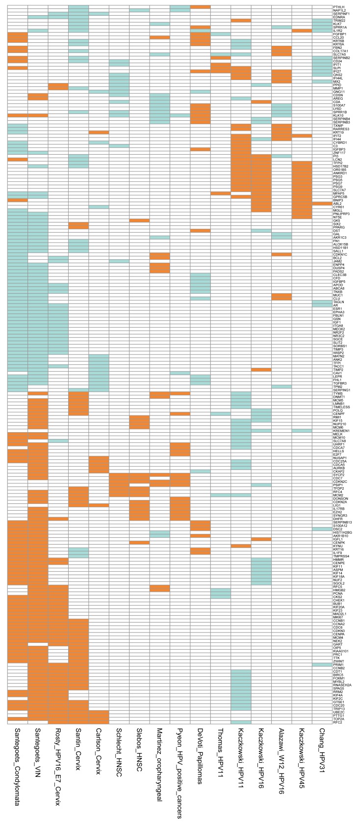
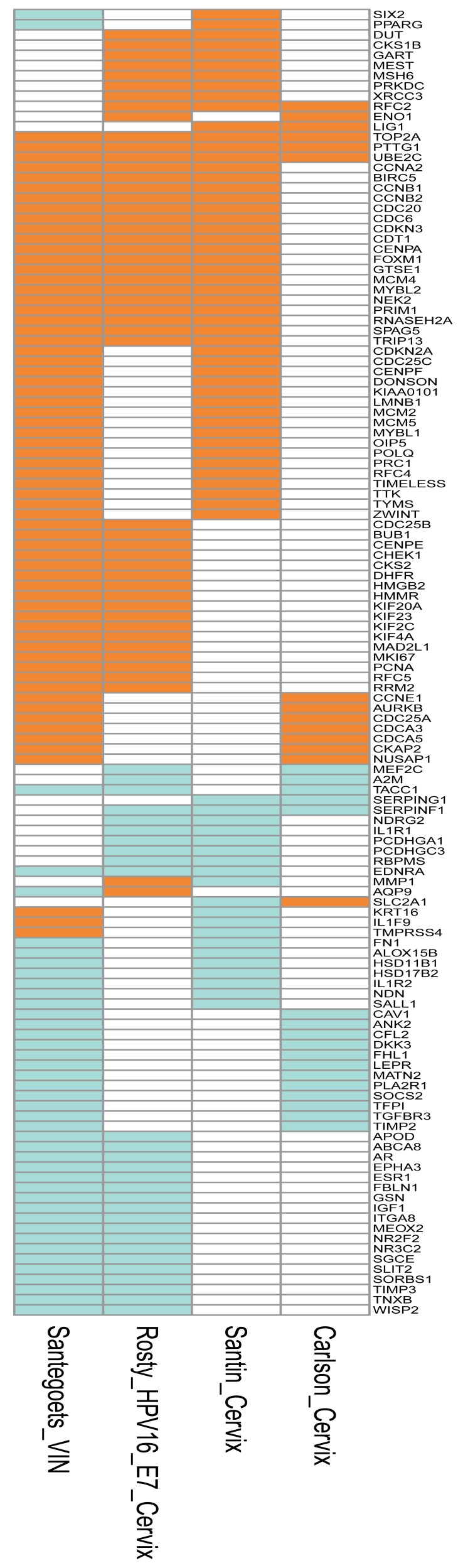
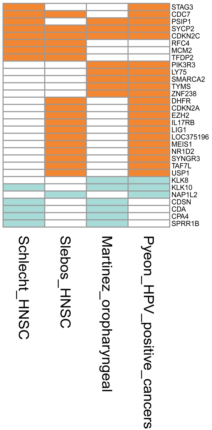
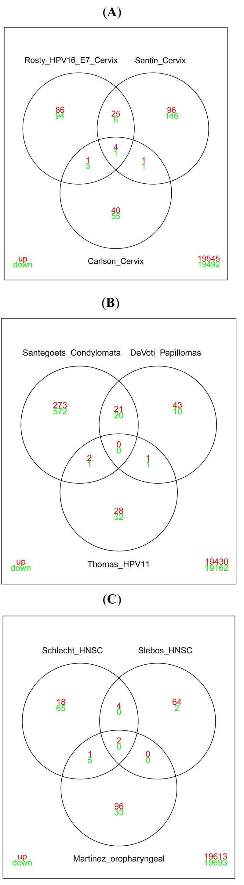
For more than a decade, global gene expression profiling has been extensively used to elucidate the biology of human papillomaviruses (HPV) and their role in cervical- and head-and-neck cancers. Since 2008, the expression profiling of miRNAs has been reported in multiple HPV studies. Two major strategies have been employed in the gene and miRNA profiling studies: In the first approach, HPV positive tumors were compared to normal tissues or to HPV negative tumors. The second strategy relied on analysis of cell cultures transfected with single HPV oncogenes or with HPV genomes compared to untransfected cells considered as models for the development of premalignant and malignant transformations.In this review, we summarize what we have learned from a decade of global expression profiling studies. We performed comprehensive analysis of the overlap of the lists of differentially expressed genes and microRNAs, in both tissue samples and cell culture based studies. The review focuses mainly on HPV16, however reports from other HPV species are used as references. We discuss the low degree of consensus among different studies and the limitation of differential expression analysis as well as the fragmented miRNA-mRNA target correlation evidence. Furthermore, we propose an approach for future research to include more comprehensive miRNA-mRNA target correlation analysis and to apply systems biology/gene networks methodology.

Keywords: Cervical cancer; HPV; head-and-neck cancer; messenger RNA profiling; miRNA..


Figures

Fig. (1)
Fig. (1)
Graphical representation of the genes that were differentially expressed in at least two cell culture studies. In five of these studies, in vitro cell cultures were transfected with genomes of different HPV types: Thomas_HPV11 [37], normal human keratinocytes transfected with HPV11; Kaczkowski_HPV11, Kaczkowski_HPV16 and Kaczkowski_HPV45 [29], HaCaT cells transfected with HPV11, HPV16 and HPV45, respectively; Chang_HPV31 [53], normal human keratinocytes from foreskin tissue transfected with wild type HPV31 genome. W12 was obtained from a low-grade cervical lesion and was naturally infected with HPV16 [34].The upregulated genes are depicted in orange, downregulated in green, and not differentially expressed in white. Interestingly, oncogene ABL2 was found upregulated in high-risk types HPV16, HPV31 and HPV45. The annotations of the genes are included as Supplementary Table 1.
Fig. (2)
Fig. (2)
Graphical representation of the differentially expressed genes from 14 comparisons reported in 12 studies. Genes differentially expressed in at least three comparisons are shown. The comparisons represent genes differentially expressed in benign and malignant growth caused by HPV infection. The signatures represent the differentially expressed genes as follows: Santegoets_Condylomata and Santegoets_VIN [51], condylomata and vulval intraepithelial neoplasia (VIN) versus normal tissue; Rosty_HPV16_E7_Cervix [46], genes correlated to HPV16 E7 expression in cervical cancer; Santin_Cervix [45] and Carlson_Cervix [38], cervical cancer versus normal cervical keratinocytes; Schlecht_HNSC [49] and Slebos_HNSC [47], HPV-positive versus HPV-negative head-and-neck squamous cell carcinoma; Martinez_oropharyngeal [48], HPV-positive versus HPV-negative oropharyngeal squamous cell carcinoma; Pyeon_HPV_positive_cancers [50], HPV-positive head-and-neck cancer (HNC) and cervical cancer versus HPV-negative HNC; DeVoti_Papillomas [52], respiratory papillomas versus adjacent tissue; Thomas_HPV11 [37], normal human keratinocytes transfected with HPV11; Kaczkowski_HPV11, Kaczkowski_HPV16 and Kaczkowski_HPV45 [29], HaCaT cells transfected with HPV11, HPV16 and HPV45, respectively; Alazawi_W12_HPV16 [34], signature from cell line generated from a low-grade cervical lesion naturally infected with HPV16; Chang_HPV31 [38, 53], normal human keratinocytes from foreskin tissue transfected with wild type HPV31 genome. The upregulated genes are depicted in orange, downregulated in green, and not differentially expressed in white. The annotations of the genes are included as Supplementary Table 1.
Fig. (3)
Fig. (3)
Depiction of the genes that were differentially expressed in at least two studies based on samples from genital lesions. The signatures represent the differentially expressed genes as follows: Santegoets_VIN [51], vulval intraepithelial neoplasia (VIN) versus normal tissue; Rosty_HPV16_E7_Cervix [46], genes correlated to HPV16 E7 expression in cervical cancer; Santin_Cervix [45] and Carlson_Cervix [38], cervical cancer versus normal cervical keratinocytes. The upregulated genes are depicted in orange, downregulated in green, and not differentially expressed in white. The annotations of the genes are included as Supplementary Table 1.
Fig. (4)
Fig. (4)
Graphical representation of the genes that were differentially expressed in at least two studies reporting the differential expression between HPV-positive versus HPV-negative head-and-neck cancers. The signatures represent the differentially expressed genes as follows: Schlecht_HNSC [49] and Slebos_HNSC [47], HPV-positive versus HPV-negative head-and-neck squamous cell carcinoma; Martinez_oropharyngeal [48], HPV-positive versus HPV-negative oropharyngeal squamous cell carcinoma; Pyeon_HPV_positive_cancers [50], HPV-positive head-and- neck cancer (HNC) and cervical cancer versus HPV-negative HNC. The upregulated genes are depicted in orange, downregulated in green, and not differentially expressed in white. The annotations of the genes are included as Supplementary Table 1.
Fig. (5)
Fig. (5)
Venn diagrams illustrating the overlap in differentially expressed genes reported in: A) studies based on cervical cancer tissues. The signatures reported by Rosty et al. [46] and Santin et al. [45, 47] show significant overlap. This is in contrast to the list of genes published by Carlson et al. [38] that shows only negligible overlap with the other studies. The genes present in all three studies are RFC2, TOP2A, PTTG1, UBE2C (upregulated) and SERPINF1 (downregulated) (also seen in Fig. 3). B) Studies of low-risk HPV. Santegoets et al. [51] and DeVoti et al. [52] reported the differentially expressed genes from condyloma and papilloma samples, respectively. Thomas et al. [37] reported the differential expression based on a cell culture model in which normal human keratinocytes were transfected with the HPV11 genome. The Venn diagram demonstrates a notable overlap between the tissue-based studies, especially concerning downregulated genes. However, little overlap can be seen between the tissue-based studies and the HPV11 cell culture model (Thomas_HPV11). C) Studies reporting the differential expression between HPV-positive versus HPV-negative head-and-neck cancers. These studies show very little overlap of the signatures. The two genes upregulated in all the studies are SYCP2 and CDKN2C (also seen in Fig. 4).

Similar articles

Cited by

References

    1. Hausen zur H. Human papillomaviruses and their possible role in squamous cell carcinomas. Curr Top Microbiol Immunol. 1977;78:1–30. - PubMed
    1. Bosch FX, Burchell AN, Schiffman M, et al. Epidemiology and natural history of human papillomavirus infections and type-specific implications in cervical neoplasia. Vaccine. 2008;26(Suppl 10 ):K1–16. - PubMed
    1. Du J, Nasman A, Carlson JW, Ramqvist T, Dalianis T. Prevalence of human papillomavirus (HPV) types in cervical cancer 2003- 2008 in Stockholm, Sweden, before public HPV vaccination. Acta Oncol. 2011;50(8 ):1215–9. - PubMed
    1. Lajer CB, von BC. The role of human papillomavirus in head and neck cancer. APMIS. 2010; 118(6-7 ):510–9. - PubMed
    1. Forslund O, Lindelöf B, Hradil E, et al. High prevalence of cutaneous human papillomavirus DNA on the top of skin tumors but not in “Stripped” biopsies from the same tumors. J Invest Dermatol. 2004;123(2 ):388–94. - PMC - PubMed

LinkOut - more resources