. 2013;8(1):e50598.
doi: 10.1371/journal.pone.0050598. Epub 2013 Jan 14.

Mutations in the fusion protein cleavage site of avian paramyxovirus serotype 4 confer increased replication and syncytium formation in vitro but not increased replication and pathogenicity in chickens and ducks

Affiliations

Mutations in the fusion protein cleavage site of avian paramyxovirus serotype 4 confer increased replication and syncytium formation in vitro but not increased replication and pathogenicity in chickens and ducks

Shin-Hee Kim et al. PLoS One. 2013.

Retraction in

Abstract

To evaluate the role of the F protein cleavage site in the replication and pathogenicity of avian paramyxoviruses (APMVs), we constructed a reverse genetics system for recovery of infectious recombinant APMV-4 from cloned cDNA. The recovered recombinant APMV-4 resembled the biological virus in growth characteristics in vitro and in pathogenicity in vivo. The F cleavage site sequence of APMV-4 (DIQPR↓F) contains a single basic amino acid, at the -1 position. Six mutant APMV-4 viruses were recovered in which the F protein cleavage site was mutated to contain increased numbers of basic amino acids or to mimic the naturally occurring cleavage sites of several paramyxoviruses, including neurovirulent and avirulent strains of NDV. The presence of a glutamine residue at the -3 position was found to be important for mutant virus recovery. In addition, cleavage sites containing the furin protease motif conferred increased replication and syncytium formation in vitro. However, analysis of viral pathogenicity in 9-day-old embryonated chicken eggs, 1-day-old and 2-week-old chickens, and 3-week-old ducks showed that none the F protein cleavage site mutations altered the replication, tropism, and pathogenicity of APMV-4, and no significant differences were observed among the parental and mutant APMV-4 viruses in vivo. Although parental and mutant viruses replicated somewhat better in ducks than in chickens, they all were highly restricted and avirulent in both species. These results suggested that the cleavage site sequence of the F protein is not a limiting determinant of APMV-4 pathogenicity in chickens and ducks.


Conflict of interest statement

Competing Interests: The authors have declared that no competing interests exist.

Figures

Figure 1
Figure 1. Construction of a cDNA clone encoding a full-length antigenomic RNA of APMV-4, and the introduction of modified F genes.
The APMV-4 cDNA was assembled between the T7 promoter (to the left) and the hepatitis delta virus antigenomic ribozyme sequence followed by a T7 RNA polymerase transcription-termination signal (to the right). Assembly of the cDNA employed SbfI, SnaBI, MluI, NotI, and PvuII sites that were introduced into untranslated regions. To generate F protein cleavage site mutant viruses, the cDNA fragment containing the F gene was swapped using the MluI and NotI sites.
Figure 2
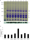
Figure 2. Analysis of proteins present in virions of parental and F protein cleavage site mutant APMV-4 viruses, and cell-surface expression of the viral F protein.
(A) Virus that was partially purified from infected chicken egg allantoic fluids by sucrose step gradients was separated by electrophoresis, and the gel was stained with Coomassie brilliant blue. Lanes: 1. Biologically-derived APMV-4, 2. rAPMV-4, 3. rAPMV-4/Fc type 3-Q, 4. rAPMV-4/Fc type 5-Q, 5. rAMPV-4/Fc BC, 6. rAMPV-4/Fc Las, 7. rAMPV-4/Fc SV, and 8. rAMPV-4/Fc PIV-1 (B) Incorporation of wild-type and mutated F proteins into the virions was further analyzed by Western blot. The separated proteins in the gel (8%) under reducing condition (in panel A) were transferred into a membrane, and the F protein was detected by using an antiserum raised against a synthetic peptide from the F protein of APMV-4. (C) Surface expression of the F protein on infected DF1 cells. DF1 cells were infected with each virus (MOI of 0.1) and, at 24 h post-infection, were stained with anti-peptide antiserum against the F protein followed by anti-Alexa Fluor 488 antibody, and were analyzed by flow cytometry.
Figure 3
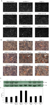
Figure 3. Production of cytopathic effect and proteolytic cleavage of the F0 proteins of parental and F protein cleavage site mutant APMV-4 viruses.
(A) DF1 cells in six-well plates were infected with the indicated viruses at a multiplicity of infection (MOI) of 0.1 PFU/cell and incubated for 72 h. (B) The viral plaques in the infected cells were visualized by immunoperoxidase staining using antiserum against the N protein of APMV-4; viral antigen is stained red. (C) Proteolytic cleavage of the F0 proteins of parental and mutant viruses in infected DF1 cells was analyzed by Western blot (I) in triplicate. The positions of the precursor protein F0 and the cleavage product F1 are indicated. The relative levels of the F0 and F1 proteins in the Western blot images were measured by Bio-Rad Gel Image analysis, and the efficiency of cleavage was determined by dividing the amount of F1 by the amount of F1 plus F0 (II).
Figure 4
Figure 4. Replication of parental and F protein cleavage site mutant APMV-4 viruses in DF1 cells.
The growth kinetics of parental and recombinant APMV-4 viruses was determined by infecting DF1 cells with each virus at an MOI of 0.01 (A) and 1 (B). The viral titers were determined by limiting dilution on DF1 cells and immunostaining with antiserum raised against the N protein of APMV-4.
Figure 5
Figure 5. Replication of parental and F protein cleavage site mutant APMV-4 viruses in primary chicken neuronal cells.
Neuronal cells were infected with APMVs at an MOI of 0.1. At 48 hpi, the cells were fixed with 4% paraformaldehyde, permeabilized with 0.2% Triton X-100, stained with a neuronal marker (anti-neuron specific beta III tubulin polyclonal antibodies) and antiserum against the N protein of APMV-4 followed by anti-Alexa Fluor 488 and 596, and then analyzed by confocal microscopy. The neuronal marker and viral N protein stained red and green, respectively.
Figure 6
Figure 6. Growth kinetics of parental and F protein cleavage site mutant APMV-4 viruses in the brains of 1-day-old chicks.
Ten 1-day-old SPF chicks were inoculated with the indicated parental or mutant viruses via the intracerebral route. Two birds in each group were sacrificed daily until 5 dpi. Brain tissue samples were harvested and virus titers were determined by limiting dilution in DF1 cells and immunostaining with antiserum against the N protein of APMV-4. Each bar represents mean and standard error of the mean of duplicate samples.
Figure 7
Figure 7. Induction of serum antibodies in response to infection of 1-day-old and 2-week-old chickens with parental and F protein cleavage site mutant APMV-4 viruses.
Chickens were inoculated with each virus (256 HA units) by the intranasal route in the same experiment as Table 2. Sera were collected at 14 dpi and evaluated for virus-specific antibodies by a hemagglutination inhibition assay using chicken erythrocytes.
Figure 8
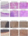
Figure 8. Histopathology and immunohistochemistry in sections of collected tissues from 3-week-old ducks infected with parental and F protein cleavage site mutant APMV-4 viruses.
As described in Table 3, ducks were inoculated with each virus (256 HA units) by the combined intranasal and intratracheal routes, and tissue samples were harvested on 4 dpi. The tissues were fixed with formalin and sections were prepared and stained with hematoxylin and eosin for histopathology (A) or with antiserum against the N protein of APMV-4 for immunohistochemistry (B). (A) Histopathological examination of tissue samples revealed similar microscopic findings in parent and mutant APMV-4 viruses. This is illustrated with representative virus, rAPMV-4. The trachea showed mild to moderate lymphocytic tracheitis with mild to moderate multifocal mucosal attenuation and reduction of tracheal mucous glands. Lung sections exhibited moderate to marked multifocal, lymphohistiocytic bronchointerstitial pneumonia with mild to moderate perivascular cuffing in the pulmonary interstitium. Spleen sections showed moderate reactive lymphoid hyperplasia characterized by expansion of the white pulp by reactive lymphocytes and increase size and cell density of lymphoid follicles. (B) The presence of antigen (stained red) was detected for parental and mutant APMV-4 in the epithelial lining of trachea, in the epithelium surrounding the medium and small bronchi of the lungs and in the spleen.

Comment in

  • Findings of Research Misconduct.
    [No authors listed] [No authors listed] Fed Regist. 2020 May 13;85(93):28643-28645. Fed Regist. 2020. PMID: 32435075 Free PMC article. No abstract available.

Similar articles

Cited by

References

    1. Lamb R, Parks G (2007) Paramyxoviridae: the viruses and their replication. In: Knipe DM, Howley PM, Griffin DE, Lamb RA, Martin MA, Roizman B, Straus SE, editors. Philadelphia: Lippincott Williams & Wilkins. pp. 1449–1496.
    1. Alexander DJ (2003) Avian paramyxoviruses 2–9. In: Saif, Y.M, (Ed.), Diseases of Poultry, 11th ed. Iowa State University Press, Ames 88–92.
    1. Miller PJ, Afonso CL, Spackman E, Scott MA, Pedersen JC, et al. (2010) Evidence for a new avian paramyxovirus serotype 10 detected in rockhopper penguins from the Falkland Islands. J Virol 84: 11496–11504. - PMC - PubMed
    1. Hu S, Ma H, Wu Y, Liu W, Wang X, et al. (2009) A vaccine candidate of attenuated genotype VII Newcastle disease virus generated by reverse genetics. Vaccine 27: 904–910. - PubMed
    1. Huang Z, Krisnamurthy S, Panda A, Samal SK (2001) High-level expression of a foreign gene from the 3′ proximal first locus of a recombinant Newcastle disease virus. J Gen Virol 82: 1729–1736. - PubMed

Publication types

MeSH terms