. 2013;8(1):e53287.
doi: 10.1371/journal.pone.0053287. Epub 2013 Jan 14.

Down-regulation of vitamin D receptor in mammospheres: implications for vitamin D resistance in breast cancer and potential for combination therapy

Affiliations

Down-regulation of vitamin D receptor in mammospheres: implications for vitamin D resistance in breast cancer and potential for combination therapy

Shehla Pervin et al. PLoS One. 2013.

Erratum in

  • PLoS One. 2013;8(10). doi:10.1371/annotation/5326d117-3f31-4e43-a5c4-9e1fb41719e9

Abstract

Vitamin D signaling in mammary cancer stem cells (MCSCs), which are implicated in the initiation and progression of breast cancer, is poorly understood. In this study, we examined vitamin D signaling in mammospheres which are enriched in MCSCs from established breast cancer cell lines. Breast cancer cells positive for aldehyde dehydrogenase (ALDH(+)) had increased ability to form mammospheres compared to ALDH(-) cells. These mammospheres expressed MCSC-specific markers and generated transplantable xenografts in nude mice. Vitamin D receptor (VDR) was significantly down-regulated in mammospheres, as well as in ALDH(+) breast cancer cells. TN aggressive human breast tumors as well as transplantable xenografts obtained from SKBR3 expressed significantly lower levels of VDR but higher levels of CD44 expression. Snail was up-regulated in mammospheres isolated from breast cancer cells. Inhibition of VDR expression by siRNA led to a significant change in key EMT-specific transcription factors and increased the ability of these cells to form mammospheres. On the other hand, over-expression of VDR led to a down-regulation of Snail but increased expression of E-cad and significantly compromised the ability of cells to form mammospheres. Mammospheres were relatively insensitive to treatment with 1,25-dihydroxyvitamin D (1,25D), the active form of vitamin D, compared to more differentiated cancer cells grown in presence of serum. Treatment of H-Ras transformed HMLE(HRas) cells with DETA NONOate, a nitric oxide (NO)-donor led to induction of MAP-kinase phosphatase -1 (MKP-1) and dephosphorylation of ERK1/2 in the mammospheres. Combined treatment of these cells with 1,25D and a low-concentration of DETA NONOate led to a significant decrease in the overall size of mammospheres and reduced tumor volume in nude mice. Our findings therefore, suggest that combination therapy using 1,25D with drugs specifically targeting key survival pathways in MCSCs warrant testing in prospective clinical trial for treatment of aggressive breast cancer.


Conflict of interest statement

Competing Interests: The authors have declared that no competing interests exist.

Figures

Figure 1
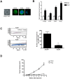
Figure 1. A: Left Panel: Bright field photomicrograph of mammospheres isolated from SKBR3 CELLS.
Right Panel: Fluorescent micrographs of SKBR3 mammospheres stained with CD44, ESA, and CD24 antigens (green) and DAPI (blue). B, Real-time quantitative PCR analysis of c-Myc, Klf4, Oct4, and Sox2 in SKBR3 cells grown under high attachment, plastic (plas) or mammosphere (mam) conditions (*, p≤0.05; **, p≤0.01). C, Isolation of ALDH+ and ALDH population from SKBR3 cells and analysis of their mammosphere forming capacity. D, Analysis of tumor volume in nude mice by SKBR3 mammospheres and plastic cells. 1×10⁵ cells obtained from dissociated mam or plas were injected into the humanized mammary fat pads of female nude mice and tumor volumes were analyzed at various time points (1–8 weeks).
Figure 2
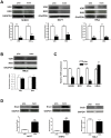
Figure 2. Selective down-regulation of VDR/RXR expression in mammospheres isolated from breast cancer cells A, Top panel: 50 µg of total cell lysates isolated from breast cancer cell lines SKBR3 (left), MCF7 (middle) and HRas (right) cells grown under plas or mam conditions and were analyzed for VDR and RXR protein expression by Western blot analysis.
Bottom panel: Quantitative densitometric analysis of VDR and RXR expression in SKBR3 (left), MCF7 (middle) and HRas (right) cell normalized to GAPDH (*, p≤0.05; **, p≤0.01). B, Top panel: 50 µg of total cell lysates isolated from mammary epithelial HMLE cells grown under plas or mam conditions were analyzed for VDR and RXR protein expression by Western blot analysis. Bottom panel: Quantitative densitometric analysis of VDR and RXR expression normalized to GAPDH (*, p≤0.05; **, p≤0.01). C, Real-time quantitative PCR analysis of VDR expression in SKBR3, MCF7 and HRas as well as in HMLE cells grown under plas or mam conditions (*, p≤0.05; **, p≤0.01). D, Selective up-regulation of Snail in MCF7 and SKBR3 cells. Top panel; 50 µg of total cell lysates isolated from MCF7 (left panel), SKBR3 (middle panel) or HMLE cells (right panel) grown under plas or mam conditions were analyzed for Snail expression by Western blot analysis. Bottom panel: Quantitative densitometric analysis of Snail expression normalized to GAPDH (**, p≤0.01).
Figure 3
Figure 3. A, PCR Array Analysis of EMT-specific genes in SKBR3 cells grown under plas and mams conditions after 4 days.
B, Validation of PCR Array data by quantitative real time PCR analysis. Experiment was performed in triplicates from three different sets of experiments (*, p≤0.05; **, p≤0.01).
Figure 4
Figure 4. A, Inhibition of VDR expression in SKBR3 cells by siRNA and analysis of VDR, Snail and E-cad protein expression.
SKBR3 cells were transfected either with scrambled siRNA (Scram) or On-target smart pool VDR siRNA (siRNA) using standard techniques and protein expression was analyzed by western blot analysis. B, Analysis of EMT-specific gene signature in Scam and VDR siRNA transfected cells (*, p≤0.05; **, p≤0.01). C, Analysis of mammospheres forming capability in Scam and VDR siRNA transfected cells. 5,000 cells after transfection were plated on 24 well ultra-low attachment plates and total numbers of mammospheres formed were analyzed (**, p≤0.01). D, Over-expression of full length human VDR gene and analysis of VDR, Snail and E-cad protein expression by western blot. E, Analysis of EMT-specific gene signature in cells transfected either with control vector or full-length human VDR (*, p≤0.05; **, p≤0.01). F, Analysis of mammospheres forming capability in cells transfected either with control vector or VDR over-expressing (VDR OE) plasmid. 5,000 cells after transfection were plated on 24 well ultra-low attachment plates and total numbers of mammospheres formed were analyzed (**, p≤0.01).
Figure 5
Figure 5. A, Analysis of VDR expression in SKBR3 cells grown under mam or plas conditions with or without growth factors (GF: EGF plus FGF).
30 µg of cell lysates were electrophoretically separated and analyzed by western blot analysis using anti-VDR antibody. Membranes were stripped and re-probed using anti-GAPDH antibody for loading controls. B (Left Panel), Analysis of VDR and CD44 protein expression in ALDH and ALDH+ populations isolated from SKBR3 cells. 30 µg of cell lysates obtained from ALDH+ and ALDH cells were electrophoretically separated and analyzed by western blot analysis using anti-VDR or anti-CD44 antibodies. Membranes were subsequently stripped and re-probed with anti-GAPDH antibody for loading controls. B (Right Panel), Normalized densitometric ratios showing relative expression levels of VDR and CD44. C (Left Panel), Analysis of VDR and CD44 protein expression in non-transplantable xenografts (NTX) and transplantable xenografts (TX). 30 µg of total cell lysates obtained from non-transplantable xenografts (NTX) and transplantable xenografts (TX) were electrophoretically separated and analyzed by western blot analysis using anti-VDR or anti-CD44 antibodies. Membranes were stripped and re-probed with anti-GAPDH antibody for loading controls (*, p≤0.05; **, p≤0.01). C (Right Panel), Normalized densitometric ratios showing relative expression levels of VDR and CD44.
Figure 6
Figure 6. A, Analysis of VDR and CD44 protein expression in tumor biopsies from triple negative (TN) and estrogen receptor positive (ER+) breast cancer patients.
50 µg lysates from 6 tumor samples each from TN and ER+ were analyzed for VDR and CD44 protein expression by Western blot analysis. B, Normalized densitometric values for VDR and CD44 expression from TN and ER+ tumor patients (*, p≤0.05; **, p≤0.01).
Figure 7
Figure 7. A, Effect of 1,25D treatment on MCF7 (left) and SKBR3 (right) cell proliferation.
Cells were treated with 1,25D (0–0.1 nM) and allowed to proliferate for 4 days under high attachment conditions and cell numbers were counted by trypan blue method (*, p≤0.05; **, p≤0.01). Medium was replaced after every 48 hrs with appropriate concentrations of 1,25D. B, SKBR3 (2×10⁴) cells were plated under mammosphere conditions on a 12-well ultra-low attachment plates in presence of different concentrations of 1,25D (0–0.1 nM) and allowed to grow under mammosphere conditions for 4 or 7 days and sphere diameters were measured. Appropriate concentrations of 1,25D were additionally supplemented in the culture medium after every 48 hours. Left Panel: Micrographs were taken at 100×magnification. Right Panel: Quantitative analysis of average diameter computed from 20 different fields from each treatment group. C, Quantitative real-time PCR analysis of Cyp27 B1 and Cyp24A1 mRNA expression from MCF-7 cells grown under plas or mam conditions after 4 days of plating (**, p≤0.01). D, HPLC analysis of 24,25D3 and 1,25D synthesis in cells grown under plastic or mammos conditions from MCF-7 cells after 4 days of plating (*, p≤0.05; **, p≤0.01).
Figure 8
Figure 8. A, Top panel: MKP-1 induction and dephosphorylation of pERK1/2 in DETA NONOate (DETA) treated HRas mammospheres.
Bottom Panel: Immunofluorescence analysis of MKP-1 and pERK1/2 in control and DETA (0.3 mM) treated mammospheres after 24 hrs. B, Top panel: Photomicrographs of HRas cells plated under mammosphere conditions and allowed to grow in medium containing either 1,25D (0.1 nM) and DETA (0.3 mM) alone or in combination for 5 days. Bottom panel; Average diameter of mammospheres computed from 20 different fields from each treatment groups (*, p≤0.05; **, p≤0.01). C, 5×10⁵ HRas cells were allowed to seed on T-25 flasks and treated with 1,25D and DETA either alone or in combination. Total number of cells were counted after 5 days (*, p≤0.05; **, p≤0.01). D, HRas cells were plated under mammospheres conditions and treated with DETA (0.3 mM), or 1,25D (0.1 nM) either alone or in combination for 3 days. Mammospheres were dissociated and 1×10⁵ cells from each treatment groups were injected into nude mice and tumor volumes were analyzed at various time points (1–8 weeks) (*, p≤0.05; **, p≤0.01 compared to the Con group).

Similar articles

Cited by

References

    1. Toner CD, Davis CD, Milner JA (2010) The vitamin D and cancer conundrum: aiming at a moving target J AM DIET ASSOC. 110: 1492–1500. - PubMed
    1. Pérez-López FR, Chedraui P, Haya J (2009) Review article: vitamin D acquisition and breast cancer risk. Reprod Sci 16: 7–19. - PubMed
    1. Trabert B, Malone KE, Daling JR, Doody DR, Bernstein L, et al. (2007) Vitamin D receptor polymorphisms and breast cancer risk in a large population-based case-control study of Caucasian and African-American women. Breast Cancer Res 9: R84. - PMC - PubMed
    1. Alimirah F, Peng X, Murillo G, Mehta RG (2011) Functional significance of vitamin D receptor FokI polymorphism in human breast cancer cells. PLoS One 6(1): e16024. - PMC - PubMed
    1. McKay JD, McCullough ML, Ziegler RG, Kraft P, Saltzman BS, et al. (2009) Vitamin D receptor polymorphisms and breast cancer risk: results from the National Cancer Institute Breast and Prostate Cancer Cohort Consortium. Cancer Epidemiol Biomarkers Prev 18: 297–305. - PubMed

Publication types

MeSH terms