

Synchrotron-generated microbeam sensorimotor cortex transections induce seizure control without disruption of neurological functions
- PMID: 23341950
- PMCID: PMC3544911
- DOI: 10.1371/journal.pone.0053549
Synchrotron-generated microbeam sensorimotor cortex transections induce seizure control without disruption of neurological functions
Abstract
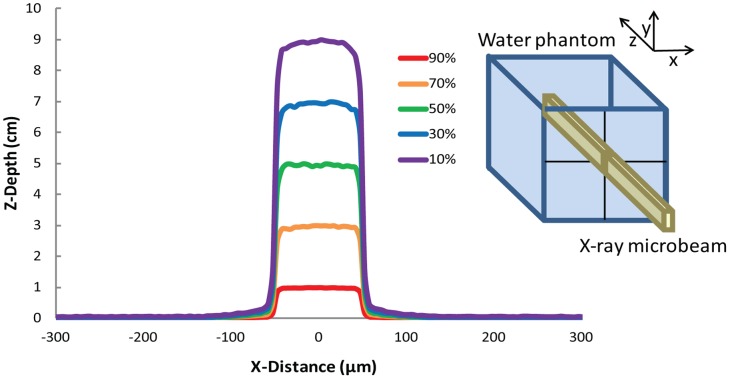
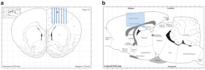
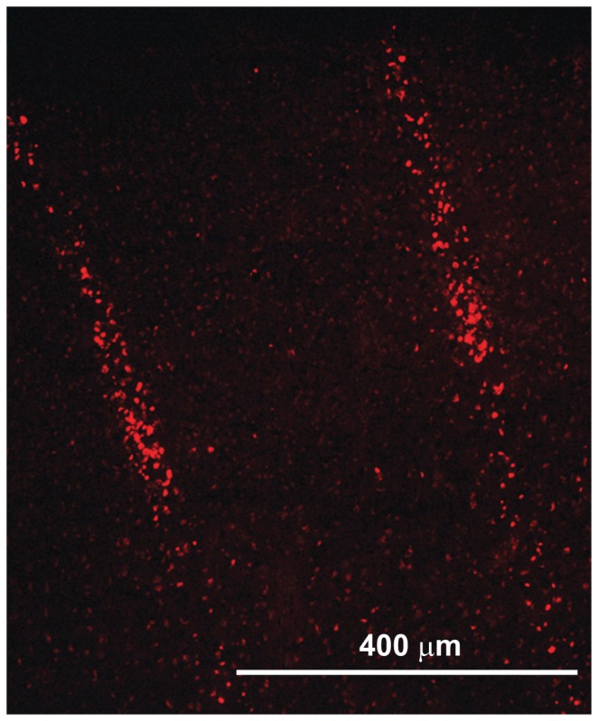
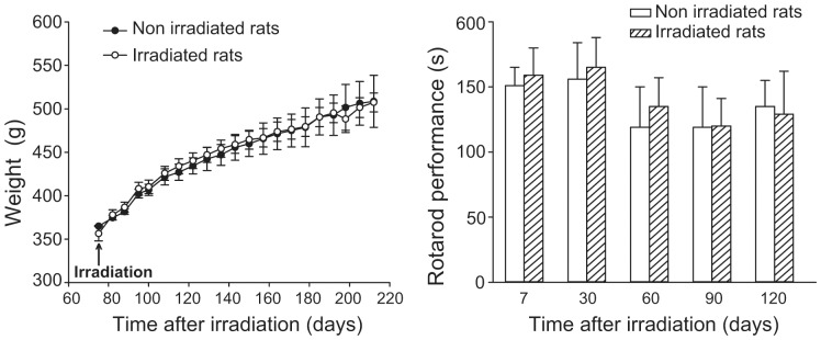
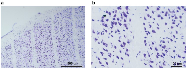
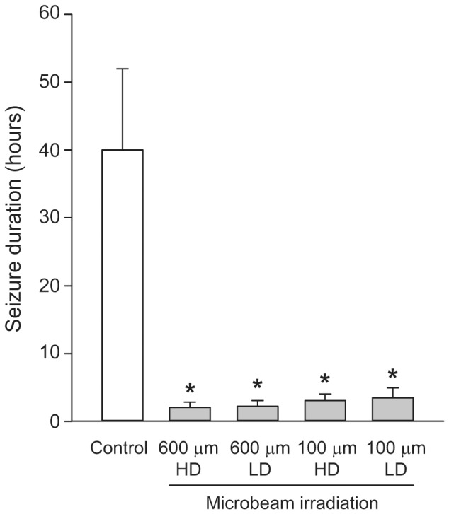
Synchrotron-generated X-ray microplanar beams (microbeams) are characterized by the ability to deliver extremely high doses of radiation to spatially restricted volumes of tissue. Minimal dose spreading outside the beam path provides an exceptional degree of protection from radio-induced damage to the neurons and glia adjacent to the microscopic slices of tissue irradiated. The preservation of cortical architecture following high-dose microbeam irradiation and the ability to induce non-invasively the equivalent of a surgical cut over the cortex is of great interest for the development of novel experimental models in neurobiology and new treatment avenues for a variety of brain disorders. Microbeams (size 100 µm/600 µm, center-to-center distance of 400 µm/1200 µm, peak entrance doses of 360-240 Gy/150-100 Gy) delivered to the sensorimotor cortex of six 2-month-old naïve rats generated histologically evident cortical transections, without modifying motor behavior and weight gain up to 7 months. Microbeam transections of the sensorimotor cortex dramatically reduced convulsive seizure duration in a further group of 12 rats receiving local infusion of kainic acid. No subsequent neurological deficit was associated with the treatment. These data provide a novel tool to study the functions of the cortex and pave the way for the development of new therapeutic strategies for epilepsy and other neurological diseases.
Conflict of interest statement
Figures
Similar articles
-
Rat sensorimotor cortex tolerance to parallel transections induced by synchrotron-generated X-ray microbeams.Sci Rep. 2017 Oct 30;7(1):14290. doi: 10.1038/s41598-017-14757-3. Sci Rep. 2017. PMID: 29085040 Free PMC article.
-
Synchrotron-generated microbeams induce hippocampal transections in rats.Sci Rep. 2018 Jan 9;8(1):184. doi: 10.1038/s41598-017-18000-x. Sci Rep. 2018. PMID: 29317649 Free PMC article.
-
Microradiosurgical cortical transections generated by synchrotron radiation.Phys Med. 2015 Sep;31(6):642-6. doi: 10.1016/j.ejmp.2015.05.007. Epub 2015 May 29. Phys Med. 2015. PMID: 26032004
-
Effects of pulsed, spatially fractionated, microscopic synchrotron X-ray beams on normal and tumoral brain tissue.Mutat Res. 2010 Apr-Jun;704(1-3):160-6. doi: 10.1016/j.mrrev.2009.12.003. Epub 2009 Dec 23. Mutat Res. 2010. PMID: 20034592 Review.
-
Synchrotron-generated microbeam radiosurgery: a novel experimental approach to modulate brain function.Neurol Res. 2011 Oct;33(8):825-31. doi: 10.1179/016164111X13123658647445. Neurol Res. 2011. PMID: 22004705 Review.
Cited by
-
Image-guided microbeam irradiation to brain tumour bearing mice using a carbon nanotube x-ray source array.Phys Med Biol. 2014 Mar 7;59(5):1283-303. doi: 10.1088/0031-9155/59/5/1283. Epub 2014 Feb 20. Phys Med Biol. 2014. PMID: 24556798 Free PMC article.
-
Synchrotron X ray induced axonal transections in the brain of rats assessed by high-field diffusion tensor imaging tractography.PLoS One. 2014 Feb 5;9(2):e88244. doi: 10.1371/journal.pone.0088244. eCollection 2014. PLoS One. 2014. PMID: 24505446 Free PMC article.
-
Rat sensorimotor cortex tolerance to parallel transections induced by synchrotron-generated X-ray microbeams.Sci Rep. 2017 Oct 30;7(1):14290. doi: 10.1038/s41598-017-14757-3. Sci Rep. 2017. PMID: 29085040 Free PMC article.
-
Synchrotron-generated microbeams induce hippocampal transections in rats.Sci Rep. 2018 Jan 9;8(1):184. doi: 10.1038/s41598-017-18000-x. Sci Rep. 2018. PMID: 29317649 Free PMC article.
-
Dose-rate plays a significant role in synchrotron radiation X-ray-induced damage of rodent testes.Int J Physiol Pathophysiol Pharmacol. 2016 Dec 25;8(4):140-145. eCollection 2016. Int J Physiol Pathophysiol Pharmacol. 2016. PMID: 28078052 Free PMC article.
References
-
- Morrell F, Whisler WW, Bleck TP (1989) Multiple subpial transection: a new approach to the surgical treatment of focal epilepsy. J Neurosurg 70: 231–239. - PubMed
-
- Morrell F, Whisler WW, Smith MC, Hoeppner TJ, de Toledo-Morrell L, et al. (1995) Landau-Kleffner syndrome. Treatment with subpial intracortical transection. Brain 118: 1529–1546. - PubMed
-
- Chervin RD, Pierce PA, Connors BW (1988) Periodicity and directionality in the propagation of epileptiform discharges across neocortex. J Neurophysiol 60: 1695–1713. - PubMed
-
- Telfeian AE, Connors BW (1998) Layer-specific pathways for the horizontal propagation of epileptiform discharges in neocortex. Epilepsia 39: 700–708. - PubMed
-
- Wadman WJ, Gutnick MJ (1993) Non-uniform propagation of epileptiform discharge in brain slices of rat neocortex. Neuroscience 52 2 255–262. - PubMed
Publication types
MeSH terms
Substances
LinkOut - more resources
Full Text Sources
Other Literature Sources
Medical
