. 2013;8(1):e53638.
doi: 10.1371/journal.pone.0053638. Epub 2013 Jan 14.

N-acetylglucosamine kinase, HXK1 is involved in morphogenetic transition and metabolic gene expression in Candida albicans

Affiliations

N-acetylglucosamine kinase, HXK1 is involved in morphogenetic transition and metabolic gene expression in Candida albicans

Kongara Hanumantha Rao et al. PLoS One. 2013.

Abstract

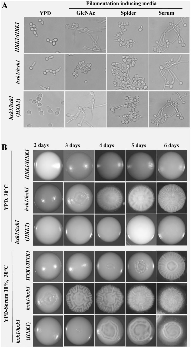
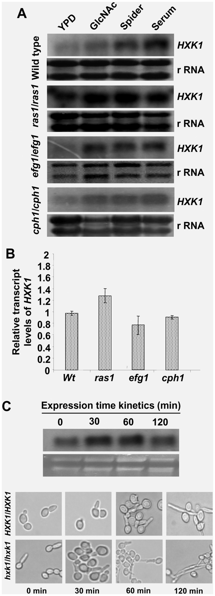
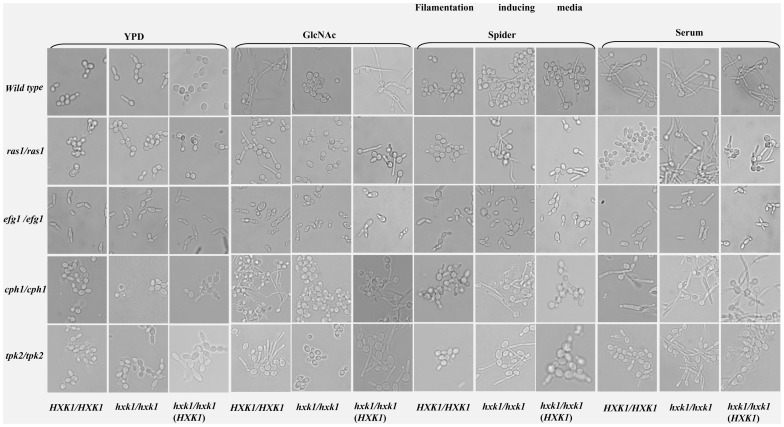
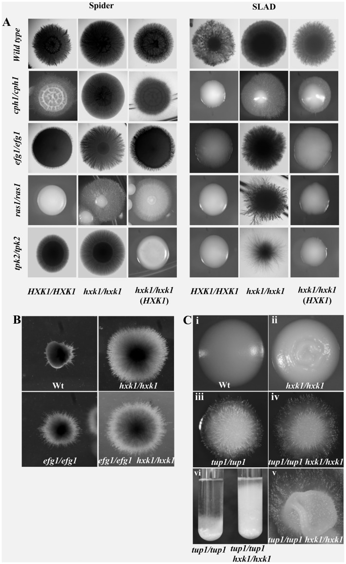
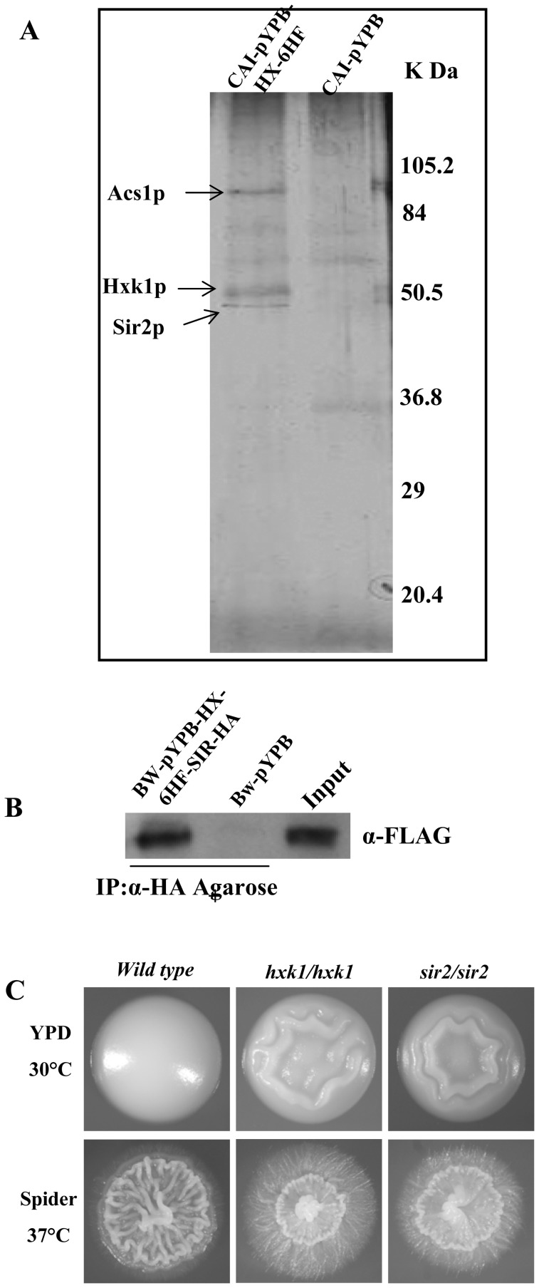
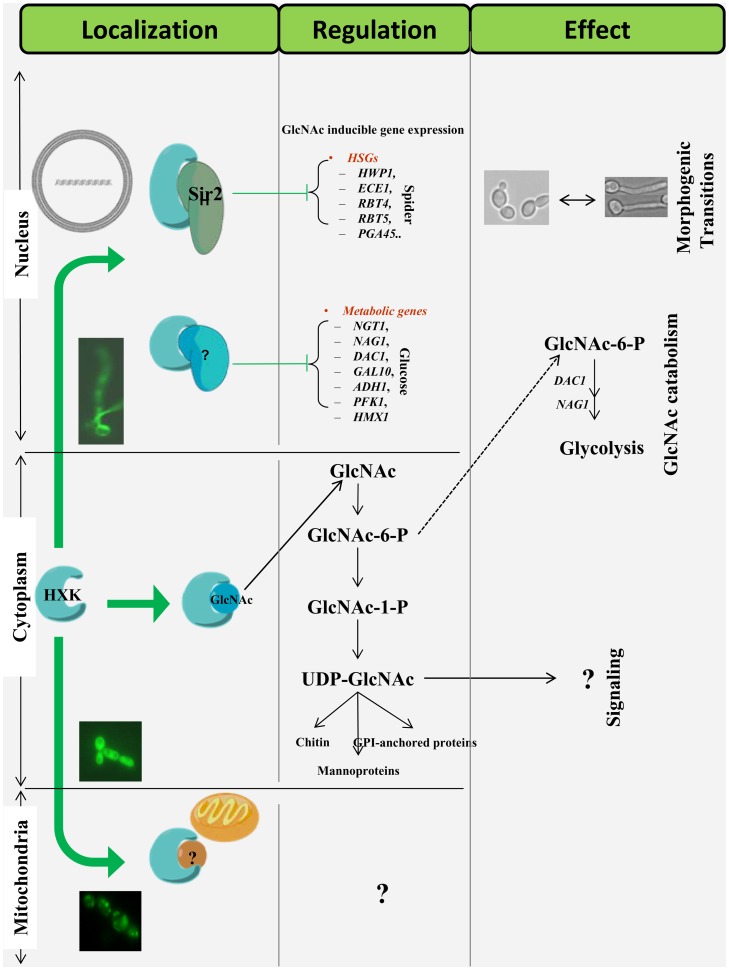
Candida albicans, a common fungal pathogen which diverged from the baker's yeast Saccharomyces cerevisiae has the unique ability to utilise N-acetylglucosamine, an amino sugar and exhibits phenotypic differences. It has acquired intricate regulatory mechanisms at different levels in accordance with its life style. N-acetylglucosamine kinase, a component of the N-acetylglucosamine catabolic cascade is an understudied gene since Saccharomyces cerevisiae lacks it. We report HXK1 to act as both positive and negative regulator of transcription of genes involved in maintaining cellular homeostasis. It is involved in repression of hyphal specific genes in addition to metabolic genes. Its regulation of filamentation and GlcNAc metabolism is independent of the known classical regulators like EFG1, CPH1, RAS1, TPK2 or TUP1. Moreover, Hxk1-GFP is localised to cytoplasm, nucleus and mitochondria in a condition specific manner. By employing two-step affinity purification, we report the interaction of HXK1 with SIR2 under filamentation inducing conditions. Our work highlights a novel regulatory mechanism involved in filamentation repression and attempts to decipher the GlcNAc catabolic regulatory cascade in eukaryotes.


Conflict of interest statement

Competing Interests: The authors have declared that no competing interests exist.

Figures

Figure 1
Figure 1. hxk1 mutant is constitutively filamentous and hyperfilamentous in filamentation inducing conditions.
A) hxk1 mutant is hyperfilamentous in liquid filamentation inducing media like Spider and Serum as compared to wild type strain (CAF2–1) and showed germ tube like protuberances (28–35%) in YPD liquid medium grown at 30°C (details are given in Text S1). B) hxk1 mutant is hyper filamentous and showed filamentation early in YPD and YPD+serum solid plates at 30°C after 3 and 2 days respectively whereas wild type and revertant colonies grown under similar conditions were completely smooth.
Figure 2
Figure 2. Expression pattern of HXK1 in filamentation inducing media.
A) Hxk1 transcript levels are upregulated in response to filamentation conditions in wild type and mutants for filamentation regulators. Wild-type strain and mutants were grown in YPD for 6 hr at 30°, washed, starved for 10 hrs and induced in YPD, GlcNAc, Spider and Serum (Experimental procedures). Cells were harvested at 2 hrs time point in case of YPD, Spider or Serum and at 4 hrs time point in case of GlcNAc and the total RNA was prepared and northern blot analyses were carried out. Ribosomal (r RNA) has been included as loading control. B) Quantitative RT-PCR of HXK1 basal transcript levels for the wild type and mutant cells grown in YPD. Almost unaltered HXK1 mRNA levels were observed in all strains. Error bars represent the coefficient of variation (n = 3). C) Northern blot analysis for the time kinetics of HXK1. Transscript levels are in response to Spider at 37°C. Ribosomal (r RNA) has been included as loading control. Corresponding cell shots representing hyperfilamentous morphology of hxk1 mutant in comparison with wild type strain have also been shown at the bottom.
Figure 3
Figure 3. Morphological and expression pattern analyses among wild type, hxk1 and different filamentation pathway mutants.
A) Morphology of wild type, filamentation pathway mutants under different hypha-inducing conditions. The starved cells of wild type (HXK1/HXK1 ) and hxk1 mutant(hxk1/hxk1), ras1/ras1, ras1/ras1 hxk1/hxk1, efg1/efg1, efg1/efg1 hxk1/hxk1, cph1/cph1, cph1/cph1 hxk1/hxk1, tpk2/tpk2, tpk2/tpk2 hxk1/hxk1l and their respective complemented strains in which one functional copy of HXK1 is reintroduced in the native locus were resuspended in non-inducing and various induction media such as 2.5 mM GlcNAc in salt base, Spider pH 7.2 and Serum (20%). Cells were induced at 37°C for 4 hours in case of GlcNAc, for 2 hours at 37°C in case of Spider and Serum and in case of YPD for 2 hours at 30°C. hxk1 mutants except efg1/efg1 hxk1/hxk1 were hyperfilamentous in filamentation inducing conditions like Spider and Serum. In response to GlcNAc, all hxk1 mutants failed to form germ tubes due to its inability to catabolize the sugar, but the complemented strains were hyper filamentous. B) Expression of hyphal specific genes in different filamentation inducing media in filamentation pathway mutants. Northern blot analysis to show the expression of Hyphal Specific Genes (HSGs) like HWP1, ECE1 or RBT4 for the cells grown in various filamentation inducing media. Total RNA was isolated from wild type, CAF2–1 (HXK1+/HXK1 ) and hxk1 mutant (hxk1/hxk1), ras1/ras1, ras1/ras1 hxk1/hxk1, efg1/efg1, efg1/efg1 hxk1/hxk1, cph1/cph1, cph1/cph1 hxk1/hxk1, tpk2/tpk2, tpk2/tpk2 hxk1/hxk1 strains induced in YPD, GlcNAc, Spider and 20% Serum. 20 µg of RNA was loaded in each lane and a Northern blotting was performed. As an internal control methylene blue staining of rRNA was used to ensure equal loading of RNA. hxk1 mutants except efg1/efg1 hxk1/hxk1 showed upregulation of HSGs in filamentation inducing conditions and upregulation was more prominent in Spider medium. C) Quantification of HXK1 mediated filamentation : q-RT PCR analysis of HSGs (ECE1, HWP1and RBT4) in hxk1 single and double mutants (ras1 hxk1, efg1hxk1, cph1hxk1 and tpk2 hxk1) in Spider grown cells compared to their wild type and respective single mutants (ras1, efg1, cph1 and tpk2). Comparative expression levels of HSGs for cells grown in YPD have also been shown. ACT1 has been selected as the endogenous control. The error bars represent co-efficient of variation.
Figure 4
Figure 4. hxk1 mutants are hyperfilamentous on filamentation inducing solid media.
A) Hyphae formation on Spider and SLAD plates. All hxk1 mutants including efg1/hxk1 showed hyperfilamentation. Strains were incubated at 37°C for 7 days in case of spider and 10 days in case of SLAD plates. Wild-type CAF2–1 (HXK1/HXK1); hxk1 mutant, H8–1–103 (hxk1/hxk1); cph1 mutant, A11–1 (cph1/cph1); double mutant of cph1/hxk1, AN8–1–16 (cph1/cph1 hxk1/hxk1); efg1 mutant, HLC52 (efg1/efg1); double mutant of efg1/hxk1 HLC67–16–1–9 (efg1/efg1 hxk1/hxk1); tpk2 mutant, TPO7.4 (tpk2/tpk2); double mutant of tpk2/hxk1, AS1–3–1–8 (tpk2/tpk2 hxk1/hxk1) and their respective complemented strains in which one functional copy of HXK1 was reintroduced in the native locus. B) efg1/efg1 hxk1/hxk1 double mutant showed hyperfilamentation under embedded conditions. Cells of wild-type, CAF2–1, hxk1/hxk1, efg1/efg1, efg1/efg1hxk1/hxk1, mutants were grown in YPD for 5 hrs at 30°, washed in sterile water and mixed with molten CM Agar (with 1% Tween-80) plated and grown for 3 days at 25°C. efg1 hxk1 double mutant showed hyperfilamentation when compared to hxk1 or efg1 single mutants (EFG1 is reported to be a negative regulator of filamentation under micro-aerophillic/embedded conditions). C) tup1/hxk1 double mutant showed growth pattern slightly different from tup1 mutant. Wild type (i), hxk1 mutant (ii), tup1(iii) and tup1/hxk1(iv) mutants colonies were grown on YPD solid and liquid medium for 3 and 2 days respectively at 30°C. tup1/hxk1double mutant shared most of the features with a tup1 single mutant (iii, iv) on YPD plates, but in some colonies of the double mutant a wavy, afilamentous fringe could be observed at the centre(v). In YPD broth the tup1 single mutant grew in clumps having the tendency to settle at the bottom, the tup1/hxk1 double mutant showed uniform turbidity throughout the culture with clumps in the bottom (vi).
Figure 5
Figure 5. Hxk1p interacts with Histone Deacetylase (Sir2) to repress filament specific genes.
A) The Hxk1p complex was purified by tandem affinity purification (TAP-tag) using anti- FLAG and Ni-nitrilotriacetic acid (Ni-NTA) agarose. Protein fractions of purified samples from 1st round with Anti-FLAG Agarose and 2nd round with Ni-NTA Agarose were separated by 12% sodium dodecyl sulfate-polyacrylamide gel electrophoresis and visualized by silver staining. Open arrowheads indicate the component of the Hxk1p complex identified by matrix-assisted laser desorption ionization–time of flight mass spectrometry. CAI-pYPB (control, CAI-4 transformed with pYPB-ADH1-pt, empty vector) and CAI-pYPB-HX-6HF (TAP tagged HXK1expressed under ADH1 p in CAI-4) are Spider medium induced cell extracts. B) Hxk1 interaction with Sir2 confirmed by Co-Immunoprecipitation. BW-pYPB-HX-6HF-SIR-HA and BW-pYPB strain cells were induced in Spider and crude extract isolated as described in Experimental procedures. Immunoprecipitation was carried out in two reactions with anti-HA-Agarose. The crude extract was used as an input. Immunoblotting was performed with anti-FLAG antibody. C) sir2 mutant showed hyperfilamentation similar to that of hxk1 mutant in inducing and non-inducing media. Upper panel shows the morphology of wild type, hxk1 mutant and sir2 mutant grown on YPD plates at 30°C for 3days and lower panel shows the morphology of wild type, hxk1 mutant and sir2 mutant grown on Spider plates at 37°C for 5 days.
Figure 6
Figure 6. Response of hxk1 response to cell wall perturbing agent and high temperature.
Wild-type strain, hxk1 mutant and revertant strains were grown in YPD for 8 hr at 30°C and 10-fold dilution series was spotted on YPD, YPD+ Nikkomycin Z (20 µg ml−1), YPD+ Congo Red (100 µg ml−1) and YPD+Calcofluor white (30 µg ml−1) plates at indicated concentrations(A and B). For temperature shift experiments, spotted plates were kept at 30°C and 42°C.
Figure 7
Figure 7. HXK1 localized in the cytoplasm, nucleus and mitochondria in condition specific manner.
Sub-cellular localization of Hxk1p-GFP in wild-type cells induced in GlcNAc 2.5 mM or 2% and non-fermentative carbon sources like ethanol or glycerol, 5%. iHXK1-GFP along with wild type (control) grown for 6 h in YPD at 30°C and then, washed in water and induced in GlcNAc 2.5 mM (salt base) or 2%. Cells were visualized either by bright field or by epifluorescence at 1 hr, 2.5 hr and 4 hr in 2.5 mM GlcNAc (A), and 4 hr in 2% GlcNAc (B) after induction. The nuclear localization of Hxk1p was further confirmed by western blotting analysis. iHXK1-Myc strain was induced in 2.5 mM GlcNAc (C) or 2% glucose (D), nuclear and total cellular fractions were checked for presence of hxk1p with help of anti c-myc antibody. The purity of nuclear fraction (free of cytoplasmic contamination) was checked by using Anti-GAPDH antibody specific for cytoplasmic fraction. To check localization in non-fermentable carbon sources, cells were grown for 6 h in YPD at 30°C, washed in water and induced in YNB w/o amino acid with 5% ethanol (E). Hxk1 localization is marked with white arrowheads.
Figure 8
Figure 8. Transcriptome analysis of HXK1 regulated genes.
A) Partial Heat map of over-represented genes (p-values <0.05, False Discovery Rate <1% in GO term analysis) differentially expressed in a HXK1 dependent manner in response to glucose. Two-color microarray data expressed as hxk1/HXK1 ratio (1 and 2 are biological replicates with dye swap, average of 1 and 2, Ave) is plotted as heat map. The color scale at the bottom indicates the log2 ratio. B) Heat map of Hyphal specific genes (HSGs) differentially expressed in a HXK1 dependent manner in response to glucose. C) Modulation of expression levels of GlcNAc catabolic genes by HXK1. Quantitative RT-PCR of GlcNAc catabolic gene transcripts NGT1, NAG1 and DAC1 in C. albicans wild type (CAF2–1) and hxk1 mutant, cells in response to glycerol-6%, Glucose-2%, Glucose-5 mM or GlcNAc-5 mM. ACT1 has been selected as the endogenous control. The error bars represent co-efficient of variation.
Figure 9
Figure 9. A brief overview of mode of action, localization, targets and downstream effects of Hxk1.
The protein capable of multifarious localization migrates to nucleus where it possibly interacts with a Histone Deacetylase Sir2 to keep the HSGs in a repressed state. It probably partners again with Sir2 or some other proteins to repress the GlcNAc metabolic genes while still in nucleus. In cytoplasm Hxk1 phosphorylates GlcNAc to carry out the catabolism of this amino sugar and contributing probably to the overall UDP-GlcNAc pool within the cell. In presence of non-fermentable carbon sources like ethanol or glycerol the protein migrates to mitochondria where its mode of functioning is still not known.

Similar articles

Cited by

References

    1. Biswas M, Singh B, Datta A (1979) Induction of N-acetylmannosamine catabolic pathway in yeast. Biochim Biophys Acta 585: 535–542. - PubMed
    1. Shepherd MG, Yin CY, Ram SP, Sullivan PA (1980) Germ tube induction in Candida albicans . Can J Microbiol 26: 21–26. - PubMed
    1. Sudbery P, Gow N, Berman J (2004) The distinct morphogenic states of Candida albicans. . Trends Microbiol 12: 317. - PubMed
    1. Whiteway M, Bachewich C (2007) Morphogenesis in Candida albicans. . Annu Rev Microbiol 61: 529–553. - PMC - PubMed
    1. Liu H (2001) Transcriptional control of dimorphism in Candida albicans. . Curr Opin Microbiol 4: 278–235. - PubMed

Publication types

MeSH terms

LinkOut - more resources