Comparative Study
. 2013;8(1):e53818.
doi: 10.1371/journal.pone.0053818. Epub 2013 Jan 14.

The pan-genome of the animal pathogen Corynebacterium pseudotuberculosis reveals differences in genome plasticity between the biovar ovis and equi strains

Affiliations
Comparative Study

The pan-genome of the animal pathogen Corynebacterium pseudotuberculosis reveals differences in genome plasticity between the biovar ovis and equi strains

Siomar C Soares et al. PLoS One. 2013.

Abstract

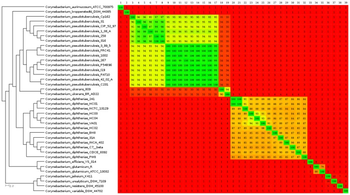
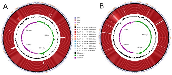
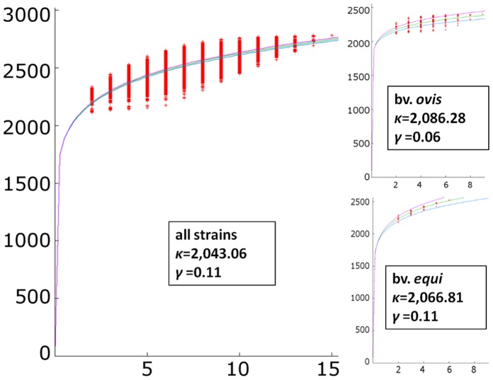
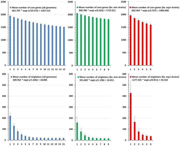
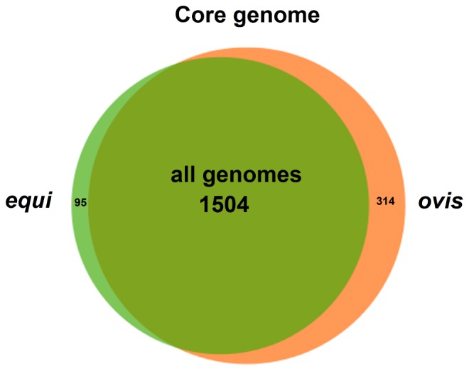
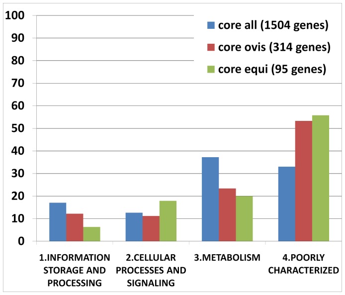
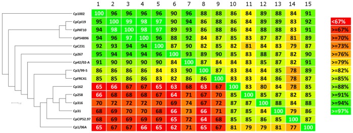
Corynebacterium pseudotuberculosis is a facultative intracellular pathogen and the causative agent of several infectious and contagious chronic diseases, including caseous lymphadenitis, ulcerative lymphangitis, mastitis, and edematous skin disease, in a broad spectrum of hosts. In addition, Corynebacterium pseudotuberculosis infections pose a rising worldwide economic problem in ruminants. The complete genome sequences of 15 C. pseudotuberculosis strains isolated from different hosts and countries were comparatively analyzed using a pan-genomic strategy. Phylogenomic, pan-genomic, core genomic, and singleton analyses revealed close relationships among pathogenic corynebacteria, the clonal-like behavior of C. pseudotuberculosis and slow increases in the sizes of pan-genomes. According to extrapolations based on the pan-genomes, core genomes and singletons, the C. pseudotuberculosis biovar ovis shows a more clonal-like behavior than the C. pseudotuberculosis biovar equi. Most of the variable genes of the biovar ovis strains were acquired in a block through horizontal gene transfer and are highly conserved, whereas the biovar equi strains contain great variability, both intra- and inter-biovar, in the 16 detected pathogenicity islands (PAIs). With respect to the gene content of the PAIs, the most interesting finding is the high similarity of the pilus genes in the biovar ovis strains compared with the great variability of these genes in the biovar equi strains. Concluding, the polymerization of complete pilus structures in biovar ovis could be responsible for a remarkable ability of these strains to spread throughout host tissues and penetrate cells to live intracellularly, in contrast with the biovar equi, which rarely attacks visceral organs. Intracellularly, the biovar ovis strains are expected to have less contact with other organisms than the biovar equi strains, thereby explaining the significant clonal-like behavior of the biovar ovis strains.


Conflict of interest statement

Competing Interests: The authors have declared that no competing interests exist.

Figures

Figure 1
Figure 1. Phylogenomic tree and heatmap analyses of the genus Corynebacterium.
All the complete genomes from the genus Corynebacterium were retrieved from the NCBI ftp site. Comparisons between the variable content of all the strains were plotted as percentages of similarity on the heatmap using Gegenees (version 1.1.4). The percentage of similarity was used to generate a phylogenomic tree with SplitsTree (version 4.12.6). Numbers from 1 to 39 (upper-left to upper-right corner) represent species from Corynebacterium aurimucosum ATCC 70097 to Corynebacterium variable DSM 44702 (upper-left to lower-left corner). Percentages were plotted with a spectrum ranging from red (low similarity) to green (high similarity). On the heatmap, the upper portion is not symmetrical to the lower portion because the variable contents of all genomes present different sizes. Therefore, considering a scenario where the variable content from genomes A and B are composed of 100 and 80 genes, respectively, with a common repertoire of 40 genes, genome A will present 40% of similarity to genome B and genome B will present 50% of similarity to genome A.
Figure 2
Figure 2. Comparative genomic maps of the C. pseudotuberculosis biovar equi and ovis strains.
A, all the C. pseudotuberculosis strains were aligned using C. pseudotuberculosis strain 1002 as a reference. From the inner to outer circle on A: the biovar equi strains Cp31, Cp1/06-A, CpCp162, Cp258, Cp316 and CpCIP52.97; and, the biovar ovis strains CpC231, CpP54B96, Cp267, CpPAT10, CpI19, Cp42/02-A, Cp3/99-5, CpFRC41 and Cp1002. B, all the C. pseudotuberculosis strains were aligned using C. pseudotuberculosis strain CIP52.97 as a reference. From the inner to outer circle on B: the biovar ovis strains CpC231, Cp1002, CpPAT10, Cp267, CpP54B96, CpI19, Cp42/02-A, CpFRC41, Cp3/99-5; and, the biovar equi strains Cp1/06-A Cp31, CpCp162, Cp316, Cp258 and CpCIP52.97. CDS, coding sequences; tRNA, transfer RNA; rRNA, ribosomal RNA; and PAI, pathogenicity island.
Figure 3
Figure 3. Pan-genome development of C. pseudotuberculosis.
Center chart, the pan-genome development using permutations of all 15 strains of C. pseudotuberculosis; upper-right chart, the pan-genome development of the C. pseudotuberculosis biovar ovis strains; lower-right chart, the pan-genome development of the C. pseudotuberculosis biovar equi strains.
Figure 4
Figure 4. Core genome and singleton development of C. pseudotuberculosis.
Upper-left, the core genome development using permutations of all 15 strains of C. pseudotuberculosis; upper-center, the core genome development of the C. pseudotuberculosis biovar ovis strains; upper-right, the core genome development of the C. pseudotuberculosis biovar equi strains; lower-left, the singleton development using permutations of all 15 strains of C. pseudotuberculosis; lower-center, the singleton development of the C. pseudotuberculosis biovar ovis strains; lower-right, the singleton development of the C. pseudotuberculosis biovar equi strains.
Figure 5
Figure 5. Venn diagram representing the core genomes of the C. pseudotuberculosis strains.
All genomes, the number of genes composing the core genome of all the strains; equi, the number of genes of the core genome of the C. pseudotuberculosis biovar equi strains, which were absent in one or more of the C. pseudotuberculosis biovar ovis strains; ovis, the number of genes of the core genome of the C. pseudotuberculosis biovar ovis strains, which were absent in one or more of the C. pseudotuberculosis biovar equi strains.
Figure 6
Figure 6. Core genes of the C. pseudotuberculosis strains classified by COG functional category.
Core all, the genes composing the core genome of all the strains; core ovis, the genes of the core genome of the C. pseudotuberculosis biovar ovis strains, which were absent in one or more of the C. pseudotuberculosis biovar equi strains; core equi, the genes of the core genome of the C. pseudotuberculosis biovar equi strains, which were absent in one or more of the C. pseudotuberculosis biovar ovis strains.
Figure 7
Figure 7. Phylogenomic tree and heatmap analyses of the Corynebacterium pseudotuberculosis strains based on pathogenicity island plasticity.
Comparisons between the PAI contents of all the strains were plotted as percentages of similarity on the heatmap using Gegenees (version 1.1.4). The percentages of similarity were used to generate a phylogenomic tree with SplitsTree (version 4.12.6). Numbers from 1 to 15 (upper-left to upper-right corner) represent the strains from Cp1002 to Cp1/06-A (upper-left to lower-left corner). On the heatmap, the upper portion is not symmetrical to the lower portion because the pathogenicity islands contents of all genomes present different sizes. Therefore, considering a scenario where the pathogenicity islands content from genomes A and B are composed of 100 and 80 genes, respectively, with a common repertoire of 40 genes, genome A will present 40% of similarity to genome B and genome B will present 50% of similarity to genome A.
Figure 8
Figure 8. Plasticity of the pilus gene clusters spaA and spaD in C. pseudotuberculosis.
A1 and B1, PiCp15 harboring the spaA cluster of genes; A2 and B2, PiCp7 harboring the spaD cluster of genes. A, all the C. pseudotuberculosis strains were aligned using C. pseudotuberculosis strain 1002 as a reference. From the inner to outer circle on A1 and A2: the biovar equi strains Cp31, Cp1/06-A, CpCp162, Cp258, Cp316, CpCIP52.97; and, the biovar ovis strains CpC231, CpP54B96, Cp267, CpPAT10, CpI19, Cp42/02-A, Cp3/99-5, CpFRC41 and Cp1002. B, all the C. pseudotuberculosis strains were aligned using C. pseudotuberculosis strain CIP52.97 as a reference. From the inner to outer circle on B1 and B2: the biovar ovis strains CpC231, Cp1002, CpPAT10, Cp267, CpP54B96, CpI19, Cp42/02-A, CpFRC41, Cp3/99-5, Cp1/06-A; and, the biovar equi strains Cp31, CpCp162, Cp316, Cp258 and CpCIP52.97. CDS, coding sequences; tRNA, transfer RNA; rRNA, ribosomal RNA; and PAI, pathogenicity island.

Similar articles

Cited by

References

    1. Dorella FA, Pacheco LGC, Oliveira SC, Miyoshi A, Azevedo V (2006) Corynebacterium pseudotuberculosis: microbiology, biochemical properties, pathogenesis and molecular studies of virulence. Vet Res 37: 201–218. - PubMed
    1. Lehman KB, Neumann R (1896) Atlas und grundriss der bakeriologie und lehrbuch der speziellen bakteriologischen diagnositk. 1st ed. J.F. Lehmann, Munchen.
    1. Pascual C, Lawson PA, Farrow JA, Gimenez MN, Collins MD (1995) Phylogenetic analysis of the genus Corynebacterium based on 16S rRNA gene sequences. Int J Syst Bacteriol 45: 724–728. - PubMed
    1. Cerdeño-Tárraga AM, Efstratiou A, Dover LG, Holden MTG, Pallen M, et al. (2003) The complete genome sequence and analysis of Corynebacterium diphtheriae NCTC13129. Nucleic Acids Res 31: 6516–6523. - PMC - PubMed
    1. Tauch A, Kaiser O, Hain T, Goesmann A, Weisshaar B, et al. (2005) Complete genome sequence and analysis of the multiresistant nosocomial pathogen Corynebacterium jeikeium K411, a lipid-requiring bacterium of the human skin flora. J Bacteriol 187: 4671–4682. - PMC - PubMed

Publication types

Substances

LinkOut - more resources