

Drosophila clueless is highly expressed in larval neuroblasts, affects mitochondrial localization and suppresses mitochondrial oxidative damage
- PMID: 23342118
- PMCID: PMC3547001
- DOI: 10.1371/journal.pone.0054283
Drosophila clueless is highly expressed in larval neuroblasts, affects mitochondrial localization and suppresses mitochondrial oxidative damage
Abstract
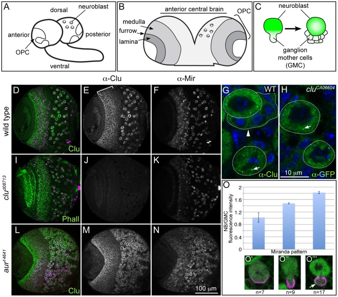
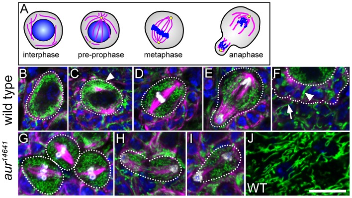
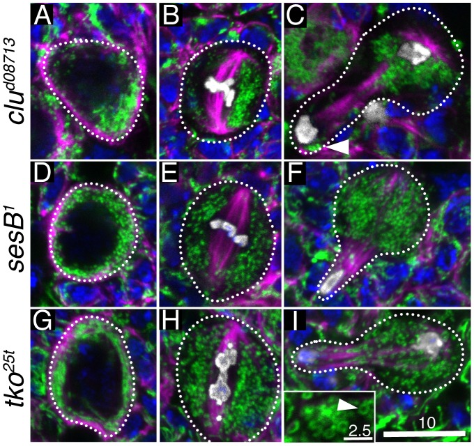
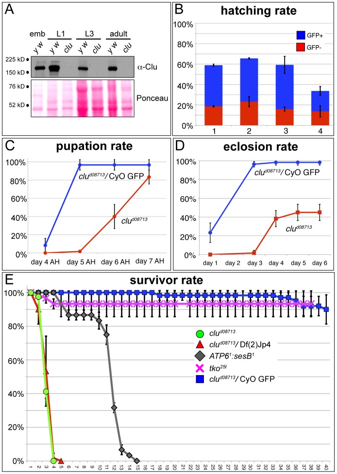
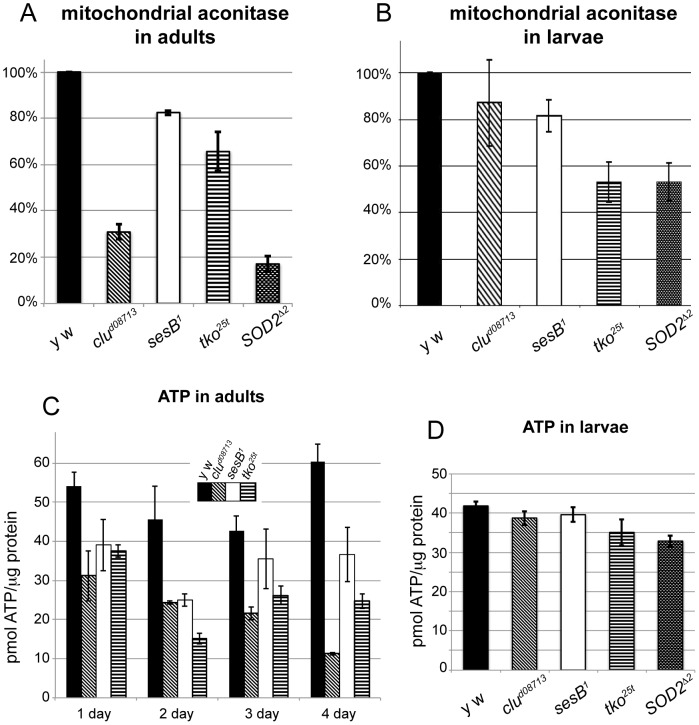
Mitochondria are critical for neuronal function due to the high demand of ATP in these cell types. During Drosophila development, neuroblasts in the larval brain divide asymmetrically to populate the adult central nervous system. While many of the proteins responsible for maintaining neuroblast cell fate and asymmetric cell divisions are known, little is know about the role of metabolism and mitochondria in neuroblast division and maintenance. The gene clueless (clu) has been previously shown to be important for mitochondrial function. clu mutant adults have severely shortened lifespans and are highly uncoordinated. Part of their lack of coordination is due to defects in muscle, however, in this study we have identified high levels of Clu expression in larval neuroblasts and other regions of the dividing larval brain. We show while mitochondria in clu mutant neuroblasts are mislocalized during the cell cycle, surprisingly, overall brain morphology appears to be normal. This is explained by our observation that clu mutant larvae have normal levels of ATP and do not suffer oxidative damage, in sharp contrast to clu mutant adults. Mutations in two other genes encoding mitochondrial proteins, technical knockout and stress sensitive B, do not cause neuroblast mitochondrial mislocalization, even though technical knockout mutant larvae suffer oxidative damage. These results suggest Clu functions upstream of electron transport and oxidative phosphorylation, has a role in suppressing oxidative damage in the cell, and that lack of Clu's specific function causes mitochondria to mislocalize. These results also support the previous observation that larval development relies on aerobic glycolysis, rather than oxidative phosphorylation. Thus Clu's role in mitochondrial function is not critical during larval development, but is important for pupae and adults.
Conflict of interest statement
Figures
Similar articles
-
Clueless forms dynamic, insulin-responsive bliss particles sensitive to stress.Dev Biol. 2020 Mar 15;459(2):149-160. doi: 10.1016/j.ydbio.2019.12.004. Epub 2019 Dec 16. Dev Biol. 2020. PMID: 31837288 Free PMC article.
-
Clueless regulates aPKC activity and promotes self-renewal cell fate in Drosophila lgl mutant larval brains.Dev Biol. 2013 Sep 15;381(2):353-64. doi: 10.1016/j.ydbio.2013.06.031. Epub 2013 Jul 5. Dev Biol. 2013. PMID: 23835532
-
Clueless, a protein required for mitochondrial function, interacts with the PINK1-Parkin complex in Drosophila.Dis Model Mech. 2015 Jun;8(6):577-89. doi: 10.1242/dmm.019208. Epub 2015 Apr 20. Dis Model Mech. 2015. PMID: 26035866 Free PMC article.
-
Drosophila neuroblast asymmetric divisions: cell cycle regulators, asymmetric protein localization, and tumorigenesis.J Cell Biol. 2008 Jan 28;180(2):267-72. doi: 10.1083/jcb.200708159. Epub 2008 Jan 21. J Cell Biol. 2008. PMID: 18209103 Free PMC article. Review.
-
Analysis of mitochondrial structure and function in the Drosophila larval musculature.Mitochondrion. 2016 Jan;26:33-42. doi: 10.1016/j.mito.2015.11.005. Epub 2015 Dec 1. Mitochondrion. 2016. PMID: 26611999 Free PMC article. Review.
Cited by
-
Characterization of Factors Involved in Localized Translation Near Mitochondria by Ribosome-Proximity Labeling.Front Cell Dev Biol. 2019 Nov 26;7:305. doi: 10.3389/fcell.2019.00305. eCollection 2019. Front Cell Dev Biol. 2019. PMID: 31929983 Free PMC article.
-
Modulation of flagellum attachment zone protein FLAM3 and regulation of the cell shape in Trypanosoma brucei life cycle transitions.J Cell Sci. 2015 Aug 15;128(16):3117-30. doi: 10.1242/jcs.171645. Epub 2015 Jul 6. J Cell Sci. 2015. PMID: 26148511 Free PMC article.
-
Drosophila Clueless ribonucleoprotein particles display novel dynamics that rely on the availability of functional protein and polysome equilibrium.bioRxiv [Preprint]. 2024 Aug 22:2024.08.21.609023. doi: 10.1101/2024.08.21.609023. bioRxiv. 2024. Update in: J Cell Sci. 2025 May 1;138(9):jcs263730. doi: 10.1242/jcs.263730. PMID: 39229069 Free PMC article. Updated. Preprint.
-
Clueless forms dynamic, insulin-responsive bliss particles sensitive to stress.Dev Biol. 2020 Mar 15;459(2):149-160. doi: 10.1016/j.ydbio.2019.12.004. Epub 2019 Dec 16. Dev Biol. 2020. PMID: 31837288 Free PMC article.
-
Nutrient-dependent regulation of a stable intron modulates germline mitochondrial quality control.Nat Commun. 2024 Feb 10;15(1):1252. doi: 10.1038/s41467-024-45651-y. Nat Commun. 2024. PMID: 38341415 Free PMC article.
References
-
- Bereiter-Hahn J, Voth M (1994) Dynamics of mitochondria in living cells: shape changes, dislocations, fusion, and fission of mitochondria. Microsc Res Tech 27: 198–219. - PubMed
-
- Chan DC (2006) Mitochondria: dynamic organelles in disease, aging, and development. Cell 125: 1241–1252. - PubMed
-
- Chai PC, Chia W, Cai Y (2012) A niche for Drosophila neuroblasts? WIREs Developmental Biology 1: 307–314. - PubMed
Publication types
MeSH terms
Substances
LinkOut - more resources
Full Text Sources
Other Literature Sources
Molecular Biology Databases
Research Materials
Miscellaneous
