

Advanced imaging in limb salvage
- PMID: 23342185
- PMCID: PMC3549647
- DOI: 10.14797/mdcj-8-4-28
Advanced imaging in limb salvage
Abstract
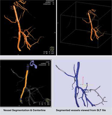
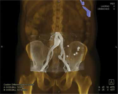
The evaluation of patients at risk for limb loss secondary to peripheral arterial disease begins with a complete history and physical exam, and noninvasive studies in the vascular lab, including duplex ultrasonography. However, successful revascularization depends on high-quality, accurate imaging of the lower extremity vasculature. The traditional gold standard for vascular imaging, digital subtraction angiography, has been improved upon as technologic advances have enabled high-quality alternatives for preoperative (i.e., computed tomography [CT] angiography and magnetic resonance angiography [MRA]) and intraoperative imaging (i.e., intravascular ultrasound [IVUS], cone beam CT, and CO(2) angiography). Here we describe these advanced invasive and noninvasive imaging alternatives and their utility in limb salvage procedures.
Keywords: Imaging; Limb Salvage; Peripheral Artery Disease.
Figures


References
-
- Schoenhagen P, White RD, Nissen SE, Tuzcu EM. Coronary imaging: angiography shows the stenosis but IVUS, CT, and MRI show the plaque. Cleve Clin J Med. 2003;70(8):713–9. - PubMed
-
- Baba R, Konno Y, Ueda K, Ikeda S. Comparison of flat-panel detector and image-intensifier detector for cone-beam CT. Comput Med Imaging Graph. 2002;26(3):153–8. - PubMed
-
- Bandyk DF. Surveillance after lower extremity arterial bypass. Perspect Vasc Surg Endovasc Ther. 2007;19(4):376–83. - PubMed
Publication types
MeSH terms
LinkOut - more resources
Full Text Sources
Medical
Miscellaneous
