. 2012 Oct;1(2):218-29.
doi: 10.1002/cam4.32. Epub 2012 Sep 13.

Expression of RET finger protein predicts chemoresistance in epithelial ovarian cancer

Affiliations

Expression of RET finger protein predicts chemoresistance in epithelial ovarian cancer

Maiko Horio et al. Cancer Med. 2012 Oct.

Abstract

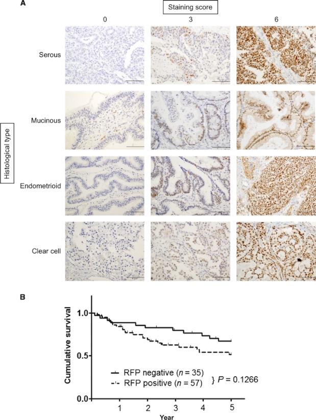
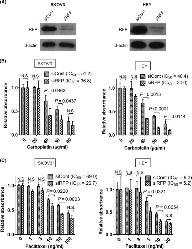
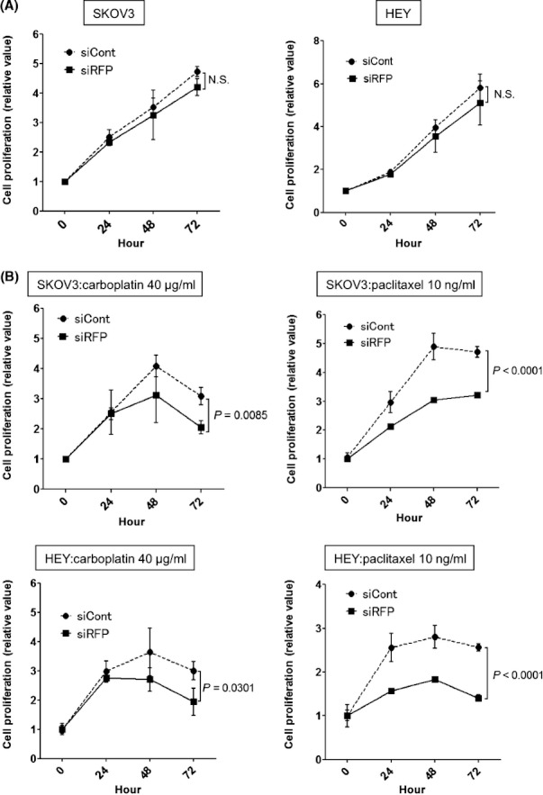
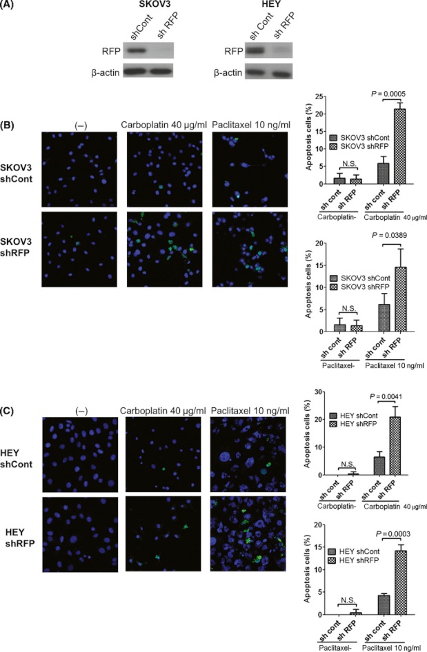
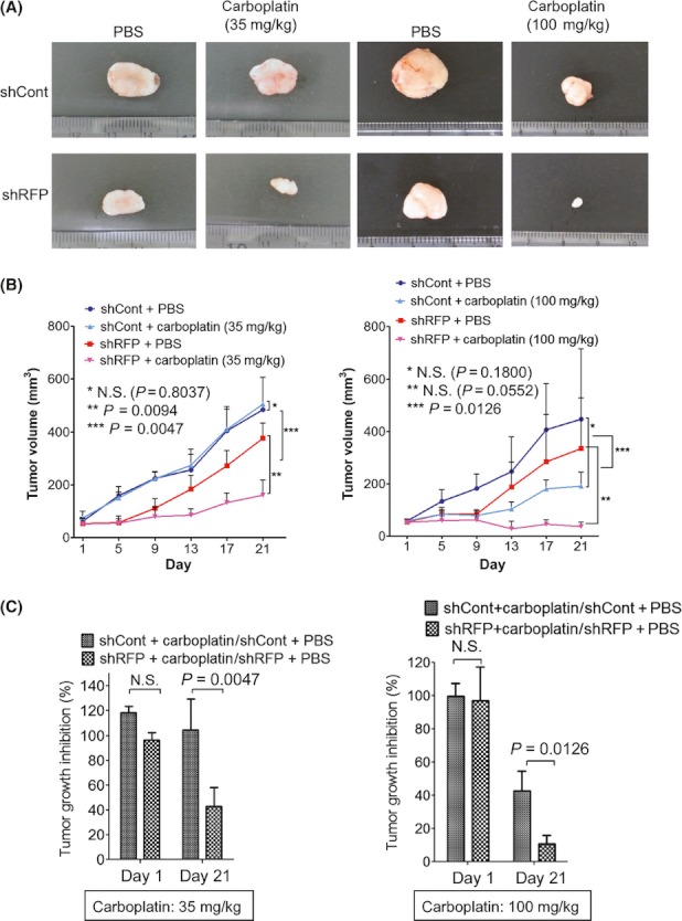
Resistance to platinum- and taxane-based chemotherapy is a major cause of treatment failure in ovarian cancer. Thus, it is necessary to develop a predictive marker and molecular target for overcoming drug resistance in ovarian cancer treatment. In a previous report, using an in vitro model, we found that the RET finger protein (RFP) (also known as tripartite motif-containing protein 27, TRIM27) confers cancer cell resistance to anticancer drugs. However, the significance of RFP expression in cancer patients remains elusive. In this study, we showed that RFP was expressed in 62% of ovarian cancer patients and its positivity significantly correlated with drug resistance. Consistent with clinical data, depletion of RFP by RNA interference (RNAi) in ovarian cancer cell lines, SKOV3 and HEY, significantly increased carboplatin- or paclitaxel-induced apoptosis and resulted in reduced anticancer drug resistance. In a nude mouse tumor xenograft model, inoculated RFP-knockdown ovarian cancer cells exhibited lower carboplatin resistance than control cells. These findings suggest that RFP could be a predictive marker for chemoresistance in ovarian cancer patients and also a candidate for a molecular-targeted agent.

Keywords: Carboplatin; RET finger protein; chemoresistance; epithelial ovarian cancer; paclitaxel.


Figures

Figure 1
Figure 1
Immunohistochemical analysis of RFP in epithelial ovarian cancer. (A) RFP expression in serous, mucinous, endometrioid, and clear cell adenocarcinomas. To evaluate RFP expression, the staining intensity was scored as 0 (negative), 1 (weak), 2 (medium), or 3 (strong). The staining extent was scored as 0 (<10%), 1 (10–30%), 2 (30–50%), or 3 (>50%) in relation to the entire cancer area. The sum of scores for the staining intensity and staining extent was used as the staining score (0–6) for RFP. Staining score 0–2, negative; 3–6, positive. Representative images with staining scores 0 (left), 3 (middle), and 6 (right) are shown. Scale bars: 200 μm. (B) Kaplan–Meier survival curves of patients with epithelial ovarian cancer stratified by RFP expression. The 5-year overall survival rate: all cases (n = 92).
Figure 2
Figure 2
RFP knockdown enhances chemosensitivity in ovarian cancer cell lines. (A) Control siRNA (siCont) and RFP siRNA (siRFP) were transfected into SKOV3 and HEY cells 72 h before the assays. Total cell lysates from each cell line were subjected to Western blotting with anti-RFP or anti-β-actin antibody. (B) and (C) SKOV3 and HEY cells were transfected with siCont or siRFP and incubated for 48 h. The cells were treated with the indicated doses of carboplatin (B) for 48 h or paclitaxel (C) for 72 h. Cell viability was measured using the MTS assay.
Figure 3
Figure 3
Effects of RFP knockdown on cancer cell proliferation. Time point MTS assay of SKOV3 and HEY cells transfected with each siRNA. (A) The assays were performed without anticancer drugs. (B) SKOV3 cells were cultured in RPMI containing 40 μg/mL of carboplatin (left) or 10 ng/mL of paclitaxel (right) (top panels); HEY cells were cultured in RPMI containing 40 μg/mL of carboplatin (left) or 10 ng/mL of paclitaxel (right) (bottom panels).
Figure 4
Figure 4
Effect of RFP knockdown on apoptosis of carboplatin- and paclitaxel-treated ovarian cancer cells. (A) SKOV3 or HEY cells were infected with retroviruses carrying control or RFP-targeting shRNA. Total cell lysates from each cell line were subjected to Western blotting with anti-RFP or anti-β-actin antibody. Representative images of SKOV3 (B) and HEY (C) cells infected with retroviruses carrying shCont or shRFP. These cells were treated with carboplatin or paclitaxel at the indicated concentration for 48 h. The cells were stained by anti-cleaved caspase 3, and positive cells were counted under ×200 magnification. Percentages of cleaved caspase 3-positive cells are shown in right panels. Total 200 cells were counted. Bars, SD. P < 0.05 was considered statistically significant. N.S., not significant.
Figure 5
Figure 5
RFP knockdown enhances the sensitivity of tumors to carboplatin in vivo; 5 × 10⁶ SKOV3 ovarian cancer cells infected with retroviruses carrying shCont or shRFP were inoculated subcutaneously (s.c.) into nude mice. When tumors reached approximately 50 mm3, 10 mice were treated with phosphate-buffered saline (PBS) or carboplatin (35 or 100 mg/kg) i.p. four times every 2 days. (A) Representative transplanted tumors excised at day 21 are shown. (B) Tumors were measured at different days after PBS or carboplatin treatment, and growth curves were drawn. Tumor volume of the carboplatin-treated shRFP group was significantly smaller than that of the control group. Left, 35 mg/kg carboplatin treatment group; right, 100 mg/kg carboplatin treatment group. (C) The mean tumor volume of the PBS-treated group was defined as 100% and compared with that of the treated group on days 1 and 21. Statistic analysis was performed by Student's t-test. Carboplatin treatment group, n = 5; PBS group, n = 3; bars, SD.

Similar articles

Cited by

References

    1. Jemal A, Siegel R, Xu J, Ward E. Cancer statistics, 2010. CA Cancer J. Clin. 2010;60:277–300. - PubMed
    1. Ushijima K. Current status of gynecologic cancer in Japan. J. Gynecol. Oncol. 2009;20:67–71. - PMC - PubMed
    1. Ferlay J, Shin HR, Bray F, Forman D, Mathers C, Parkin DM. Estimates of worldwide burden of cancer in 2008: GLOBOCAN 2008. Int. J. Cancer. 2010;127:2893–2917. - PubMed
    1. Engel J, Eckel R, Schubert-Fritschle G, Kerr J, Kuhn W, Diebold J, et al. Moderate progress for ovarian cancer in the last 20 years: prolongation of survival, but no improvement in the cure rate. Eur. J. Cancer. 2002;38:2435–2445. - PubMed
    1. Averette HE, Janicek MF, Menck HR. The National Cancer Data Base report on ovarian cancer. American College of Surgeons Commission on Cancer and the American Cancer Society. Cancer. 1995;76:1096–1103. - PubMed

Publication types

MeSH terms