Review
. 2012 Oct;1(2):237-60.
doi: 10.1002/cam4.22. Epub 2012 Sep 14.

Strategies for discovery and validation of methylated and hydroxymethylated DNA biomarkers

Affiliations
Review

Strategies for discovery and validation of methylated and hydroxymethylated DNA biomarkers

Ekaterina Olkhov-Mitsel et al. Cancer Med. 2012 Oct.

Abstract

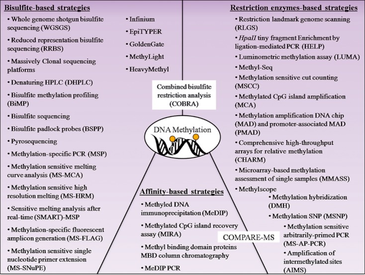
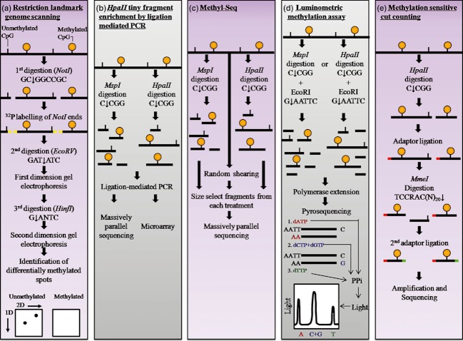
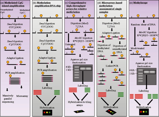
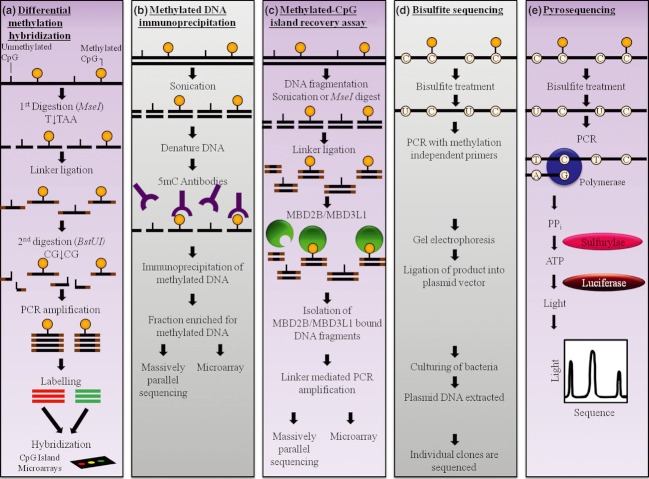
DNA methylation, consisting of the addition of a methyl group at the fifth-position of cytosine in a CpG dinucleotide, is one of the most well-studied epigenetic mechanisms in mammals with important functions in normal and disease biology. Disease-specific aberrant DNA methylation is a well-recognized hallmark of many complex diseases. Accordingly, various studies have focused on characterizing unique DNA methylation marks associated with distinct stages of disease development as they may serve as useful biomarkers for diagnosis, prognosis, prediction of response to therapy, or disease monitoring. Recently, novel CpG dinucleotide modifications with potential regulatory roles such as 5-hydroxymethylcytosine, 5-formylcytosine, and 5-carboxylcytosine have been described. These potential epigenetic marks cannot be distinguished from 5-methylcytosine by many current strategies and may potentially compromise assessment and interpretation of methylation data. A large number of strategies have been described for the discovery and validation of DNA methylation-based biomarkers, each with its own advantages and limitations. These strategies can be classified into three main categories: restriction enzyme digestion, affinity-based analysis, and bisulfite modification. In general, candidate biomarkers are discovered using large-scale, genome-wide, methylation sequencing, and/or microarray-based profiling strategies. Following discovery, biomarker performance is validated in large independent cohorts using highly targeted locus-specific assays. There are still many challenges to the effective implementation of DNA methylation-based biomarkers. Emerging innovative methylation and hydroxymethylation detection strategies are focused on addressing these gaps in the field of epigenetics. The development of DNA methylation- and hydroxymethylation-based biomarkers is an exciting and rapidly evolving area of research that holds promise for potential applications in diverse clinical settings.

Keywords: Affinity-based methylation analysis; bisulfite modification; hydroxymethylation; methylation-sensitive restriction enzymes; microarrays; next-generation sequencing.


Figures

Figure 1
Figure 1
Main strategies for DNA methylation analysis classified into three categories: restriction enzymes-based, affinity-based, and bisulfite-based strategies. The COBRA approach has been placed between bisulfate-based and restriction enzymes-based strategies, while the COMPARE-MS approach has been placed between restriction enzymes-based and affinity-based strategies because these combine two approaches. COMPARE-MS, combination of methylated-DNA precipitation and methylation-sensitive restriction enzymes.
Figure 2
Figure 2
A panel of methylation-sensitive restriction enzyme-based strategies for DNA methylation analysis including (a) restriction landmark genomic scanning (RLGS), (b) HpaII tiny fragment enrichment by ligation-mediated PCR (HELP), (c) Methyl-Seq, (d) luminometric methylation assay (LUMA), and (e) methylation-sensitive cut counting (MSCC). (a) In RLGS, genomic DNA is digested with a methylation-sensitive enzyme such as NotI, radioactive nucleotides are incorporated into the NotI half-sites, and size-fractionation is achieved using gel electrophoresis. The digestion products are further digested with two more restriction enzymes and the fragments are separated by two-dimensional electrophoresis. On the gel, unmethylated DNA is indicated by a spot on the gel, whereas methylated DNA has no corresponding spot on the gel. (b) In HELP, DNA is digested with the methylation-sensitive enzyme HpaII. In parallel, a second aliquot of DNA is digested with the methylation-insensitive isoschizomer, MspI. The digestion products are PCR amplified and analyzed by microarrays or sequencing. (c) In Methyl-Seq, DNA is either digested with MspI, HpaII, or randomly sheared. The digestion products are size fractioned and the selected fragments are sequenced. (d) In LUMA, DNA is digested with HpaII or MspI followed by digestion with EcoRI, bioluminometric polymerase extension, and pyrosequencing. (e) In MSCC, DNA is digested by HpaII, followed by adaptor ligation, MmeI digestion, second adaptor ligation, PCR amplification and sequencing.
Figure 3
Figure 3
A panel of methylation-dependent and methylation-sensitive restriction enzyme-based strategies for DNA methylation analysis including (a) methylated CpG island amplification (MCA), (b) methylation amplification DNA chip (MAD), (c) comprehensive high-throughput arrays for relative methylation (CHARM), (d) microarray-based methylation assessment of single samples (MMASS), and (e) MethylScope. (a) In MCA, genomic DNA undergoes digestion with SmaI followed by XmaI, adaptor ligation, and PCR amplification. Methylation is then assessed by microarrays or sequencing. (b) In MAD, DNA digested with SmaI and XmaI is PCR amplified, labeled, and cohybridized to microarrays specifically developed for CpG island methylation analysis. (c) In CHARM, MseI digested DNA is separated into two: one-half is digested with McrBC to cut methylated sequences and the other is undigested. Digestion products are size fractioned by gel electrophoresis, and fragments of selected size are purified from the gel, labeled, and cohybridized to tiling arrays. (d) In MMASS, MseI-digested DNA is separated into two: one-half is digested with McrBC to cut methylated sequences and the other is cut with methylation-sensitive enzymes to cut unmethylated sequences. The fragments are then PCR amplified, labeled, and cohybridized to microarrays. (e) In MethylScope, randomly sheared DNA is separated to aliquots: one is digested with McrBC, while the other is untreated. Digestion products are size fractioned by gel electrophoresis, and fragments of selected size are purified from the gel, labeled, and cohybridized to tiling arrays.
Figure 4
Figure 4
A panel of strategies for DNA methylation analysis including (a) differential methylation hybridization (DMH), (b) methyled DNA immunoprecipitation (MeDIP), (c) methylated CpG island recovery assay (MIRA), (d) bisulfite sequencing, and (e) pyrosequencing. (a) In DMH, genomic DNA is fragmented with a methylation-independent restriction enzyme and undergoes adaptor ligation. Next, DNA is digested with the methylation-sensitive enzyme BstUI, PCR amplified, labeled, and cohybridized to CpG island microarrays. (b) In MeDIP, DNA is sheared through sonication, denatured, and immunoprecipitated with antibody against 5-methylcytidine. Methylated DNA is then analyzed using microarrays or sequencing. (c) In MIRA, DNA sheared by sonication or MseI digestion undergoes adaptor ligation followed by incubation with MBD2b/MBD3L1 proteins. The MIRA captured DNA is then PCR amplified and analyzed using microarrays or sequencing. (d) In bisulfite sequencing, bisulfite-treated DNA is PCR amplified with methylation-independent primers and size fractioned using gel electrophoresis. The purified PCR products are then cloned into E. coli and individual clones (usually 5–10) are sequenced. (e) In pyrosequencing, bisulfite-modified DNA is amplified with DNA polymerase and sequencing primers. As the complementary DNA strand is synthesized, PPi is released and converted into ATP. The ATP provides the energy to form a luciferase–luciferin–AMP complex, which in the presence of oxygen results in the release of light in a proportional amount to the available ATP and thus PPi.
Figure 5
Figure 5
Schematic diagram of the bisulfite padlock probes approach to DNA methylation analysis. Bisulfite-modified DNA is combined with thousands of padlock probes that contain a common linker sequence represented in green. The library of padlock probes is hybridized to the bisulfite-converted DNA, circularized, and PCR amplified. The probes contain an enzyme digestion site such as MmeI-recognition site for uniform size selection. Next, the PCR-amplified DNA is digested and processed for next-generation bisulfite sequencing analysis.
Figure 6
Figure 6
The basic principle of EpiTYPER analysis. Bisulfite-modified DNA is PCR amplified with T7 promoter-tagged reverse primer. Next, in vitro RNA transcription is performed, followed by digestion with RNase A. The digestion products are analyzed by MALDI-TOF MS. Methylated cytosines are transcribed to guanine, whereas unmethylated cytosines are converted to uracils and transcribed to adenines. This is represented in the mass spectrum by signal pairs separate by 16 m/z (or multiples thereof).
Figure 7
Figure 7
A panel of quantitative variations of methylation-specific PCR strategies including (a) MethyLight, (b) methylation-sensitive melting curve analysis (MS-MCA), (c) sensitive melting analysis after real-time SMART-MSP, (d) HeavyMethyl, and (e) methylation-specific fluorescent amplicon generation (MS-FLAG). (a) MethyLight utilizes methylation-specific primers and probe contains a fluorophore (F) and a quencher (Q) for specific amplification of methylated genomic DNA. During the PCR reaction, the probe is cleaved by the exonuclease activity of DNA polymerase, causing the fluorophore to be released from the quencher and light to be emitted. The emitted light signal is proportional to the amount of methylated DNA present in the sample. (b) In MS-MCA, bisulfite-treated DNA is PCR amplified with methylation-independent primers and double-stranded intercalating dye such as SYBR green (represented by green circles). Following PCR, the reaction temperature is increased and DNA melting properties are examined. Methylated DNA is C and G rich and consequently more resistant to melting. Therefore, more fluorescent signal is recorded at higher melting temperatures. (c) In SMART-MSP, bisulfite-modified DNA undergoes methylation-specific amplification in the presence of double-stranded intercalating dye such as SYBR green (represented by green circles) and the amount of signal detected is proportional to the amount of methylated DNA. Following PCR, the reaction temperature is increased and DNA melting properties are examined. (d) HeavyMethyl utilizes blocker oligonucleotides that specifically bind to unmethylated DNA and prevent its amplification. Alternatively, methylated DNA is amplified using methylation-independent primers and a methylation-specific probe that contains a fluorophore (F) and a quencher (Q). During the PCR reaction, the probe is cleaved by the exonuclease activity of DNA polymerase, causing the fluorophore to be released from the quencher and light to be emitted. The emitted light signal is proportional to the amount of methylated DNA present in the sample. (e) In MS-FLAG, bisulfite-treated DNA is amplified with methylation-specific primers that contain a cleavage site for PspGI. Additionally, the primers contain a fluorophore (F) and a quencher (Q). The cleavage of the primers by PspGI enables the release of the quencher from the fluorophore and light to be emitted, which is proportional to amount of methylated DNA.

Similar articles

Cited by

References

    1. Waddington CH. The epigenotype. Endeavour. 1942;1:18–20.
    1. Jablonka E, Lamb MJ. The changing concept of epigenetics. Ann. N. Y. Acad. Sci. 2002;981:82–96. - PubMed
    1. Bird A, Taggart M, Frommer M, Miller OJ, Macleod D. A fraction of the mouse genome that is derived from islands of nonmethylated, CpG-rich DNA. Cell. 1985;40:91–99. - PubMed
    1. Gardiner-Garden M, Frommer M. CpG islands in vertebrate genomes. J. Mol. Biol. 1987;196:261–282. - PubMed
    1. Irizarry RA, Ladd-Acosta C, Wen B, et al. The human colon cancer methylome shows similar hypo- and hypermethylation at conserved tissue-specific CpG island shores. Nat. Genet. 2009;41:178–186. - PMC - PubMed

Publication types

LinkOut - more resources