

Modulation of CXCL-8 expression in human melanoma cells regulates tumor growth, angiogenesis, invasion, and metastasis
- PMID: 23342280
- PMCID: PMC3544458
- DOI: 10.1002/cam4.28
Modulation of CXCL-8 expression in human melanoma cells regulates tumor growth, angiogenesis, invasion, and metastasis
Abstract
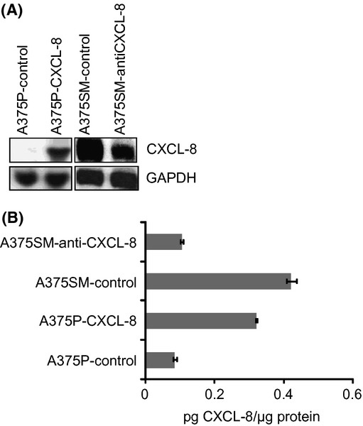
CXCL-8, a chemokine secreted by melanoma and stromal cells, serves as a growth and angiogenic factor for melanoma progression. This study evaluated how modulation of CXCL-8 levels in melanoma cell lines with different tumorigenic and metastatic potentials affected multiple tumor phenotypes. A375P cells (CXCL-8 low expressor) were stably transfected with a CXCL-8 mammalian expression vector to overexpress CXCL-8, whereas A375SM cells (CXCL-8 high expressor) were transfected with a CXCL-8 antisense expression vector to suppress CXCL-8 expression. Subsequent cell proliferation, migration, invasion, and soft-agar colony formation were analyzed, and in vivo tumor growth and metastasis were evaluated using mouse xenograft models. Our data demonstrate that overexpression of CXCL-8 significantly enhanced primary tumor growth and lung metastasis, accompanied by increased microvessel density in vivo, as compared with vector control-transfected cells. We also observed increased clonogenic ability, growth, and invasive potential of CXCL-8 overexpressing cells in vitro. Knockdown of CXCL-8 using an antisense vector resulted in increased cell death and reduced tumor growth relative to control. Taken together, these data confirm that CXCL-8 expression plays a critical role in regulating multiple cellular phenotypes associated with melanoma growth and metastasis.
Keywords: Angiogenesis; CXCL-8; melanoma; metastasis; proliferation.
Figures


Similar articles
-
Interleukin-8 is a key mediator of FKBP51-induced melanoma growth, angiogenesis and metastasis.Br J Cancer. 2015 May 26;112(11):1772-81. doi: 10.1038/bjc.2015.154. Epub 2015 May 5. Br J Cancer. 2015. PMID: 25942396 Free PMC article.
-
Interleukin 10 suppresses tumor growth and metastasis of human melanoma cells: potential inhibition of angiogenesis.Clin Cancer Res. 1996 Dec;2(12):1969-79. Clin Cancer Res. 1996. PMID: 9816156
-
Host CXCR2-dependent regulation of melanoma growth, angiogenesis, and experimental lung metastasis.Cancer Res. 2009 Jan 15;69(2):411-5. doi: 10.1158/0008-5472.CAN-08-3378. Cancer Res. 2009. PMID: 19147552 Free PMC article.
-
Role of interleukin-8 in tumor growth and metastasis of human melanoma.Pathobiology. 1999;67(1):12-8. doi: 10.1159/000028045. Pathobiology. 1999. PMID: 9873223 Review.
-
Why is melanoma so metastatic?Pigment Cell Melanoma Res. 2014 Jan;27(1):19-36. doi: 10.1111/pcmr.12172. Epub 2013 Oct 17. Pigment Cell Melanoma Res. 2014. PMID: 24106873 Review.
Cited by
-
Effect of Ionizing Radiation on Human EA.hy926 Endothelial Cells under Inflammatory Conditions and Their Interactions with A549 Tumour Cells.J Immunol Res. 2019 Sep 2;2019:9645481. doi: 10.1155/2019/9645481. eCollection 2019. J Immunol Res. 2019. PMID: 31565662 Free PMC article.
-
The Proliferation and Differentiation of Adipose-Derived Stem Cells in Neovascularization and Angiogenesis.Int J Mol Sci. 2020 May 27;21(11):3790. doi: 10.3390/ijms21113790. Int J Mol Sci. 2020. PMID: 32471255 Free PMC article. Review.
-
Multi-omics profiling of metastatic colorectal cancer reveals the transcriptional network of focal adhesion and immune suppression and the role of p-RPS6.Cancer Cell Int. 2025 Aug 7;25(1):300. doi: 10.1186/s12935-025-03924-6. Cancer Cell Int. 2025. PMID: 40770803 Free PMC article.
-
Down-regulation of CXCL11 inhibits colorectal cancer cell growth and epithelial-mesenchymal transition.Onco Targets Ther. 2018 Oct 23;11:7333-7343. doi: 10.2147/OTT.S167872. eCollection 2018. Onco Targets Ther. 2018. PMID: 30425523 Free PMC article.
-
Simultaneous blocking of IL-6 and IL-8 is sufficient to fully inhibit CAF-induced human melanoma cell invasiveness.Histochem Cell Biol. 2016 Aug;146(2):205-17. doi: 10.1007/s00418-016-1433-8. Epub 2016 Apr 21. Histochem Cell Biol. 2016. PMID: 27102177
References
-
- Rigel DS, Russak J, Friedman R. The evolution of melanoma diagnosis: 25 years beyond the ABCDs. CA Cancer J. Clin. 2010;60:301–316. - PubMed
-
- Siegel R, Ward E, Brawley O, Jemal A. Cancer statistics, 2011: the impact of eliminating socioeconomic and racial disparities on premature cancer deaths. CA Cancer J. Clin. 2011;61:212–236. - PubMed
-
- Gray-Schopfer V, Wellbrock C, Marais R. Melanoma biology and new targeted therapy. Nature. 2007;445:851–857. - PubMed
-
- Balkwill F. Cancer and the chemokine network. Nat. Rev. Cancer. 2004;4:540–550. - PubMed
Publication types
MeSH terms
Substances
Grants and funding
LinkOut - more resources
Full Text Sources
Medical
