. 2013;3(1):1-15.
Epub 2013 Jan 5.

Optical imaging of tumor microenvironment

Affiliations

Optical imaging of tumor microenvironment

Yihan Wu et al. Am J Nucl Med Mol Imaging. 2013.

Abstract

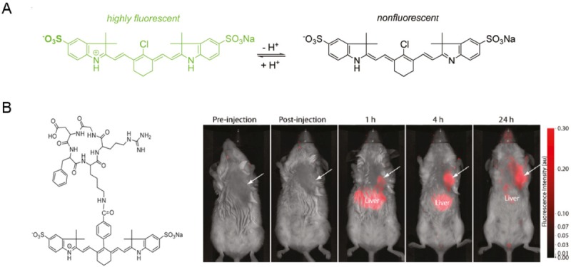
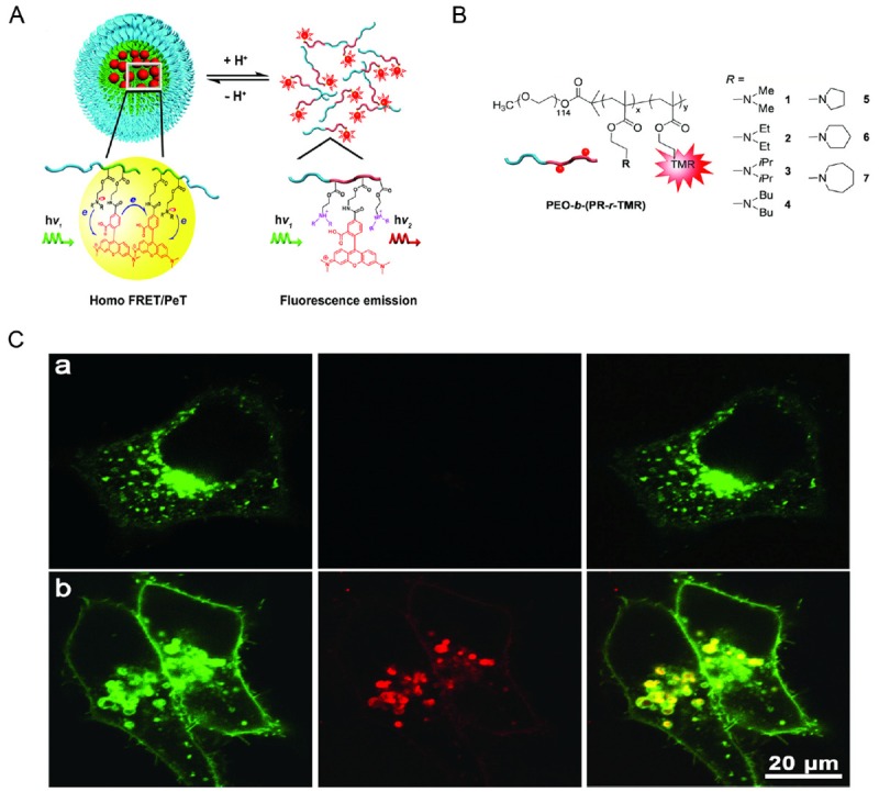
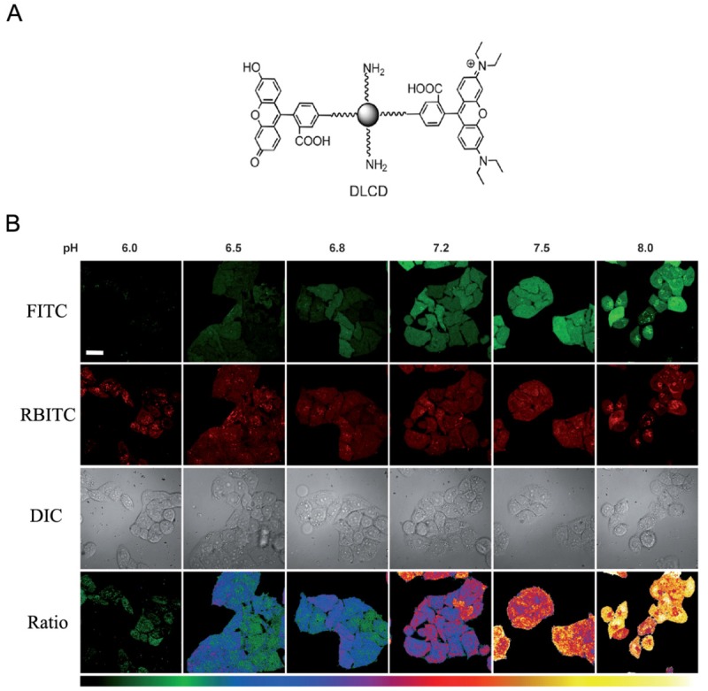
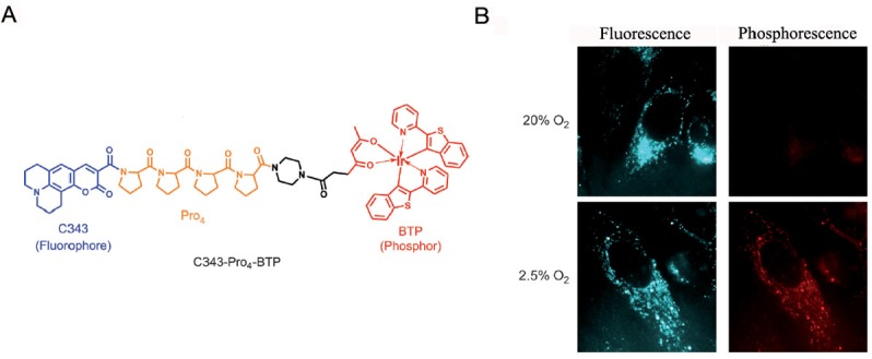
Tumor microenvironment plays important roles in tumor development and metastasis. Features of the tumor microenvironment that are significantly different from normal tissues include acidity, hypoxia, overexpressed proteases and so on. Therefore, these features can serve as not only biomarkers for tumor diagnosis but also theraputic targets for tumor treatment. Imaging modalities such as optical, positron emission tomography (PET) and magnetic resonance imaging (MRI) have been intensively applied to investigate tumor microenvironment. Various imaging probes targeting pH, hypoxia and proteases in tumor microenvironment were thus well developed. In this review, we will focus on recent examples on fluorescent probes for optical imaging of tumor microenvironment. Construction of these fluorescent probes were based on characteristic feature of pH, hypoxia and proteases in tumor microenvironment. Strategies for development of these fluorescent probes and applications of these probes in optical imaging of tumor cells or tissues will be discussed in this review paper.

Keywords: Optical imaging; hypoxia; pH; protease; tumor microenvironment.


Figures

Figure 1
Figure 1
A. Mechanism for activation of pH sensitive Cy dyes. B. Structure of cRGD linked pH activatable Cy dye and its application in optical imaging of orthotopic 4T1/luc tumor, arrows indicated the tumor region. Adapted from reference [15].
Figure 2
Figure 2
A. Design of pH-activatable micellar nanoprobes. B. Different amine groups linked to the micelle backbone to make the nanoprobes activatable at different pH upon protonation of the amine group. C. Fluorescent images of cells treated of pH-activatable nanoprobe with (top panel) or without (bottom panel) the inhibition of lysosomal acidification. Nanoprobe activation was indicated by the red fluorescence signals. Adapted from reference [30].
Figure 3
Figure 3
A. Structure of the dual-labeled carbon nanodot as ratiometric pH nanoprobe. B. Fluorescent images of HeLa cells at pH 6.0, 6.5, 6.8, 7.2, 7.5 and 8.0, respectively. Adapted from reference [32].
Figure 4
Figure 4
A. Chemical structure of ratiometric probe C343-Pro4-BTP. B. Luminescent images of HeLa cells incubated under aerobic and hypoxic conditions. Adapted from reference [39].
Figure 5
Figure 5
A. Chemical structure of 2-nitroimidazoles modified NIR dye Cye. B. In vivo optical imaging of subcutaneous tumors. Adapted from reference [44].
Figure 6
Figure 6
A. Chemical structure of activity-based fluorescent probe LP-1. B. In vivo optical imaging of active legumain using LP-1. Adapted from reference [61].
Figure 7
Figure 7
A. Chemical structure of ANPFAP. B. In vivo imaging of FAPα in C6 and U87MG tumor-bearing mice. Adapted from reference [69].

Similar articles

Cited by

References

    1. Bissell MJ, Hines WC. Why don’t we get more cancer? A proposed role of the microenvironment in restraining cancer progression. Nat Med. 2011;17:320–329. - PMC - PubMed
    1. Albini A, Sporn MB. The tumour microenvironment as a target for chemoprevention. Nat Rev Cancer. 2007;7:139–147. - PubMed
    1. Burden-Gulley SM, Qutaish MQ, Sullivant KE, Lu H, Wang J, Craig SE, Basilion JP, Wilson DL, Brady-Kalnay SM. Novel cryo-imaging of the glioma tumor microenvironment reveals migration and dispersal pathways in vivid three-dimensional detail. Cancer Res. 2011;71:5932–5940. - PMC - PubMed
    1. Vakoc BJ, Lanning RM, Tyrrell JA, Padera TP, Bartlett LA, Stylianopoulos T, Munn LL, Tearney GJ, Fukumura D, Jain RK, Bouma BE. Three-dimensional microscopy of the tumor microenvironment in vivo using optical frequency domain imaging. Nat Med. 2009;15:1219–1223. - PMC - PubMed
    1. Jansen JF, Schoder H, Lee NY, Wang Y, Pfister DG, Fury MG, Stambuk HE, Humm JL, Koutcher JA, Shukla-Dave A. Noninvasive assessment of tumor microenvironment using dynamic contrast-enhanced magnetic resonance imaging and 18F-fluoromisonidazole positron emission tomography imaging in neck nodal metastases. Int J Radiat Oncol Biol Phys. 2010;77:1403–1410. - PMC - PubMed

LinkOut - more resources