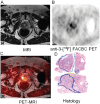
Characterization of primary prostate carcinoma by anti-1-amino-2-[(18)F] -fluorocyclobutane-1-carboxylic acid (anti-3-[(18)F] FACBC) uptake
- PMID: 23342303
- PMCID: PMC3545368
Characterization of primary prostate carcinoma by anti-1-amino-2-[(18)F] -fluorocyclobutane-1-carboxylic acid (anti-3-[(18)F] FACBC) uptake
Abstract
Anti-1-amino-3-[(18)F] fluorocyclobutane-1-carboxylic acid (anti-3-[(18)F] FACBC) is a synthetic amino acid positron emission tomography (PET) radiotracer with utility in the detection of recurrent prostate carcinoma. The aim of this study is to correlate uptake of anti-3-[(18)F] FACBC with histology of prostatectomy specimens in patients undergoing radical prostatectomy and to determine if uptake correlates to markers of tumor aggressiveness such as Gleason score. Ten patients with prostate carcinoma pre-radical prostatectomy underwent 45 minute dynamic PET-CT of the pelvis after IV injection of 347.8 ± 81.4 MBq anti-3-[(18)F] FACBC. Each prostate was co-registered to a separately acquired MR, divided into 12 sextants, and analyzed visually for abnormal focal uptake at 4, 16, 28, and 40 min post-injection by a single reader blinded to histology. SUVmax per sextant and total sextant activity (TSA) was also calculated. Histology and Gleason scores were similarly recorded by a urologic pathologist blinded to imaging. Imaging and histologic analysis were then compared. In addition, 3 representative sextants from each prostate were chosen based on highest, lowest and median SUVmax for immunohistochemical (IHC) analysis of Ki67, synaptophysin, P504s, chromogranin A, P53, androgen receptor, and prostein. 79 sextants had malignancy and 41 were benign. Highest combined sensitivity and specificity was at 28 min by visual analysis; 81.3% and 50.0% respectively. SUVmax was significantly higher (p<0.05) for malignant sextants (5.1±2.6 at 4 min; 4.5±1.6 at 16 min; 4.0±1.3 at 28 min; 3.8±1.0 at 40 min) compared to non-malignant sextants (4.0±1.9 at 4 min; 3.5±0.8 at 16 min; 3.4±0.9 at 28 min; 3.3±0.9 at 40 min), though there was overlap of activity between malignant and non-malignant sextants. SUVmax also significantly correlated (p<0.05) with Gleason score at all time points (r=0.28 at 4 min; r=0.42 at 16 min; r=0.46 at 28 min; r=0.48 at 40 min). There was no significant correlation of anti-3-[(18)F] FACBC SUVmax with Ki-67 or other IHC markers. Since there was no distinct separation between malignant and non-malignant sextants or between Gleason score levels, we believe that anti-3-[(18)F] FACBC PET should not be used alone for radiation therapy planning but may be useful to guide biopsy to the most aggressive lesion.
Keywords: Positron emission tomography (PET); anti-3-[18F] FACBC; prostate carcinoma.
Figures


Similar articles
-
Pilot study of the utility of the synthetic PET amino-acid radiotracer anti-1-amino-3-[(18)F]fluorocyclobutane-1-carboxylic acid for the noninvasive imaging of pulmonary lesions.Mol Imaging Biol. 2013 Oct;15(5):633-43. doi: 10.1007/s11307-012-0606-7. Mol Imaging Biol. 2013. PMID: 23595643 Clinical Trial.
-
Initial experience with the radiotracer anti-1-amino-3-18F-fluorocyclobutane-1-carboxylic acid with PET/CT in prostate carcinoma.J Nucl Med. 2007 Jan;48(1):56-63. J Nucl Med. 2007. PMID: 17204699
-
Pilot evaluation of anti-1-amino-2-[18F] fluorocyclopentane-1-carboxylic acid (anti-2-[18F] FACPC) PET-CT in recurrent prostate carcinoma.Mol Imaging Biol. 2011 Dec;13(6):1272-7. doi: 10.1007/s11307-010-0445-3. Mol Imaging Biol. 2011. PMID: 20976627 Free PMC article.
-
18F-Facbc in Prostate Cancer: A Systematic Review and Meta-Analysis.Cancers (Basel). 2019 Sep 11;11(9):1348. doi: 10.3390/cancers11091348. Cancers (Basel). 2019. PMID: 31514479 Free PMC article. Review.
-
Review of 18F-Fluciclovine PET for Detection of Recurrent Prostate Cancer.Radiographics. 2019 May-Jun;39(3):822-841. doi: 10.1148/rg.2019180139. Radiographics. 2019. PMID: 31059396 Review.
Cited by
-
Prospective evaluation of fluciclovine (18F) PET-CT and MRI in detection of recurrent prostate cancer in non-prostatectomy patients.Eur J Radiol. 2018 May;102:1-8. doi: 10.1016/j.ejrad.2018.02.006. Epub 2018 Feb 24. Eur J Radiol. 2018. PMID: 29685521 Free PMC article.
-
Update on 18F-Fluciclovine PET for Prostate Cancer Imaging.J Nucl Med. 2018 May;59(5):733-739. doi: 10.2967/jnumed.117.204032. Epub 2018 Mar 9. J Nucl Med. 2018. PMID: 29523631 Free PMC article. Review.
-
Emerging Role of Nuclear Medicine in Prostate Cancer: Current State and Future Perspectives.Cancers (Basel). 2023 Sep 27;15(19):4746. doi: 10.3390/cancers15194746. Cancers (Basel). 2023. PMID: 37835440 Free PMC article. Review.
-
PET Molecular Imaging-Directed Biopsy: A Review.AJR Am J Roentgenol. 2017 Aug;209(2):255-269. doi: 10.2214/AJR.17.18047. Epub 2017 May 15. AJR Am J Roentgenol. 2017. PMID: 28504563 Free PMC article. Review.
-
Functional and molecular imaging: applications for diagnosis and staging of localised prostate cancer.Clin Oncol (R Coll Radiol). 2013 Aug;25(8):451-60. doi: 10.1016/j.clon.2013.05.001. Epub 2013 May 27. Clin Oncol (R Coll Radiol). 2013. PMID: 23722008 Free PMC article. Review.
References
-
- Siegel R, Naishadham D, Jemal A. Cancer statistics, 2012. CA Cancer J Clin. 2012;62:10–29. - PubMed
-
- Schoder H, Larson SM. Positron emission tomography for prostate, bladder, and renal cancer. Semin Nucl Med. 2004;34:274–292. - PubMed
-
- Brawer MK, Ploch NR, Bigler SA. Prostate cancer tumor location as predicted by digital rectal examination transferred to ultrasound and ultrasound-guided prostate needle biopsy. J Cell Biochem Suppl. 1992;16H:74–77. - PubMed
Grants and funding
LinkOut - more resources
Full Text Sources
Other Literature Sources
Medical
Research Materials
Miscellaneous
