Comparative Study
. 2013 Feb;4(2):161-75.
doi: 10.4155/tde.12.144.

Nanomedicine for uterine leiomyoma therapy

Affiliations
Comparative Study

Nanomedicine for uterine leiomyoma therapy

Hazem Ali et al. Ther Deliv. 2013 Feb.

Abstract

Background: The purpose of this work was to engineer polymeric nanoparticles to encapsulate and deliver 2-methoxyestradiol, a potential antitumor drug for treatment of uterine leiomyoma (fibroids), the most common hormone-dependent pathology affecting women of reproductive age.

Methods/results: Encapsulation efficiency and drug release from the nanoparticles were monitored by HPLC. Cell morphology and in vitro cytotoxicity experiments were carried out in a human leiomyoma cell line. The nanoparticles displayed high encapsulation efficiency (>86%), which was verified by differential scanning calorimetry and x-ray diffraction. Excellent long-term stability of the nanoparticles and gradual drug release without burst were also observed. Cellular uptake of fluorescent nanoparticles was confirmed by confocal imaging. The drug-loaded poly(lactic acid) and poly(lactic-co-glycolic acid) nanoparticles induced cytotoxicity in human leiomyoma cells to a significantly greater extent than the free drug at 0.35 µM.

Conclusion: This novel approach represents a potential fertility-preserving alternative to hysterectomy.


Conflict of interest statement

Disclosure

The authors declare no competing financial interests.

Figures
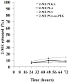
Figure 1
Figure 1. In vitro release of 2-ME-loaded nanoparticles at 37°C in PBS (pH=7.4) containing 1.0% Tween® 80 to maintain sink conditions
Scintillation vials were prepared for each time point and shaken (50 rpm) in a hot box (37°C). At each time point, 500 μL of the nanoparticle dispersion were placed in the upper chamber of the filter assembly as explained in the Materials & Methods section. The assembly was then centrifuged at 3000 rpm for 15 minutes at 25°C to separate the released 2-ME from the nanoparticles. The filtrate was diluted with the mobile phase and injected into HPLC to determine the concentration of the released 2-ME. Experiments were carried out in triplicate, and results are expressed as mean ± standard deviation. PBS: Phosphate Buffer Saline; 2-ME: 2-methoxyestradiol; HPLC: High Performance Liquid Chromatography.
Figure 2
Figure 2. DSC thermographs of 2-ME-loaded nanoparticles composed of PLA (A); PLGA (B); PSA (C); and PSA-co-PEG (D)
Samples (2–3 mg), hermetically sealed in aluminum pans, were heated at a scan rate of 10°C/min from 20°C to 200°C and then allowed to cool to 10°C to observe the exothermic phase. The generated data were analyzed with Universal Analysis 2000 software. Graphs were displaced along the ordinate for better visualization. DSC: Differential Scanning Calorimetry; PLA: polylactide; PLGA: carboxylate end group 50:50 poly(D,L-lactide-co-glycolide); PSA: poly(sebacic) acid; PSA-co-PEG: poly(sebacic acid-co-PEG); PEG: Polyethylene glycol.
Figure 3
Figure 3. X-ray diffraction patterns of 2-ME, blank nanoparticles, and 2-ME-loaded nanoparticles composed of PLA (A); PLGA (B); PSA (C); and PSA-co-PEG (D)
Patterns were obtained by wide-angle X-ray scattering (2θ=5–30°, step size = 0.04). Data were recorded at an anode voltage of 40 kV and a current of 30 mA. The generated data were processed through FIT2D software to convert the image into a two-dimensional plot. Graphs were displaced along the ordinate for better visualization. 2-ME: 2-methoxyestradiol; PEG: Polyethylene glycol; PLA: polylactide; PLGA: carboxylate end group 50:50 poly(D,L-lactide-co-glycolide); PSA: poly(sebacic) acid; PSA-co-PEG: poly(sebacic acid-co-PEG).
Figure 4
Figure 4. Cytotoxic effects on huLM cells of 0.35 μM 2-ME (freely dissolved in culture medium containing ≤ 0.03% ethanol), 2-ME-loaded nanoparticles formed of PLA, PLGA, PSA, and PSA-co-PEG (2-ME concentration = 0.35 μM), or blank/unloaded nanoparticles (with total polymer concentrations equal to that of the drug-loaded nanoparticles)
Cells were initially plated at a density of 2×10³ cells/well (n=6) in 96-well plates and exposed to formulation-supplemented media for 48 hours. Cell viability was determined using the MTT colorimetric assay. Vertical bars indicate the mean cell viability as percent of control ± standard deviation. *P < 0.05 as compared to control (cells exposed only to cell culture medium). 2-ME: 2-methoxyestradiol; PEG: Polyethylene glycol; PLA: polylactide; PLGA: carboxylate end group 50:50 poly(D,L-lactide-co-glycolide); PSA: poly(sebacic) acid; PSA-co-PEG: poly(sebacic acid-co-PEG); huLM: human leiomyoma cell line; MTT: (3-[(4,5-dimethylthiazol-2-yl]-2,5-diphenyl tetrazolium bromide).
Figure 5
Figure 5. Morphology of huLM cells after treatment with nanoparticles and controls
(A) 0.1 % Triton-X-100 (positive control), (B) culture media with ≤ 0.03% ethanol (negative control), (C, D) 2-ME freely dissolved in ethanol/culture media at 1.5 μM and 10μM, respectively, (E,F) 2-ME-loaded PLA and PLGA nanoparticles, respectively (10 μM 2-ME), and (G,H) 2-ME-loaded PSA and PSA-co-PEG nanoparticles, respectively (10 μM 2-ME). HuLM cells were seeded at a density of 7×10⁴ cells/well. Arrows represent dead cells. Photographs were taken by a Nikon Eclipse TS100 microscope at 10X magnification after 48 hours of treatment. 2-ME: 2-methoxyestradiol; PEG: Polyethylene glycol; PLA: polylactide; PLGA: carboxylate end group 50:50 poly(D,L-lactide-co-glycolide); PSA: poly(sebacic) acid; PSA-co-PEG: poly(sebacic acid-co-PEG); huLM: Human leiomyoma cell line.
Figure 6
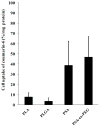
Figure 6. Quantitative cell uptake of coumarin-6-loaded nanoparticles
HuLM cells were plated at a density of 1×10⁴ cells/well (6 wells/treatment) in 24-well plates and exposed to treatments diluted with HBSS for 2 hours. Cell uptake was calculated by measuring the fluorescence at 485 nm (excitation) and 528 nm (emission) after lysing the cells with 0.5% Triton X-100 in 0.2N sodium hydroxide solution. The protein content was determined by using Pierce® BCA Protein Assay Kit according to the manufacturer’s protocol. Data are presented as the percentage of fluorescent nanoparticles administered to the cells per mg of total cellular protein. Vertical bars indicate the average ± standard deviation (n=6). huLM: Human Leiomyoma Cell line; HBSS: Hank’s Balanced Salt Solution.
Figure 7
Figure 7. Confocal laser microscopy images of huLM cells after 2-hour incubation with coumarin-6-loaded PLGA nanoparticles (A) and HBSS (negative control) (B) at 37°C
Upper left are nuclei counterstained with DAPI (blue), lower left are cell membrane counterstained with Wheat Germ Agglutinin, Alexa Fluor® 594 conjugate (red), upper right are coumarin-6-loaded nanoparticles (green), and lower right is an overlaid image. huLM: Human Leiomyoma Cell line; PLGA: carboxylate end group 50:50 poly(D,L-lactide-co-glycolide); HBSS: Hank’s Balanced Salt Solution.

References

    1. Zhang D, Al-Hendy M, Richard-Davis G, Montgomery-Rice V, Rajaratnam V, Al-Hendy A. Antiproliferative and proapoptotic effects of epigallocatechin gallate on human leiomyoma cells. Fertil Steril. 2010;94(5):1887–1893. - PMC - PubMed
    1. Stewart EA. Uterine fibroids. Lancet. 2001;357(9252):293–298. Uterine leiomyoma results in heavy bleeding during menstruation for longer periods of times. Hystrectomy is considered one of the treatment options. - PubMed
    1. Al-Hendy A, Salama SA. Catechol-O-methyltransferase polymorphism is associated with increased uterine leiomyoma risk in different ethnic groups. J Soc Gynecol Investig. 2006;13(2):136–144. - PubMed
    1. Othman EE, Al-Hendy A. Molecular genetics and racial disparities of uterine leiomyomas. Best Pract Res Clin Obstet Gynaecol. 2008;22(4):589–601. - PMC - PubMed
    1. Martel KM, Ko AC, Christman GM, Stribley JM. Apoptosis in human uterine leiomyomas. Semin Reprod Med. 2004;22(2):91–103. - PubMed

Publication types

MeSH terms

LinkOut - more resources