

Development of adenovirus capsid proteins for targeted therapeutic delivery
- PMID: 23343164
- PMCID: PMC3645976
- DOI: 10.4155/tde.12.155
Development of adenovirus capsid proteins for targeted therapeutic delivery
Abstract
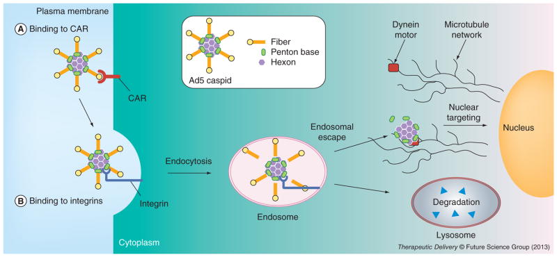
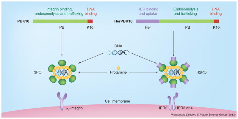
The outer shell of the adenovirus capsid comprises three major types of protein (hexon, penton base and fiber) that perform the majority of functions facilitating the early stages of adenovirus infection. These stages include initial cell-surface binding followed by receptor-mediated endocytosis, endosomal penetration and cytosolic entry, and intracellular trafficking toward the nucleus. Numerous studies have shown that the penton base contributes to several of these steps and have supported the development of this protein into a delivery agent for therapeutic molecules. Studies revealing that the fiber and hexon bear unexpected properties of cell entry and/or nuclear homing have supported the development of these capsid proteins, as well into potential delivery vehicles. This review summarizes the findings to date of the protein-cell activities of these capsid proteins in the absence of the whole virus and their potential for therapeutic application with regard to the delivery of foreign molecules.
Figures
Similar articles
-
Typical and atypical trafficking pathways of Ad5 penton base recombinant protein: implications for gene transfer.Gene Ther. 2006 May;13(10):821-36. doi: 10.1038/sj.gt.3302729. Gene Ther. 2006. PMID: 16482205
-
Endocytosis of adenovirus and adenovirus capsid proteins.Adv Drug Deliv Rev. 2003 Nov 14;55(11):1485-96. doi: 10.1016/j.addr.2003.07.010. Adv Drug Deliv Rev. 2003. PMID: 14597142 Review.
-
Symmetry types, systems and their multiplicity in the structure of adenovirus capsid. I. Symmetry networks and general symmetry motifs.Acta Microbiol Immunol Hung. 2006 Mar;53(1):1-23. doi: 10.1556/AMicr.53.2006.1.1. Acta Microbiol Immunol Hung. 2006. PMID: 16696547
-
Novel fiber-dependent entry mechanism for adenovirus serotype 5 in lacrimal acini.J Virol. 2006 Dec;80(23):11833-51. doi: 10.1128/JVI.00857-06. Epub 2006 Sep 20. J Virol. 2006. PMID: 16987972 Free PMC article.
-
Novel partner proteins of adenovirus penton.Curr Top Microbiol Immunol. 2003;272:37-55. doi: 10.1007/978-3-662-05597-7_2. Curr Top Microbiol Immunol. 2003. PMID: 12747546 Review.
Cited by
-
Genetic-Driven Druggable Target Identification and Validation.Trends Genet. 2018 Jul;34(7):558-570. doi: 10.1016/j.tig.2018.04.004. Epub 2018 May 23. Trends Genet. 2018. PMID: 29803319 Free PMC article. Review.
-
IRAM: virus capsid database and analysis resource.Database (Oxford). 2019 Jan 1;2019:baz079. doi: 10.1093/database/baz079. Database (Oxford). 2019. PMID: 31318422 Free PMC article.
-
Interaction between hexon and L4-100K determines virus rescue and growth of hexon-chimeric recombinant Ad5 vectors.Sci Rep. 2016 Mar 3;6:22464. doi: 10.1038/srep22464. Sci Rep. 2016. PMID: 26934960 Free PMC article.
-
Engineered Oncolytic Adenoviruses: An Emerging Approach for Cancer Therapy.Pathogens. 2022 Oct 4;11(10):1146. doi: 10.3390/pathogens11101146. Pathogens. 2022. PMID: 36297203 Free PMC article. Review.
-
HER3-targeted protein chimera forms endosomolytic capsomeres and self-assembles into stealth nucleocapsids for systemic tumor homing of RNA interference in vivo.Nucleic Acids Res. 2019 Dec 2;47(21):11020-11043. doi: 10.1093/nar/gkz900. Nucleic Acids Res. 2019. PMID: 31617560 Free PMC article.
References
-
- Van Oostrum J, Smith PR, Mohraz M, Burnett RM. The structure of the adenovirus capsid. III. Hexon packing determined from electron micrographs of capsid fragments. J Mol Biol. 1987;198(1):73–89. - PubMed
-
- Medina-Kauwe LK. Endocytosis of adenovirus and adenovirus capsid proteins. Adv Drug Deliv Rev. 2003;55(11):1485–1496. - PubMed
-
- Boudin ML, Boulanger P. Assembly of adenovirus penton base and fiber. Virology. 1982;116(2):589–604. - PubMed
Publication types
MeSH terms
Substances
Grants and funding
LinkOut - more resources
Full Text Sources
Other Literature Sources