

Up-regulation of COX-2/PGE2 by endothelin-1 via MAPK-dependent NF-κB pathway in mouse brain microvascular endothelial cells
- PMID: 23343326
- PMCID: PMC3560266
- DOI: 10.1186/1478-811X-11-8
Up-regulation of COX-2/PGE2 by endothelin-1 via MAPK-dependent NF-κB pathway in mouse brain microvascular endothelial cells
Abstract
Background: Endothelin-1 (ET-1) is a proinflammatory mediator and elevated in the regions of several brain injury and inflammatory diseases. The deleterious effects of ET-1 on endothelial cells may aggravate brain inflammation mediated through the regulation of cyclooxygenase-2 (COX-2)/prostaglandin E2 (PGE2) system in various cell types. However, the signaling mechanisms underlying ET-1-induced COX-2 expression in brain microvascular endothelial cells remain unclear. Herein we investigated the effects of ET-1 in COX-2 regulation in mouse brain microvascular endothelial (bEnd.3) cells.
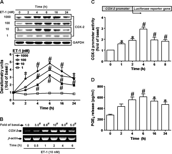
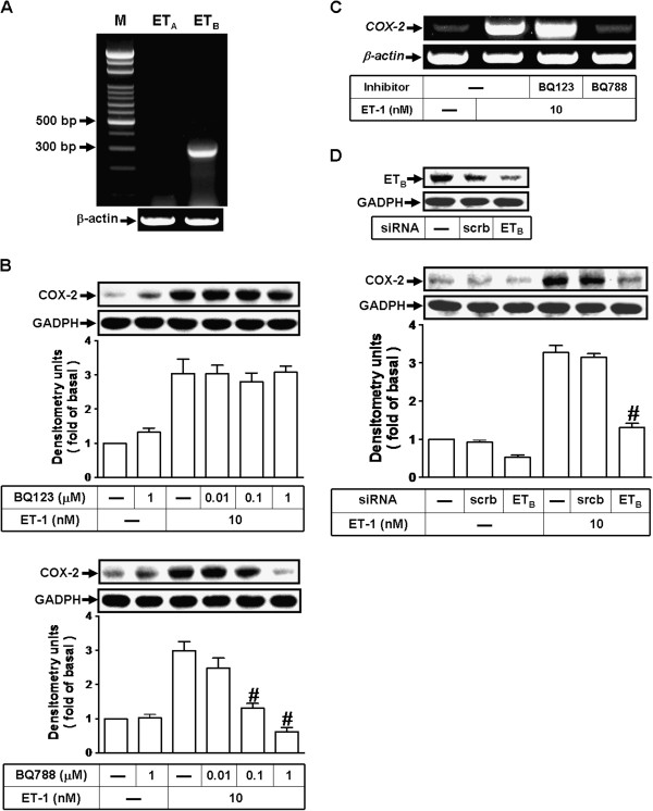
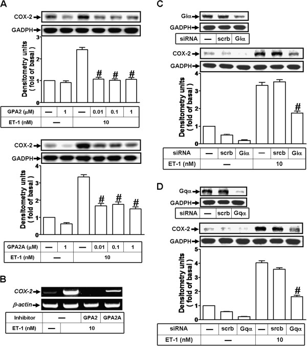
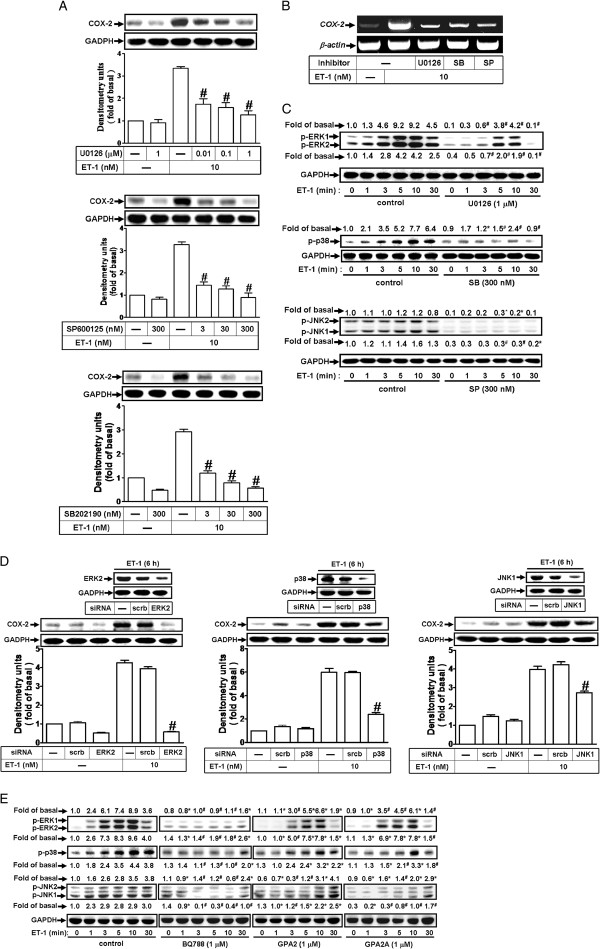
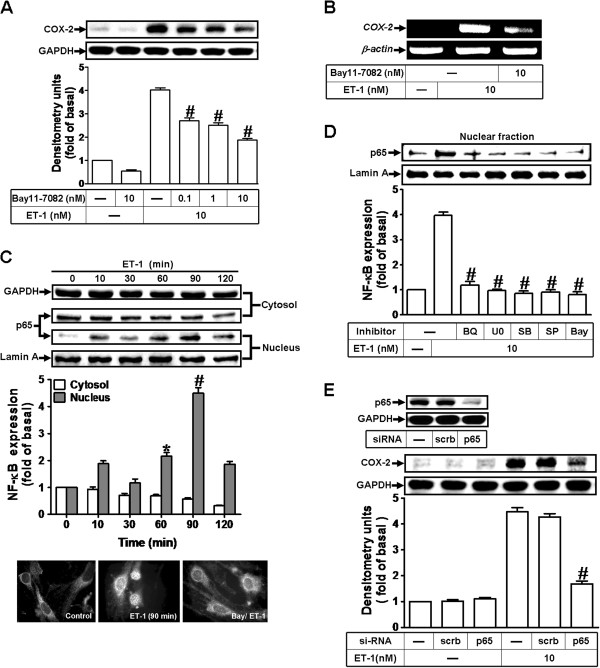
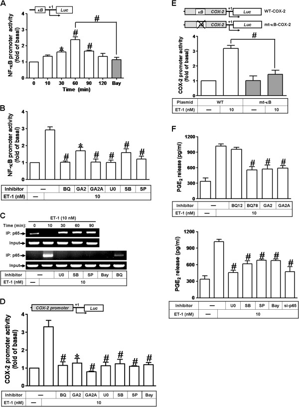
Results: The data obtained with Western blotting, RT-PCR, and immunofluorescent staining analyses showed that ET-1-induced COX-2 expression was mediated through an ETB-dependent transcriptional activation. Engagement of Gi- and Gq-protein-coupled ETB receptors by ET-1 led to phosphorylation of ERK1/2, p38 MAPK, and JNK1/2 and then activated transcription factor NF-κB. Moreover, the data of chromatin immunoprecipitation (ChIP) and promoter reporter assay demonstrated that the activated NF-κB was translocated into nucleus and bound to its corresponding binding sites in COX-2 promoter, thereby turning on COX-2 gene transcription. Finally, up-regulation of COX-2 by ET-1 promoted PGE2 release in these cells.
Conclusions: These results suggested that in mouse bEnd.3 cells, activation of NF-κB by ETB-dependent MAPK cascades is essential for ET-1-induced up-regulation of COX-2/PGE2 system. Understanding the mechanisms of COX-2 expression and PGE2 release regulated by ET-1/ETB system on brain microvascular endothelial cells may provide rationally therapeutic interventions for brain injury or inflammatory diseases.
Figures
Similar articles
-
Upregulation of COX-2/PGE2 by ET-1 mediated through Ca2+-dependent signals in mouse brain microvascular endothelial cells.Mol Neurobiol. 2014 Jun;49(3):1256-69. doi: 10.1007/s12035-013-8597-1. Epub 2013 Nov 28. Mol Neurobiol. 2014. PMID: 24287977
-
c-Src-dependent EGF receptor transactivation contributes to ET-1-induced COX-2 expression in brain microvascular endothelial cells.J Neuroinflammation. 2012 Jul 2;9:152. doi: 10.1186/1742-2094-9-152. J Neuroinflammation. 2012. PMID: 22747786 Free PMC article.
-
Sphingosine 1-Phosphate Induces Cyclooxygenase-2/Prostaglandin E2 Expression via PKCα-dependent Mitogen-Activated Protein Kinases and NF-κB Cascade in Human Cardiac Fibroblasts.Front Pharmacol. 2020 Oct 30;11:569802. doi: 10.3389/fphar.2020.569802. eCollection 2020. Front Pharmacol. 2020. PMID: 33192511 Free PMC article.
-
Thrombin Induces COX-2 and PGE2 Expression via PAR1/PKCalpha/MAPK-Dependent NF-kappaB Activation in Human Tracheal Smooth Muscle Cells.Mediators Inflamm. 2022 Apr 19;2022:4600029. doi: 10.1155/2022/4600029. eCollection 2022. Mediators Inflamm. 2022. PMID: 35497094 Free PMC article.
-
BK Induces cPLA2 Expression via an Autocrine Loop Involving COX-2-Derived PGE2 in Rat Brain Astrocytes.Mol Neurobiol. 2015;51(3):1103-15. doi: 10.1007/s12035-014-8777-7. Epub 2014 Jun 12. Mol Neurobiol. 2015. PMID: 24915969
Cited by
-
Endothelin-1 dependent expression of GAG genes involves NOX and p38 mediated Smad linker region phosphorylation.Clin Exp Pharmacol Physiol. 2022 Jul;49(7):710-718. doi: 10.1111/1440-1681.13650. Epub 2022 May 17. Clin Exp Pharmacol Physiol. 2022. PMID: 35527471 Free PMC article.
-
TLR2, TLR4 and CD14 recognize venom-associated molecular patterns from Tityus serrulatus to induce macrophage-derived inflammatory mediators.PLoS One. 2014 Feb 7;9(2):e88174. doi: 10.1371/journal.pone.0088174. eCollection 2014. PLoS One. 2014. PMID: 24516606 Free PMC article.
-
The cardiovascular effect of the uremic solute indole-3 acetic acid.J Am Soc Nephrol. 2015 Apr;26(4):876-87. doi: 10.1681/ASN.2013121283. Epub 2014 Aug 21. J Am Soc Nephrol. 2015. PMID: 25145928 Free PMC article.
-
Endothelial cell dysfunction in viral hemorrhage and edema.Front Microbiol. 2015 Jan 5;5:733. doi: 10.3389/fmicb.2014.00733. eCollection 2014. Front Microbiol. 2015. PMID: 25601858 Free PMC article.
-
Anthocyanin-rich fractions from red raspberries attenuate inflammation in both RAW264.7 macrophages and a mouse model of colitis.Sci Rep. 2014 Aug 29;4:6234. doi: 10.1038/srep06234. Sci Rep. 2014. PMID: 25167935 Free PMC article.
References
LinkOut - more resources
Full Text Sources
Other Literature Sources
Research Materials
Miscellaneous
