

Review
doi: 10.3109/17453674.2013.765626.
Epub 2013 Jan 23.
The role of the acetabular labrum in hip dysplasia. A literature overview
Affiliations
- PMID: 23343376
- PMCID: PMC3584604
- DOI: 10.3109/17453674.2013.765626
Item in Clipboard
Review
The role of the acetabular labrum in hip dysplasia. A literature overview
Acta Orthop.
2013 Feb.
Abstract
A periacetabular osteotomy (PAO) is the preferred joint preserving treatment for young adults with symptomatic hip dysplasia and no osteoarthritis. In symptomatic dysplasia of the hip, there is labral pathology in up to 90% of cases. However, no consensus exists as to whether a labral tear should be treated before the periacetabular osteotomy (PAO), treated simultaneously with the PAO, or left alone and only treated if symptoms persist after the PAO. This review is an update of aspects of labral anatomy and function, the etiology of labral tears in hip dysplasia, and diagnostic assessment of labral tears, and we discuss treatment strategies for coexisting labral tears and hip dysplasia.
Figures
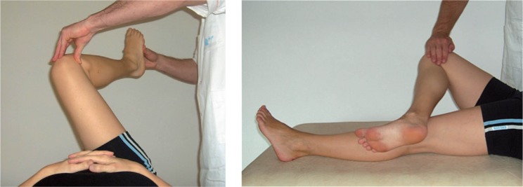
Demonstration of (a) the impingement test, performed by passively moving the hip joint into 90o of flexion, internal rotation, and adduction, and (b) the FABER test. Both tests are considered positive if groin pain is produced (Reproduced with permission from Troelsen et al., Acta Orthop 2009; 80 (3): 314-8). A: acetabulum; FH: femoral head.
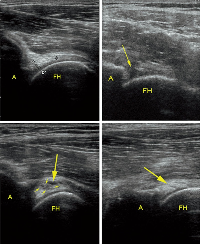
Ultrasound examination of the acetabular labrum. A. A normal labrum. The crosses mark the base and the apex of the labrum (“D1” marking is automatically supplied with the crosses). B. An acetabular labral tear with detachment (arrow). C. A labral tear with intra-substance cystic formation (large arrow) and a slightly irregular hypoechoic cleft (small arrows). D. An intra-substance linear labral tear (arrow). (Reproduced with permission from Troelsen et al., Acta Radiol 2007; 48: 1004-10).
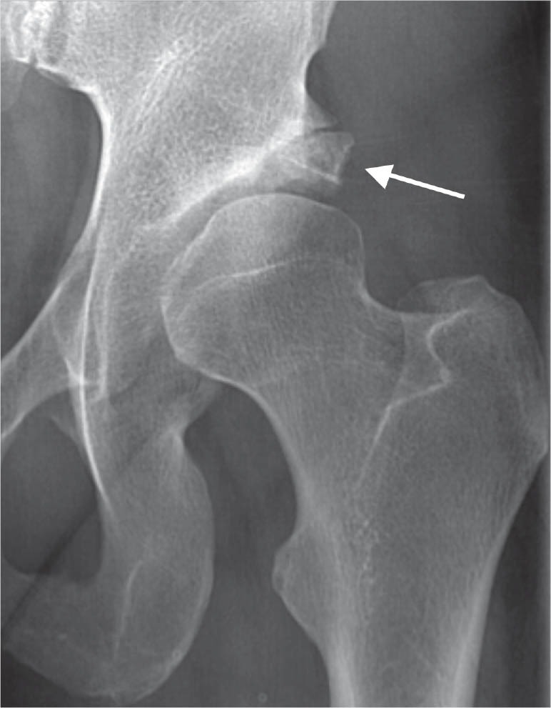
A dysplastic hip, with arrow showing a large os acetabuli or rim fractures.
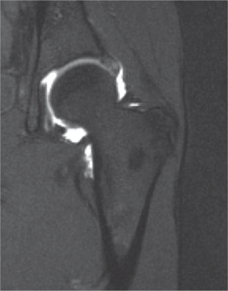
Coronal section MR arthrography of a dysplastic hip. The labrum is hypertrophic and contrast medium is running through the base of the labrum, an indication that the labrum is detached from the acetabular rim.
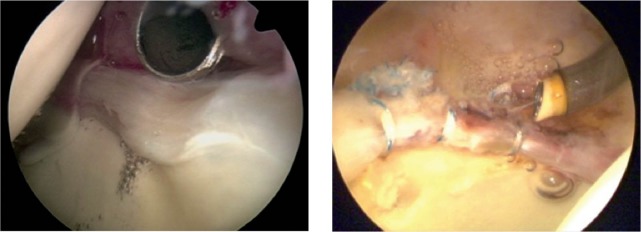
Arthroscopic pictures illustrating (a) a detached labrum with wave sign and synovitis, and (b) a labrum re-attached to the acetabular rim with 3 suture anchors. (The images were kindly provided by the Department of Sports Traumatology, Aarhus University Hospital, Denmark).
References
-
- Abe I, Harada Y, Oinuma K, et al. Acetabular labrum: Abnormal findings at MR imaging in asymptomatic hips. Radiology. 2000;216(2):576–81. - PubMed
-
- Burnett RS, Della Rocca GJ, Prather H, et al. Clinical presentation of patients with tears of the acetabular labrum. J Bone Joint Surg (Am) 2006;88(7):1448–57. - PubMed
-
- Byrd JW, Jones KS. Hip arthroscopy in the presence of dysplasia. Arthroscopy. 2003;19(10):1055–60. - PubMed
-
- Byrd JW, Jones KS. Hip arthroscopy for labral pathology: Prospective analysis with 10-year follow-up. Arthroscopy. 2009;25(4):365–8. - PubMed
-
- Cashin M, Uhthoff H, O’Neill M, Beaule PE. Embryology of the acetabular labral-chondral complex. J Bone Joint Surg (Br) 2008;90(8):1019–24. - PubMed
Publication types
MeSH terms
LinkOut - more resources
Full Text Sources
Other Literature Sources
Medical