. 2013 Feb 27;135(8):3173-85.
doi: 10.1021/ja311408y. Epub 2013 Feb 8.

Fully convergent chemical synthesis of ester insulin: determination of the high resolution X-ray structure by racemic protein crystallography

Affiliations

Fully convergent chemical synthesis of ester insulin: determination of the high resolution X-ray structure by racemic protein crystallography

Michal Avital-Shmilovici et al. J Am Chem Soc. .

Abstract

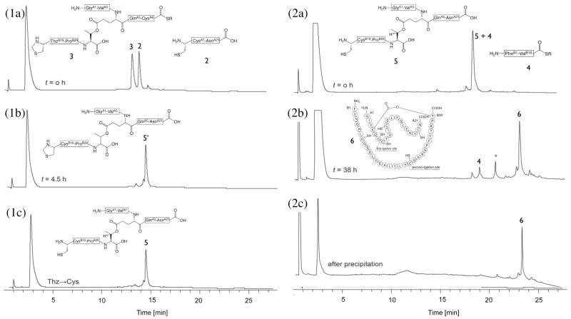
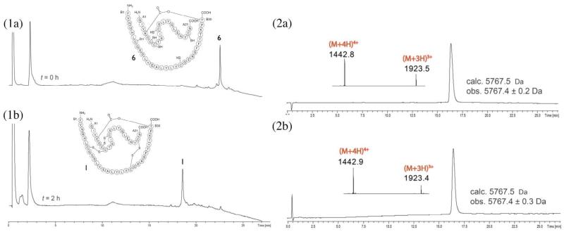
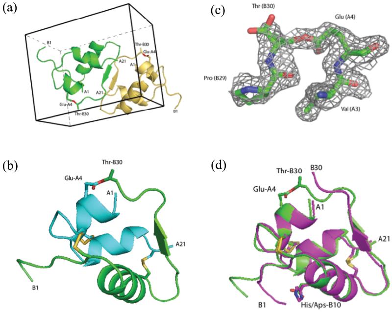
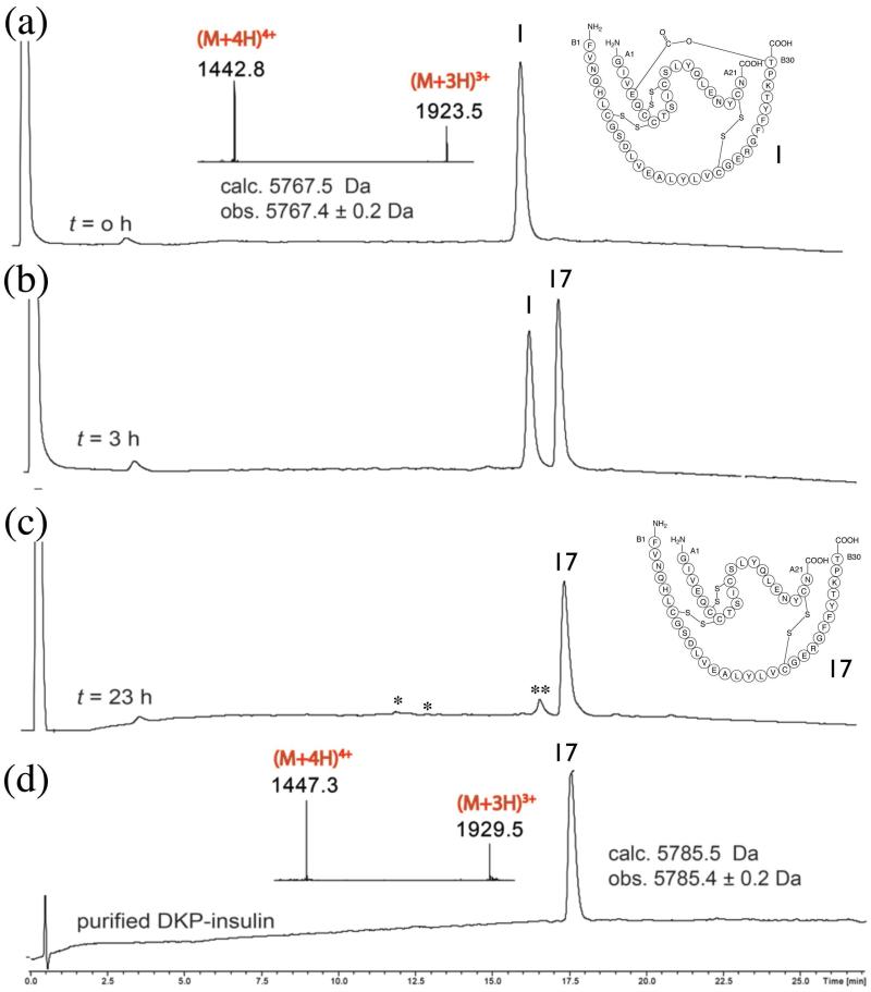
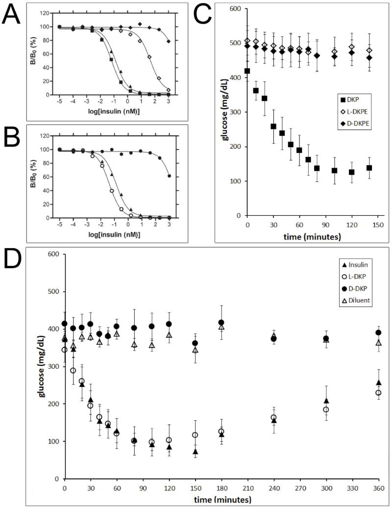
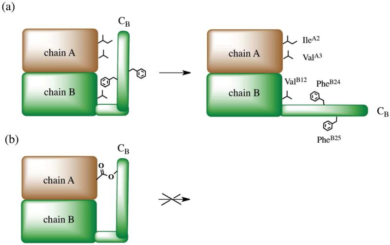
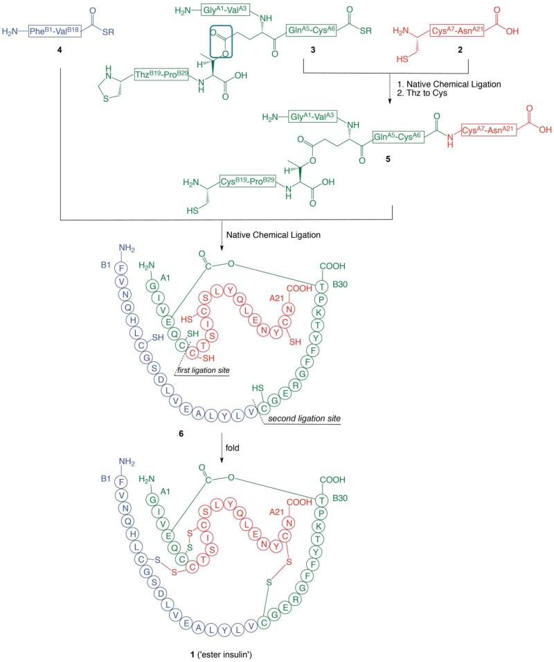
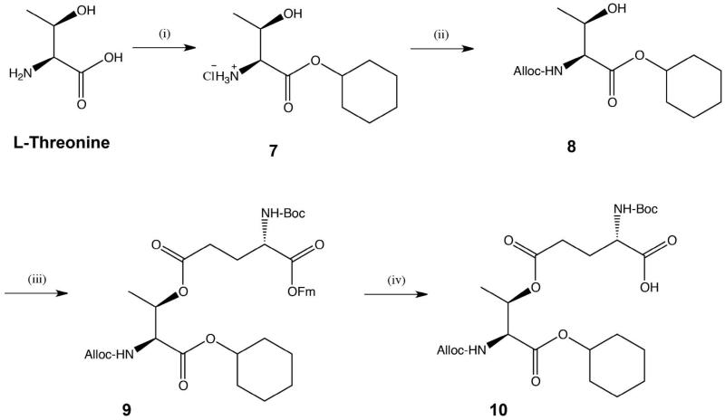
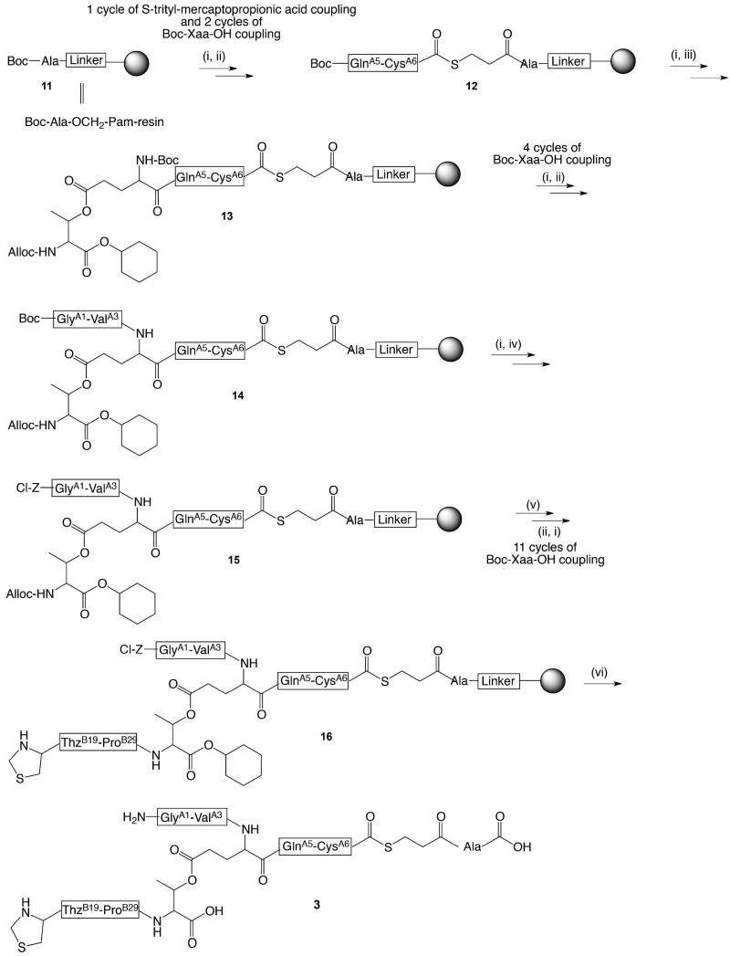
Efficient total synthesis of insulin is important to enable the application of medicinal chemistry to the optimization of the properties of this important protein molecule. Recently we described "ester insulin"--a novel form of insulin in which the function of the 35 residue C-peptide of proinsulin is replaced by a single covalent bond--as a key intermediate for the efficient total synthesis of insulin. Here we describe a fully convergent synthetic route to the ester insulin molecule from three unprotected peptide segments of approximately equal size. The synthetic ester insulin polypeptide chain folded much more rapidly than proinsulin, and at physiological pH. Both the D-protein and L-protein enantiomers of monomeric DKP ester insulin (i.e., [Asp(B10), Lys(B28), Pro(B29)]ester insulin) were prepared by total chemical synthesis. The atomic structure of the synthetic ester insulin molecule was determined by racemic protein X-ray crystallography to a resolution of 1.6 Å. Diffraction quality crystals were readily obtained from the racemic mixture of {D-DKP ester insulin + L-DKP ester insulin}, whereas crystals were not obtained from the L-ester insulin alone even after extensive trials. Both the D-protein and L-protein enantiomers of monomeric DKP ester insulin were assayed for receptor binding and in diabetic rats, before and after conversion by saponification to the corresponding DKP insulin enantiomers. L-DKP ester insulin bound weakly to the insulin receptor, while synthetic L-DKP insulin derived from the L-DKP ester insulin intermediate was fully active in binding to the insulin receptor. The D- and L-DKP ester insulins and D-DKP insulin were inactive in lowering blood glucose in diabetic rats, while synthetic L-DKP insulin was fully active in this biological assay. The structural basis of the lack of biological activity of ester insulin is discussed.


Figures

Figure 1
Figure 1. One pot reactions for the total chemical synthesis of the ester insulin polypeptde 6
(1) Native chemical ligation for the synthesis of GlyA1-GluA4[OβThrB30-CysB19]-AsnA21 (5). (1a-b) Reaction of peptide segments GlyA1-GluA4[OβThrB30-ThzB19]-CysA6-αCOSR (3) (R = –CH2CH2CO–Ala–COOH) and CysA7-AsnA21 (2). (1c) Conversion of Thz- to Cys-. The chromatographic separations were performed on a C18 column using a linear gradient (9%–53%) of buffer B in buffer A over 22 min (buffer A = 0.1% TFA in water, buffer B = 0.08% TFA in acetonitrile). (2) Native chemical ligation in the synthesis of GlyA1-GluA4[OβThrB30-PheB1]-AsnA21 (6). (2a-b) Reaction of GlyA1-GluA4[OβThrB30-CysB19])-AsnA21 (5) and PheB1-ValB18-αCOSR (4). Both peptide segments have the same retention time, as confirmed by LC-MS. Upon completion, excess PheB1-ValB18-αCOSR (4) remained. *: PheB1-ValB18-αCO MPAA thioester. (2c) Crude polypeptide (6) after precipitation from water. The chromatographic separations were performed on a C18 column using a linear gradient (4%–48%) of buffer B in buffer A over 22 min. All analyses were by LC-MS; only the HPLC data are shown.
Figure 2
Figure 2. Folding of ester insulin
Folding was carried at room temperature in a pH 7.6 buffer solution containing 1.5 M GnHCl, 20 mM Tris, 8 mM cysteine, 1 mM cystine·HCl, at a starting polypeptide concentration of 0.3 mg/mL. (1a) Crude linear polypeptide ester insulin (6) at t = 0 h. (1b) Folding reaction after 2 h. The chromatographic separations were performed on a C18 column using a linear gradient (4%–48%) of buffer B in buffer A over 22 min (buffer A = 0.1% TFA in water, buffer B = 0.08% TFA in acetonitrile). (2a) Folded L-ester insulin after prep HPLC purification(Inset: on-line ESI-MS spectra of the main peak). (2b) Folded D-ester insulin after prep HPLC purification (Inset: on-line ESI-MS spectra of the main peak). The chromatographic separations were performed on a C18 column using a linear gradient (9%–53%) of buffer B in buffer A over 22 min.
Figure 3
Figure 3. X-ray structure of ester insulin (1.6 Å), obtained from racemic crystallization of D-DKP ester insulin and L-DKP ester insulin
(a) Racemic crystal packing in centrosymmetric P1 unit cell, showing the L-DKP ester insulin molecule (green) and the D-DKP ester insulin molecule (gold). The ester linkage is shown in sticks (O, red). (b) Cartoon representation of the A chain (cyan) and B-chain (green) of L-DKP ester insulin. The three disulfide bonds of the folded protein and the ester linkage are shown as sticks (O, red; S, gold). (c) SigmaA-weighted 2Fo-Fc electron density map, contoured at 1.5σ, showing the ester linkage between GluA4-ThrB30 (C, green; O, red; N, blue). (d) Superposition of X-ray crystal structures the T-form of KP insulin (PDB ID: 1LPH) (magenta) and DKP ester insulin (green). The three disulfide bonds and the ester linkage are shown as sticks (O, red; N, blue S, gold).
Figure 4
Figure 4. Conversion of DKP ester insulin to native DKP insulin
Saponification of ester insulin 1 to give DKP insulin 17: (a) Ester insulin 1 at t = o h (Inset: on line ESI-MS spectra of the main peak). (b,c) Reaction mixture at (b) t = 3 h and (c) t = 23 h. *: derived from A chain. **: oxidized B-chain. (d) Purified DKP insulin (Inset: on-line ESI-MS spectra of the main peak). The observed increase of 18 Da for the hydrolyzed DKP insulin confirms the conversion of ester insulin into DKP insulin by addition of the elements of water. The chromatographic separations were performed on a C18 column using a linear gradient (9%–53%) of buffer B in buffer A over 22 min.
Figure 5
Figure 5. Receptor-binding and diabetic rat assays
(A) receptor-binding assay: biosynthetic DKP insulin (■), biosynthetic wild-type (wt) insulin (▲), synthetic L-DKP ester insulin (L-DKPE) (◇), synthetic D-DKP ester insulin (D-DKPE) (◆). (B) Receptor-binding assay: biosynthetic wt insulin (▲), synthetic L-DKP insulin (○), synthetic D-DKP insulin (●). (C) Diabetic rat assay: biosynthetic DKP insulin (■), synthetic L-DKPE (◇), synthetic D-DKPE (◆). The experiment presented in panel C was abbreviated since both ester insulins were not active. (D) Diabetic rat assay: biosynthetic wt insulin (▲), synthetic L-DKP insulin (○), synthetic D-DKP insulin (●).“Diluent” (△) in panel D means a buffer control (i.e., no protein). The same symbols have the same meanings throughout.
Figure 6
Figure 6. Conformation affects insulin binding to its receptor
(a) Schematic illustration of conformational change of insulin upon its binding to the receptor, after Weiss et al. Chains A and B are schematically shown as brown and green regions, respectively. Residues IleA2, ValA3, ValB12, PheB24, and PheB25 implicated in binding to the insulin receptor are schematically shown. (b) Schematic model of ester insulin ‘locked’ in an unproductive conformation by the ester tether.
Scheme 1
Scheme 1
Convergent synthetic strategy from three peptide segments of approximately equal size used for the total chemical synthesis of DKP ester insulin by ‘one pot’ native chemical ligations (R = –CH2CH2CO–Ala–COOH). The amino acid sequences are given in the single letter code. Color-coding corresponds to the peptide segments used in the synthesis. The key ester moiety is boxed in the top panel. The illustrations for ester insulin follow the convention used in Sohma et al. Angew Chem 2010.
Scheme 2
Scheme 2. Synthesis of ester-containing dipeptide Boc-L-Glu[Oβ(Alloc-L-Thr-α-O-cHex)]-OH (10)
(i) cyclohexanol, SOCl2, 100 °C, 2.5 h; (ii) allyl chloroformate, Et3N, THF, rt, overnight (94% for both steps); (iii) Boc-Glu-OFm, EDC·HCl, DMAP, DCM, rt, overnight, (78%); (iv) 20 % piperidine in DCM, rt, 3 h, (91%).
Scheme 3
Scheme 3. Synthesis of ThzB19-[A1-GluA4(Oβ ThrB30)-A6-αCOSR
(i) TFA for Boc deprotection. Trityl moiety was removed with a cocktail of 95:2.5:2.5 TFA:H2O:triisopropylsilane; (ii) Boc-Xaa-OH, HBTU, DIEA, DMF; (iii) Boc-Glu(Oβ(Alloc-Thr-α-O-cHex))-OH (10), HBTU, DIEA, DMF; (iv) DIEA, DMF, Z(2-Cl)-OSu; (v) Pd(PPh3)4, PhSiH3, DCM; (vi) HF, p-cresol.

Similar articles

Cited by

References

    1. Sanger F. Science. 1959;129:1340. - PubMed
    1. Brown H, Sanger F, Kitai R. Biochem. J. 1955;60:556. - PMC - PubMed
    1. Meienhofer J, Schnabel E, Bremer H, Brinkhoff O, Zabel R, Sroka W, Klostermeyer H, Brandenburg D, Okuda T, Zahn H. Z Naturforsch. 1963;18b:1120. - PubMed
    1. Katsoyannis PG. Science. 1966;154:1509. - PubMed
    1. Kong YT, Du YC, Huang WT, et al. Sci Sin. 1966;15:544. as cited in: Zhang, Y.S. Science China 2010, 53, 16. - PubMed

Publication types