

Regulatory light chain mutants linked to heart disease modify the cardiac myosin lever arm
- PMID: 23343568
- PMCID: PMC3587134
- DOI: 10.1021/bi301500d
Regulatory light chain mutants linked to heart disease modify the cardiac myosin lever arm
Abstract
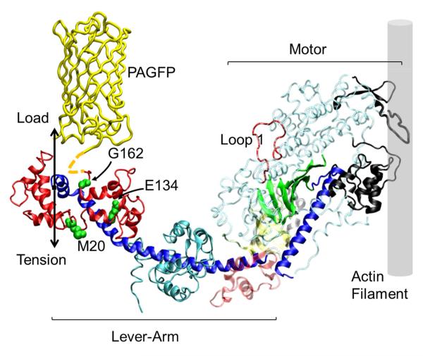
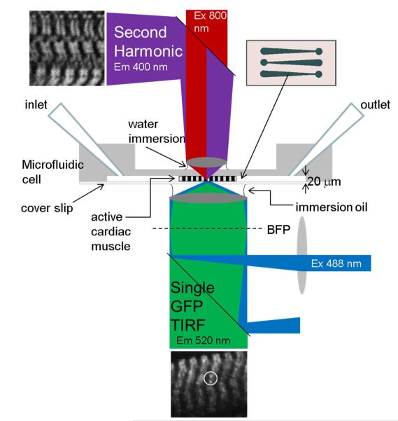
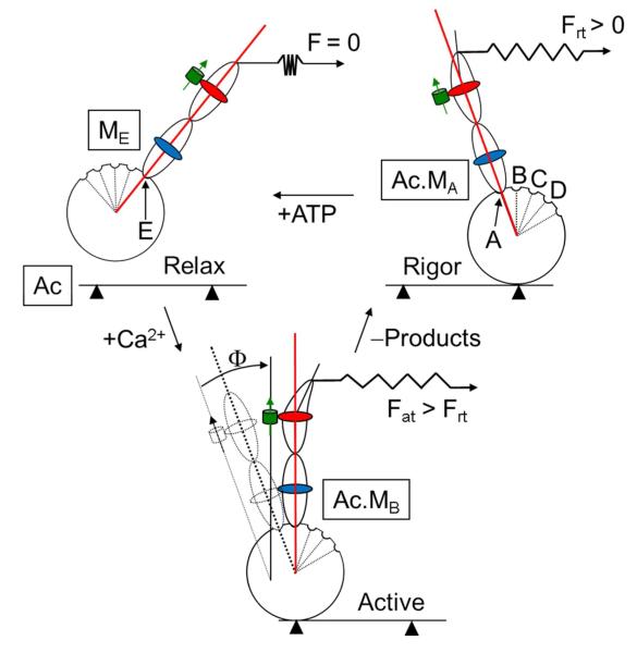
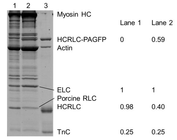
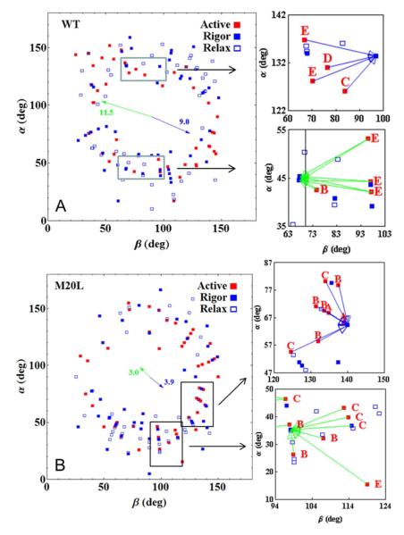
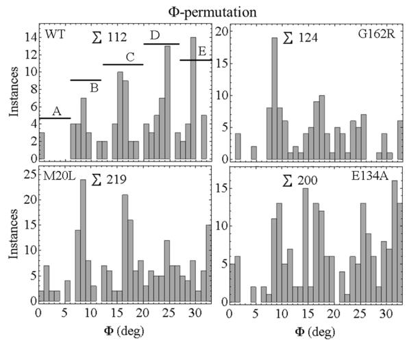
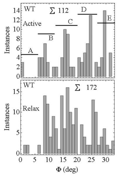
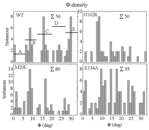
Myosin is the chemomechanical energy transducer in striated heart muscle. The myosin cross-bridge applies impulsive force to actin while consuming ATP chemical energy to propel myosin thick filaments relative to actin thin filaments in the fiber. Transduction begins with ATP hydrolysis in the cross-bridge driving rotary movement of a lever arm converting torque into linear displacement. Myosin regulatory light chain (RLC) binds to the lever arm and modifies its ability to translate actin. Gene sequencing implicated several RLC mutations in heart disease, and three of them are investigated here using photoactivatable GFP-tagged RLC (RLC-PAGFP) exchanged into permeabilized papillary muscle fibers. A single-lever arm probe orientation is detected in the crowded environment of the muscle fiber by using RLC-PAGFP with dipole orientation deduced from the three-spatial dimension fluorescence emission pattern of the single molecule. Symmetry and selection rules locate dipoles in their half-sarcomere, identify those at the minimal free energy, and specify active dipole contraction intermediates. Experiments were performed in a microfluidic chamber designed for isometric contraction, total internal reflection fluorescence detection, and two-photon excitation second harmonic generation to evaluate sarcomere length. The RLC-PAGFP reports apparently discretized lever arm orientation intermediates in active isometric fibers that on average produce the stall force. Disease-linked mutants introduced into RLC move intermediate occupancy further down the free energy gradient, implying lever arms rotate more to reach stall force because mutant RLC increases lever arm shear strain. A lower free energy intermediate occupancy involves a lower energy conversion efficiency in the fiber relating a specific myosin function modification to the disease-implicated mutant.
Figures
Similar articles
-
Single myosin cross-bridge orientation in cardiac papillary muscle detects lever-arm shear strain in transduction.Biochemistry. 2011 Sep 13;50(36):7809-21. doi: 10.1021/bi2008992. Epub 2011 Aug 18. Biochemistry. 2011. PMID: 21819137 Free PMC article.
-
GFP-tagged regulatory light chain monitors single myosin lever-arm orientation in a muscle fiber.Biophys J. 2007 Sep 15;93(6):2226-39. doi: 10.1529/biophysj.107.107433. Epub 2007 May 18. Biophys J. 2007. PMID: 17513376 Free PMC article.
-
In vivo orientation of single myosin lever arms in zebrafish skeletal muscle.Biophys J. 2014 Sep 16;107(6):1403-14. doi: 10.1016/j.bpj.2014.07.055. Biophys J. 2014. PMID: 25229148 Free PMC article.
-
Molecular mechanisms of cardiomyopathy phenotypes associated with myosin light chain mutations.J Muscle Res Cell Motil. 2015 Dec;36(6):433-45. doi: 10.1007/s10974-015-9423-3. Epub 2015 Sep 18. J Muscle Res Cell Motil. 2015. PMID: 26385864 Free PMC article. Review.
-
Signaling to myosin regulatory light chain in sarcomeres.J Biol Chem. 2011 Mar 25;286(12):9941-7. doi: 10.1074/jbc.R110.198697. Epub 2011 Jan 21. J Biol Chem. 2011. PMID: 21257758 Free PMC article. Review.
Cited by
-
Hybrid method for representing ions in implicit solvation calculations.Comput Struct Biotechnol J. 2021 Jan 20;19:801-811. doi: 10.1016/j.csbj.2021.01.020. eCollection 2021. Comput Struct Biotechnol J. 2021. PMID: 33598096 Free PMC article.
-
Phosphorylation of the regulatory light chain of myosin in striated muscle: methodological perspectives.Eur Biophys J. 2016 Dec;45(8):779-805. doi: 10.1007/s00249-016-1128-z. Epub 2016 Apr 15. Eur Biophys J. 2016. PMID: 27084718 Free PMC article. Review.
-
The Qdot-labeled actin super-resolution motility assay measures low-duty cycle muscle myosin step size.Biochemistry. 2013 Mar 5;52(9):1611-21. doi: 10.1021/bi301702p. Epub 2013 Feb 21. Biochemistry. 2013. PMID: 23383646 Free PMC article.
-
Respiratory syncytial virus-associated mortality in a healthy 3-year-old child: a case report.BMC Pediatr. 2019 Nov 27;19(1):462. doi: 10.1186/s12887-019-1847-2. BMC Pediatr. 2019. PMID: 31771554 Free PMC article.
-
Pseudophosphorylation of cardiac myosin regulatory light chain: a promising new tool for treatment of cardiomyopathy.Biophys Rev. 2017 Feb;9(1):57-64. doi: 10.1007/s12551-017-0248-8. Epub 2017 Jan 25. Biophys Rev. 2017. PMID: 28510043 Free PMC article. Review.
References
Publication types
MeSH terms
Substances
Grants and funding
LinkOut - more resources
Full Text Sources
Other Literature Sources
