

Magnetoencephalography reveals a unique neurophysiological profile of focal-onset epileptic spasms
- PMID: 23343709
- PMCID: PMC3904381
- DOI: 10.1620/tjem.229.147
Magnetoencephalography reveals a unique neurophysiological profile of focal-onset epileptic spasms
Erratum in
- Tohoku J Exp Med. 2013;229(2):171
Abstract
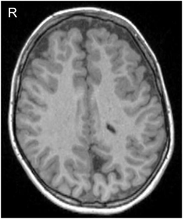
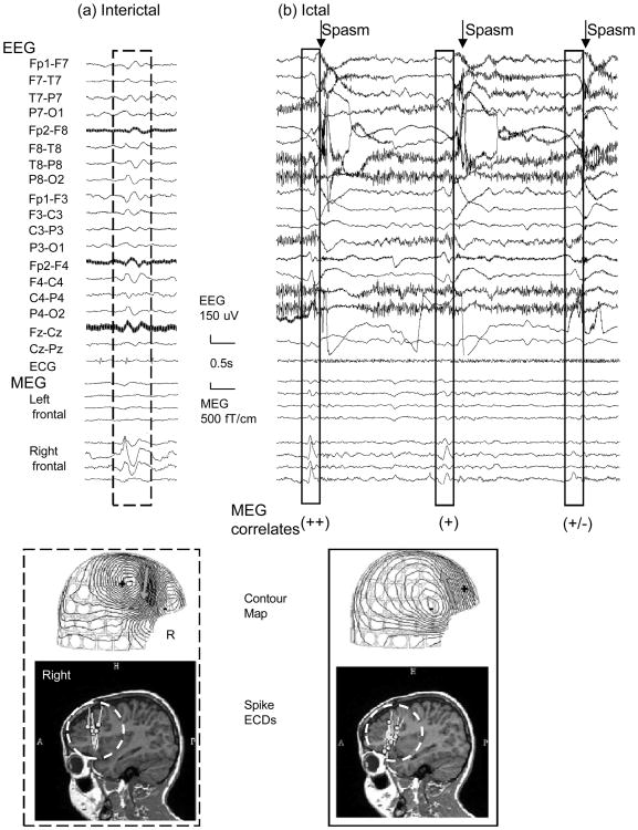
Epilepsy is defined as a disorder of the brain characterized by an enduring predisposition to experience epileptic seizures and the neurobiological, cognitive, psychological, and social difficulties relating to the condition. An epileptic spasm (ES) is a type of seizure characterized by clusters of short contractions involving axial muscles and proximal segments. However, the precise mechanism of ESs remains unknown. Despite the potential of magnetoencephalography (MEG) as a tool for investigating the neurophysiological mechanism of ESs, it has been difficult to use this methodology due to magnetic artifacts attributable to patient movement. We report on an 8-year-old girl suffering from intractable epileptic spasms from the age of 7 months. She was diagnosed with possible Aicardi syndrome [corrected] (AGS), characterized by the triad of callosal agenesis, infantile spasms, and chorioretinal lacunae. She is now intellectually delayed and suffers from intractable ES. We used both MEG and electroencephalography to investigate her epilepsy. The recording captured two series of spasm clusters. Spikes were clearly identified with MEG in about four-fifths of all spasms but were identified poorly or not at all in the remainder. MEG findings support previous studies that used intracranial electrodes to analyze patients with ESs and that showed variability in ES-associated spikes in terms of manner of cortical involvement and magnitude. Given the limitations of intracranial electrodes, such as sampling restrictions and invasiveness, MEG may be a helpful tool for non-invasively investigating the unique pathophysiological profile of focal-onset ESs.
Conflict of interest statement
Conflict of Interest: The authors declare no conflict of Interest.
Figures
Similar articles
-
The value of magnetoencephalography for seizure-onset zone localization in magnetic resonance imaging-negative partial epilepsy.Brain. 2013 Oct;136(Pt 10):3176-86. doi: 10.1093/brain/awt213. Epub 2013 Sep 6. Brain. 2013. PMID: 24014520 Free PMC article.
-
EEG and MEG source analysis of single and averaged interictal spikes reveals intrinsic epileptogenicity in focal cortical dysplasia.Epilepsia. 2004 Jun;45(6):621-31. doi: 10.1111/j.0013-9580.2004.56503.x. Epilepsia. 2004. PMID: 15144427
-
Epileptic spasms in older pediatric patients: MEG and ictal high-frequency oscillations suggest focal-onset seizures in a subset of epileptic spasms.Epilepsy Res. 2008 Feb;78(2-3):216-24. doi: 10.1016/j.eplepsyres.2007.12.007. Epub 2008 Jan 22. Epilepsy Res. 2008. PMID: 18215506
-
Magnetoencephalography in focal epilepsy.Epilepsia. 2000;41 Suppl 3:S39-47. doi: 10.1111/j.1528-1157.2000.tb01533.x. Epilepsia. 2000. PMID: 11001335 Review.
-
Multimodal localization and surgery for epileptic spasms of focal origin: a review.Neurosurg Focus. 2018 Sep;45(3):E4. doi: 10.3171/2018.6.FOCUS18217. Neurosurg Focus. 2018. PMID: 30173609 Review.
Cited by
-
Magnetoencephalography-identified preictal spiking correlates to preictal spiking on stereotactic EEG.Epilepsy Behav Rep. 2022 Mar 24;19:100538. doi: 10.1016/j.ebr.2022.100538. eCollection 2022. Epilepsy Behav Rep. 2022. PMID: 35573060 Free PMC article.
-
Classification of partial seizures based on functional connectivity: A MEG study with support vector machine.Front Neuroinform. 2022 Aug 18;16:934480. doi: 10.3389/fninf.2022.934480. eCollection 2022. Front Neuroinform. 2022. PMID: 36059865 Free PMC article.
References
-
- Aicardi J. Aicardi syndrome. Brain Dev. 2005;27:164–171. - PubMed
-
- Asano E, Chugani DC, Juhász DC, Muzik O, Chugani HT. Surgical treatment of West syndrome. Brain Dev. 2001;23:668–676. - PubMed
-
- Barkley GL, Baumgartner C. MEG and EEG in epilepsy. J Clin Neurophysiol. 2003;20:163–178. - PubMed
-
- Chugani HT, Shewmon DA, Sankar R, Chen BC, Phelps ME. Infantile spasms: II. Lenticular nuclei and brain stem activation on positron emission tomography. Ann Neurol. 1992;31:212–219. - PubMed
Publication types
MeSH terms
Supplementary concepts
Grants and funding
LinkOut - more resources
Full Text Sources
Other Literature Sources