. 2013 Feb 15;12(4):684-97.
doi: 10.4161/cc.23598. Epub 2013 Jan 23.

Phospho-ΔNp63α/microRNA feedback regulation in squamous carcinoma cells upon cisplatin exposure

Affiliations

Phospho-ΔNp63α/microRNA feedback regulation in squamous carcinoma cells upon cisplatin exposure

Yiping Huang et al. Cell Cycle. .

Abstract

Our previous reports showed that the cisplatin exposure induced the ATM-dependent phosphorylation of ΔNp63a, which is subsequently involved in transcriptional regulation of gene promoters encoding mRNAs and microRNAs in squamous cell carcinoma (SCC) cells upon cisplatin-induced cell death. We showed that phosphorylated (p)-ΔNp63a plays a role in upregulation of pro-apoptotic proteins, while non-p-ΔNp63a is implicated in pro-survival signaling. In contrast to non-p-ΔNp63a, p-ΔNp63a modulated expression of specific microRNAs in SCC cells exposed to cisplatin. These microRNAs were shown to attenuate the expression of several proteins involved in cell death/survival, suggesting the critical role for p-ΔNp63a in regulation of tumor cell resistance to cisplatin. Here, we studied the function of ΔNp63a in transcriptional activation and repression of the specific microRNA promoters whose expression is affected by cisplatin treatment of SCC cells. We quantitatively studied chromatin-associated proteins bound to tumor protein (TP) p63-responsive element, we found that p-ΔNp63a along with certain transcription coactivators (e.g., CARM1, KAT2B, TFAP2A, etc.) necessary to induce gene promoters for microRNAs (630 and 885-3p) or with transcription corepressors (e.g., EZH2, CTBP1, HDACs, etc.) needed to repress promoters for microRNAs (181a-5p, 374a-5p and 519a-3p) in SCC cells exposed to cisplatin.

Keywords: DNA/protein interactions; cisplatin; microRNAs; p53; p63; squamous cell carcinomas.


Figures

None
Figure 1. P-Δ;Np63α regulated transcription of the specific microRNA promoters in SCC-11 cells upon cisplatin exposure. SCC-11 cells (A) and SCC-11M (B) cells were transfected with an empty vector and with the Δ;Np63α-S385G-FL or Δ;Np63α-wt-FL expression cassettes for 24 h, exposed to control media or 10 μg/ml cisplatin (CIS) for 12 h and then tested for specific microRNA expression using qPCR (A and B) qPCR experiments were performed in triplicate with +SD as indicated (< 0.05). (C) Resulting SCC-11 and SCC-11M cells were used for ChIP analysis to identify the binding of Δ;Np63α to the specific microRNA promoters. The amount of immunoprecipitated-enriched DNA in each sample (ChIP) is represented as signal relative to the total amount of input chromatin DNA (input) using the same primers for the specific promoter region. (D) SCC-11 cells and SCC-11M cells were also transfected with 100 ng of the control promoter-less pLightSwitch_Prom plasmid and the pLightSwitch_Prom plasmids containing promoter sequences of specific microRNAs (as indicated) and luciferase reporter gene as indicated. Cells were exposed to control medium without cisplatin (Con) and medium with 10 μg/ml cisplatin (CIS) for 12 h. RenSP Renilla luciferase reporter activity assays were conducted in triplicate (+SD are indicated, p < 0.05). Data presented as relative to data obtained from the control untreated cells containing the promoter-less reporter plasmid designated as 1.
None
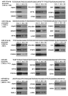
Figure 2. Various transcription factors bound to the specific microRNA promoters in SCC upon cisplatin exposure. SCC-11 cells and SCC-11M cells were exposed to 10 μg/ml cisplatin for 12 h. Nuclear lysates were incubated with the 50 bp sequences (locations are indicated in parentheses) derived from the specific microRNA promoters (miR-181a-5p, miR-519-3p, miR-374a-5p, miR-630 and miR-885-3p) containing the predicted TP63 binding site. Proteins bound to the tested sequences were analyzed by iTRAQ (LS/MS/MS) and validated by immunoblotting assays with the indicated antibodies.
None
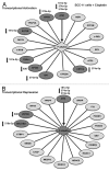
Figure 3. Schematic representation of p-Δ;Np63α-dependent protein interaction network involved in transcriptional activation (A) or repression (B) of the specific microRNA promoters in SCC-11 cells upon cisplatin exposure. Proteins that are potentially affected by specific microRNAs highlighted in darker shades and specific microRNAs indicated.
None
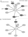
Figure 4. Schematic representation of Δ;Np63α-dependent protein interaction network involved in transcriptional activation (A) or repression (B) of the specific microRNA promoters in SCC-11M cells upon cisplatin exposure. Proteins that are potentially affected by specific microRNAs highlighted in darker shades and specific microRNAs indicated.
None
Figure 5. Specific microRNA mimics and inhibitors modulated the expression of Δ;Np63α via its 3′-UTR sequences upon cisplatin exposure. (A) Predicted “seed” sequences for specific miRs in the TP63 3′-UTR with the target prediction scores in parentheses. SCC-11 cells (B) and SCC-11M cells (C) were transfected with the LightSwitch_3UTR vector for the TP63 3′-UTR along with the scrambled microRNA, or mimics and inhibitors for miR-181a-5p, miR-374a-5p, miR-519-3p, miR-630 and miR-885-3p for 36 h. Cells were treated with control medium without cisplatin (Con) or medium with 10 μg/ml cisplatin (CIS) for additional 12 h and then tested for the RenSP Renilla luciferase reporter activity. Measurements (in triplicate) for the luciferase activity presented as relative units (RU). Values obtained from cells transfected with the scrambled RNA and treated with control medium were designated as 1. (D) Total lysates from the resulting SCC-11M cells from (C) treated with control medium were subsequently analyzed by immunoblotting with antibodies to Δ;Np63α and β-actin. Relative levels of Δ;Np63α normalized for β-actin levels were quantified and shown above immunoblot images. Levels of Δ;Np63α in cells with the scrambled microRNA were designated as 1.
None
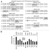
Figure 6. Specific microRNA mimics modulated the expression of the Δ;Np63α protein interacting targets via their 3′-UTR sequences. (A) Predicted “seed” sequences for specific microRNAs in the protein target 3′-UTRs with the target prediction scores in parentheses. (B) SCC-11 cells were transfected with the LightSwitch_3UTR plasmids for the indicated protein 3′-UTRs along with the scrambled microRNA and mimics for miR-181a-5p, miR-374a-5p, miR-519-3p, miR-630 and miR-885-3p for 36 h. Cells were then tested for the RenSP Renilla luciferase reporter activity. Measurements (in triplicate) for the luciferase activity presented as relative units (RU). Values from cells transfected with the scrambled RNA were designated as 1.
None
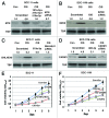
Figure 7. Specific microRNA mimics modulated expression of the Δ;Np63α protein interacting targets in SCC cells exposed to cisplatin and affected cell viability. Immunoblotting assay. SCC-11 cells (A and C) and SCC-11M cells (B and D) were transfected with the scrambled microRNA and indicated microRNA mimics for 36 h. Cells were then treated with control medium without cisplatin (Con) or medium with 10 μg/ml cisplatin (CIS) for additional 12 h and nuclear lysates were tested for indicated endogenous proteins. Loading levels were tested using a TBP antibody. Relative protein levels normalized for the TBP levels were quantified and shown above immunoblot images. Protein levels in cells with the scrambled miR were designated as 1. Cell viability assay. (E) SCC-11 cells were transfected with the scrambled siRNA (Scr) and siRNAs against ATM (si-ATM) or BHLHE41 (si-BHLHE41). (F) SCC-11M cells were transfected with the scrambled siRNA (Scr) and siRNAs against CARM1 (si-CARM1) or EZH2 (si-EZH2). Resulting cells were cultured in the presence (CIS) or absence (Con) of the 10 μg/ml cisplatin for indicated times. 104 cells/well in 96-well plates were then incubated in serum-free medium with 5 μg/ml of the 3-(4,5-dimethyl thiazol-2-yl)-2,5-diphenyl tetrazolium bromide in the dark for 4 h at 37°C. Cells were lysed and incubated for 2 h at 37°C, and the measurements were obtained on a Spectra Max-250 plate reader. Each assay was repeated at three times in triplicate. The bars are the mean ± SD of triplicate; p < 0.05, t-test.

Similar articles

Cited by

References

    1. Helmbach H, Kern MA, Rossmann E, Renz K, Kissel C, Gschwendt B, et al. Drug resistance towards etoposide and cisplatin in human melanoma cells is associated with drug-dependent apoptosis deficiency. J Invest Dermatol. 2002;118:923–32. doi: 10.1046/j.1523-1747.2002.01786.x. - DOI - PubMed
    1. Zangen R, Ratovitski EA, Sidransky D. DeltaNp63α levels correlate with clinical tumor response to cisplatin. Cell Cycle. 2005;4:1313–5. doi: 10.4161/cc.4.10.2066. - DOI - PubMed
    1. Galluzzi L, Senovilla L, Vitale I, Michels J, Martins I, Kepp O, et al. Molecular mechanisms of cisplatin resistance. Oncogene. 2012;31:1869–83. doi: 10.1038/onc.2011.384. - DOI - PubMed
    1. Galluzzi L, Morselli E, Vitale I, Kepp O, Senovilla L, Criollo A, et al. miR-181a and miR-630 regulate cisplatin-induced cancer cell death. Cancer Res. 2010;70:1793–803. doi: 10.1158/0008-5472.CAN-09-3112. - DOI - PubMed
    1. Ory B, Ellisen LW. A microRNA-dependent circuit controlling p63/p73 homeostasis: p53 family cross-talk meets therapeutic opportunity. Oncotarget. 2011;2:259–64. - PMC - PubMed

Publication types

MeSH terms