. 2013 Apr;109(8):2118-28.
doi: 10.1152/jn.00783.2012. Epub 2013 Jan 23.

Early postnatal development of GABAergic presynaptic inhibition of Ia proprioceptive afferent connections in mouse spinal cord

Affiliations

Early postnatal development of GABAergic presynaptic inhibition of Ia proprioceptive afferent connections in mouse spinal cord

Patrick M Sonner et al. J Neurophysiol. 2013 Apr.

Abstract

Sensory feedback is critical for normal locomotion and adaptation to external perturbations during movement. Feedback provided by group Ia afferents influences motor output both directly through monosynaptic connections and indirectly through spinal interneuronal circuits. For example, the circuit responsible for reciprocal inhibition, which acts to prevent co-contraction of antagonist flexor and extensor muscles, is driven by Ia afferent feedback. Additionally, circuits mediating presynaptic inhibition can limit Ia afferent synaptic transmission onto central neuronal targets in a task-specific manner. These circuits can also be activated by stimulation of proprioceptive afferents. Rodent locomotion rapidly matures during postnatal development; therefore, we assayed the functional status of reciprocal and presynaptic inhibitory circuits of mice at birth and compared responses with observations made after 1 wk of postnatal development. Using extracellular physiological techniques from isolated and hemisected spinal cord preparations, we demonstrate that Ia afferent-evoked reciprocal inhibition is as effective at blocking antagonist motor neuron activation at birth as at 1 wk postnatally. In contrast, at birth conditioning stimulation of muscle nerve afferents failed to evoke presynaptic inhibition sufficient to block functional transmission at synapses between Ia afferents and motor neurons, even though dorsal root potentials could be evoked by stimulating the neighboring dorsal root. Presynaptic inhibition at this synapse was readily observed, however, at the end of the first postnatal week. These results indicate Ia afferent feedback from the periphery to central spinal circuits is only weakly gated at birth, which may provide enhanced sensitivity to peripheral feedback during early postnatal experiences.


Figures

Fig. 1.
Fig. 1.
Conditioning stimulation of afferents innervating the knee extensor quadriceps muscle group (Quad afferents) produces 2 phases of inhibition of posterior biceps and semitendinosus (PBST) responses that are pharmacologically distinct. A: diagram of experimental model used demonstrates dorsal root L5 (DRL5) afferents were stimulated via a matrix electrode and the response in PBST motor neurons (MNs), termed the “test” response (T), could be recorded in the periphery via a suction electrode. A conditioning stimulus (C) could also be applied to Quad afferents via a separate suction electrode. B: representative average traces illustrating the effect of conditioning stimulation of Quad afferents on PBST response (postnatal day 7; P7). Black trace illustrates compound action potential recorded on the PBST nerve in the periphery following test pulse stimulation of DRL5 afferents (2-mA DRL5 stimulation). Red trace illustrates PBST response when a conditioning pulse of quadriceps nerve is given prior to test pulse (C+T; 200-μA quadriceps stimulation; 22-ms conditioning stimulus interval). The portion of compound action potential (CAP) in the shaded box was rectified and integrated to quantify the effects of the conditioning pulse and to generate a response ratio. C: response ratios measured at different conditioning stimulus intervals from a representative isolated spinal cord preparation from a P8 mouse. Intervals represent time (ms) between the Quad conditioning pulse and the test pulse stimulation on DRL5. Note 2 phases of inhibition were observed, a short- and long-interval inhibitory period. D: response ratios for another representative P7 preparation. E: response ratios for same preparation as in C following addition of 0.4 μM strychnine. F: response ratios for same preparation as in D following addition of 5 μM bicuculline. Test (T; black) and condition + test pulse (C+T; red) example traces are shown at right of C–F. Black circles on response ratio graphs indicate the specific conditioning stimulus interval depicted in example traces. In C–F, data points represent mean response ratio ± SD.
Fig. 2.
Fig. 2.
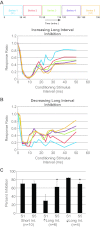
Long-interval inhibition at P7 is variably modulated by long-term repetitive proprioceptive stimulation. A and B: response ratio graphs for 2 representative P7 preparations. Note the colored boxes at top of A indicate the respective time course of each series and their respective response ratios in A and B. In both preparations (A and B), short-interval inhibition was consistent throughout all series. In some preparations (A), long-interval inhibition increased over the course of the series presented. In other preparations (B), the degree of long-interval inhibition decreased. C: quantitation of short- and long-interval percent inhibition from multiple preparations. Average responses for long-interval inhibition are presented in 2 groups: those with increasing (↑) inhibition over time (as illustrated by example in A) and those with decreasing (↓) inhibition over time (as illustrated by example in B). *P ≤ 0.05. Int., interval; S1, series 1; S5, series 5.
Fig. 3.
Fig. 3.
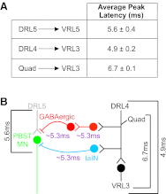
Predicted durations of individual stages of proposed pathways mediating inhibition of PBST responses based on measurements of monosynaptic connectivity between Ia afferents and MNs at P7/8. A: latency-to-peak values (means ± SE) of various monosynaptic connections measured in lumbar spinal cord. B: schematic of predicted proprioceptive pathways that mediate Quad afferent effects on PBST MN responses. Average measured durations (black brackets) are shown for various stimulus and recording combinations. Estimated duration of 1 central synaptic relay (purple) was used to predict central latencies of the Ia interneuron (IaIN)-mediated reciprocal inhibitory pathway (blue) and GABAergic presynaptic inhibitory pathway (red). VR, ventral root.
Fig. 4.
Fig. 4.
Sensory afferent action potentials in peripheral nerves evoked by primary afferent depolarization at P7/P8 are blocked by bicuculline. A: schematic diagram of experiment to record action potentials in primary sensory afferents antidromically conducted into the periphery following stimulation. A suction electrode was placed on the Quad nerve for stimulation to elicit primary afferent depolarization in the spinal cord. Afferent responses were recorded via a second suction electrode placed on the peripheral PBST nerve. All ventral roots were cut to prevent stimulation or recording of motor axon responses. B: representative trace illustrating action potential responses recorded from PBST afferents following stimulation of Quad afferents in a P7 preparation (top trace; n = 5). These responses were blocked by 5 μM bicuculline (bottom trace; n = 3).
Fig. 5.
Fig. 5.
Conditioning stimulation of Quad afferents produces only 1 phase of inhibition of PBST responses at birth. A: response ratios from representative preparation at P1. T (black) and C+T (red) example traces from preparation are displayed at right (22-ms conditioning intervals as indicated by black circle in graph). B: response ratios from representative preparation at P0. Example traces from 22-ms conditioning latency are shown at right. C: response ratios from same preparation as in A, but after addition of 0.4 μM strychnine. Inhibition was blocked and replaced by facilitation of PBST response at some conditioning intervals. Example traces (as in A) from the same preparation following addition of 0.4 μM strychnine are displayed at right. D: response ratios from same preparation as in B, but after addition of 5 μM bicuculline. Note that the latency and degree of inhibition are not altered. Example traces from the same preparation following addition of 5 μM bicuculline are displayed at right. Data points represent mean response ratio ± SD.
Fig. 6.
Fig. 6.
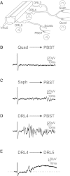
Sensory afferent action potentials in peripheral nerves evoked by primary afferent depolarization at birth. A: schematic diagram of experimental setup similar to that described in Fig. 4. Afferent responses were recorded via a suction electrode placed on the peripheral PBST nerve or on DRL5. Stimulation pulses were presented via a suction electrode on the Quad or saphenous (Saph) nerve or on DRL4. All ventral roots were cut to prevent stimulation or recording of motor axon responses. B–D: recordings from PBST afferents following stimulation of Quad (B) or Saph nerves (C) or the entire DRL4 (D). E: dorsal root potential recorded from DRL5 following stimulation of DRL4. Data in B–E were recorded from the same preparation.

Similar articles

Cited by

References

    1. Alvarez FJ. Anatomical basis for presynaptic inhibition of primary sensory fibers. In: Presynaptic Inhibition and Neural Control, edited by Rudomin P, Romo R, Mendell L. New York: Oxford University Press, 1998, p. 13–49
    1. Arber S, Ladle DR, Lin JH, Frank E, Jessell TM. ETS gene Er81 controls the formation of functional connections between group Ia sensory afferents and motor neurons. Cell 101: 485–498, 2000 - PubMed
    1. Baldissera F, Hultborn H, Illert M. Integration in spinal neuronal systems. In: Handbook of Physiology. The Nervous System. Bethesda, MD: Am. Physiol. Soc., 1981, p. 509–596
    1. Banks RW. The muscle spindle. In: Peripheral Neuropathy, edited by Dyck PJ, Thomas PK. Philadelphia, PA: Saunders, 2005, p. 131–150
    1. Bautista W, Nagy JI, Dai Y, McCrea DA. Requirement of neuronal connexin36 in pathways mediating presynaptic inhibition of primary afferents in functionally mature mouse spinal cord. J Physiol 590: 3821–3839, 2012 - PMC - PubMed

Publication types

LinkOut - more resources