. 2012 Dec 21;14(1):205-25.
doi: 10.3390/ijms14010205.

Expression and functional studies on the noncoding RNA, PRINS

Affiliations

Expression and functional studies on the noncoding RNA, PRINS

Krisztina Szegedi et al. Int J Mol Sci. .

Abstract

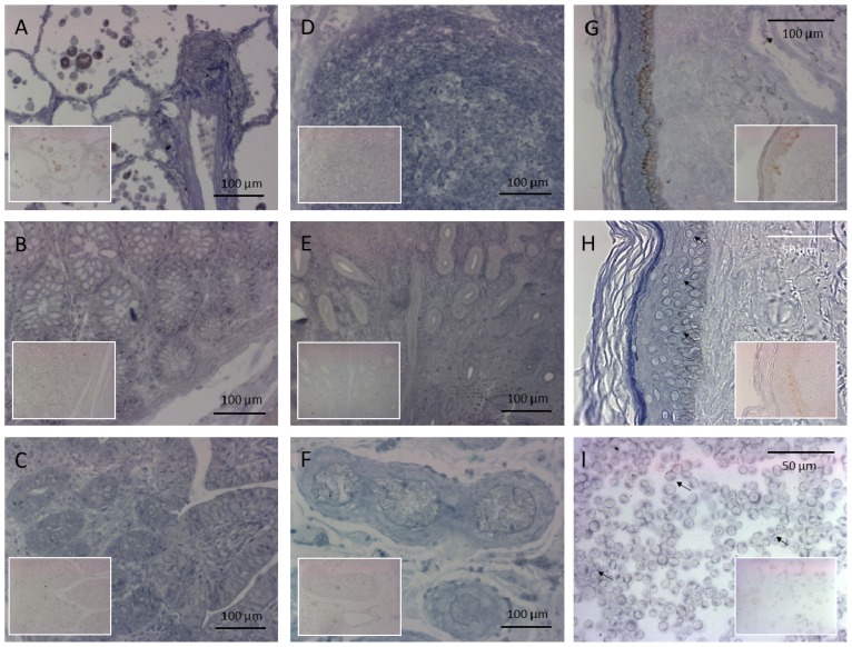
PRINS, a noncoding RNA identified earlier by our research group, contributes to psoriasis susceptibility and cellular stress response. We have now studied the cellular and histological distribution of PRINS by using in situ hybridization and demonstrated variable expressions in different human tissues and a consistent staining pattern in epidermal keratinocytes and in vitro cultured keratinocytes. To identify the cellular function(s) of PRINS, we searched for a direct interacting partner(s) of this stress-induced molecule. In HaCaT and NHEK cell lysates, the protein proved to be nucleophosmin (NPM) protein as a potential physical interactor with PRINS. Immunohistochemical experiments revealed an elevated expression of NPM in the dividing cells of the basal layers of psoriatic involved skin samples as compared with healthy and psoriatic uninvolved samples. Others have previously shown that NPM is a ubiquitously expressed nucleolar phosphoprotein which shuttles to the nucleoplasm after UV-B irradiation in fibroblasts and cancer cells. We detected a similar translocation of NPM in UV-B-irradiated cultured keratinocytes. The gene-specific silencing of PRINS resulted in the retention of NPM in the nucleolus of UV-B-irradiated keratinocytes; suggesting that PRINS may play a role in the NPM-mediated cellular stress response in the skin.


Figures

Figure 1
Figure 1
Detection of psoriasis susceptibility-related RNA gene induced by stress (PRINS) expression in various human tissue samples by ISH. The sections were incubated with an LNA RNA detection probe for PRINS and for the control staining (see insets at the same magnification) we used a scrambled control sequence. Relatively strong PRINS positivity was seen in the (A) lungs; (B) the large bowel; (C) the small bowel; (D) the lymph nodes; (E) the uterus; (F) the testicles; (G,H) the skin; and (I) cultured keratinocytes.
Figure 2
Figure 2
Detection of PRINS expression in psoriasis. ISH for PRINS expression was performed on healthy normal (n = 10), psoriatic uninvolved (n = 6) and psoriatic involved (n = 6) skin specimens. Sections were incubated with the PRINS-specific LNA probe or with the control scrambled (see insets at the same magnification) LNA probe. Representative stainings for each tissue types: (A) normal healthy skin; (B) psoriatic uninvolved skin; and (C) psoriatic involved skin. Bar = 100 μm.
Figure 3
Figure 3
Identification of the direct interacting partner of PRINS. The in vitro binding assay and the consecutive experiments with the three independent cell lysates (two from HaCaT keratinocytes and one from cultured NHEK) were performed as depicted in the Figure. A 39mer RNA transcript of PRINS was used. The elutes obtained with the RNP-complex purification kit were run on SDS-PAGE. Bands (arrows) that appeared in the PRINS binding fraction but not for the control samples, were cut out from the gel and further analyzed with MALDI-TOF. (A) Flow chart of the in vitro experiments. (B) Following the binding assay in HaCaT cells, the proteins in the eluates were separated on 7.5% SDS-PAGE and stained with Coomassie Brilliant Blue. NPM from a ~250-kD band was identified by MALDI-TOF (shown by black arrows). S, elute from the 1st affinity matrix (tobramycin) bound with the in vitro transcribed PRINS and cellular extract. F3, elute from the 2nd affinity matrix (streptavidin) incubated with elute “S”. TF, flow-through from the tobramycin affinity matrix incubated with the cellular extract. SF, flow-through from the streptavidin affinity matrix incubated with the cellular extract. Tend, elute from the tobramycin affinity matrix incubated with the cellular extract. Send, elute from the streptavidin affinity matrix incubated with the cellular extract
Figure 4
Figure 4
Immunohistochemical analysis of nucleophosmin (NPM) protein expressions in healthy and psoriatic skin samples. Paraffin-embedded tissues of healthy, non-lesional and lesional psoriatic skin samples were used for NPM-specific immunohistochemistry. After nonspecific antigen blocking, sections were incubated with anti-NPM mouse monoclonal antibody at a dilution of 1:500. The incubation with secondary antibody was followed by the development of a DAB color reaction, the intensity of which reaction was controlled under a light microscope. Sections were counterstained with hematoxylin. In the healthy and psoriatic uninvolved skin, the keratinocytes presented nuclear staining throughout all of the epidermal layers. In the healthy epidermis, some subgranular nuclei showed a high density of NPM, while in the uninvolved epidermis, a higher rate of spinous suprabasal nuclear staining was observed. In the psoriatic involved skin, the various layers of the epidermis exhibited a different NPM protein expression pattern. The keratinocytes presented pronounced cytoplasmic immunopositivity besides nuclear staining. Bar = 100 μm; (a) healthy epidermis; (b) isotype control for healthy epidermis; (c) psoriatic non-lesional; (d) isotype control for psoriatic non-lesional; (e) psoriatic lesional epidermis; (f) isotype control for lesional psoriatic epidermis.
Figure 5
Figure 5
Expression of NPM protein during keratinocyte proliferation/differentiation in vitro. Cultured normal human epidermal keratinocyte (NHEK) cultures (n = 3) were grown until subconfluency, when the 0-h sample was taken. (A) Changes in the expression of NPM mRNA were analyzed by real-time RT-PCR at the indicated times. Relative expression is shown compared to the 0-h sample. (B) At the indicated times, cells were harvested and lysed, then subjected to denaturing SDS-PAGE analysis and electroblotted to nitrocellulose. NPM protein was detected with a mouse monoclonal anti-NPM antibody and visualized with the NBT/BCIP system.
Figure 6
Figure 6
UV-B radiation induces the intracellular trafficking of nucleoplasmic NPM in HPV-Ker cells. Unirradiated and UV-B-irradiated (312 nm, 40 mJ/cm2) HPV-Ker cells were followed for the indicated periods of time, fixed and immunostained for NPM. DNA was stained with 4,6-diamidino-2-phenylindole (DAPI).
Figure 7
Figure 7
Silencing of the PRINS expression modifies the UV-B-induced trafficking of NPM. HPV-Ker cells were transfected with a PRINS gene-specific silencing vector (AK696). Control cells were transfected with a vector containing a scrambled sequence (SC1313). One day after the transfection, the unirradiated (A) and the UV-B-irradiated (B) HPV-Ker cells were followed for the indicated periods of time, fixed and immunostained for NPM (red). DNA was stained with DAPI (blue). Following the immunostaining, we performed a semiquantitative analysis of the cells. In every group, 25 fields of view were counted. Mean values are plotted on the chart (C).
Figure 7
Figure 7
Silencing of the PRINS expression modifies the UV-B-induced trafficking of NPM. HPV-Ker cells were transfected with a PRINS gene-specific silencing vector (AK696). Control cells were transfected with a vector containing a scrambled sequence (SC1313). One day after the transfection, the unirradiated (A) and the UV-B-irradiated (B) HPV-Ker cells were followed for the indicated periods of time, fixed and immunostained for NPM (red). DNA was stained with DAPI (blue). Following the immunostaining, we performed a semiquantitative analysis of the cells. In every group, 25 fields of view were counted. Mean values are plotted on the chart (C).

Similar articles

Cited by

References

    1. Lebwohl M. Psoriasis. Lancet. 2003;361:1197–1204. - PubMed
    1. Bowcock A.M., Krueger J.G. Getting under the skin: The immunogenetics of psoriasis. Nat. Rev. Immunol. 2005;5:699–711. - PubMed
    1. Duffy D.L., Spelman L.S., Martin N.G. Psoriasis in Australian twins. J. Am. Acad. Dermatol. 1993;29:428–434. - PubMed
    1. Bos J.D., De Rie M.A., Teunissen M.B., Piskin G. Psoriasis: Ddysregulation of innate immunity. Br. J. Dermatol. 2005;152:1098–1107. - PubMed
    1. Bos J.D., Hulsebosch H.J., Krieg S.R., Bakker P.M., Cormane R.H. Immunocompetent cells in psoriasis. In situ immunophenotyping by monoclonal antibodies. Arch. Dermatol. Res. 1983;275:181–189. - PubMed

Publication types

MeSH terms

LinkOut - more resources