. 2012 Dec 21;14(1):286-307.
doi: 10.3390/ijms14010286.

Quantitative profiling of DNA damage and apoptotic pathways in UV damaged cells using PTMScan Direct

Affiliations

Quantitative profiling of DNA damage and apoptotic pathways in UV damaged cells using PTMScan Direct

Matthew P Stokes et al. Int J Mol Sci. .

Abstract

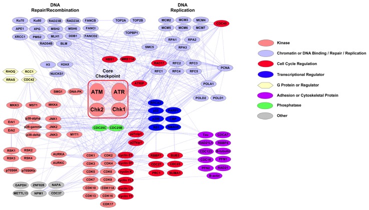
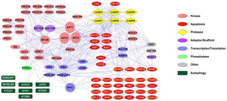
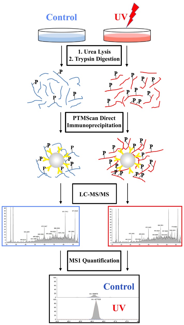
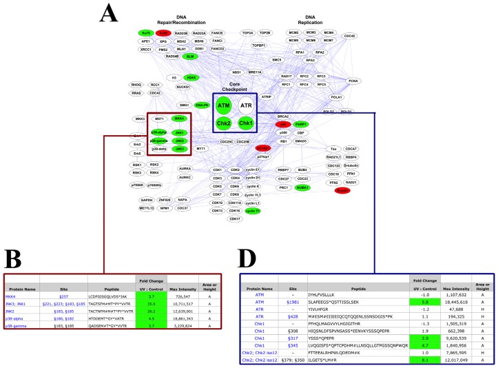
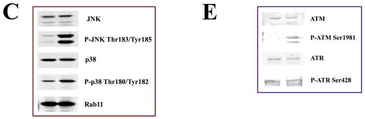
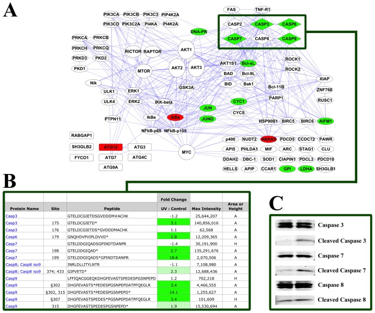
Traditional methods for analysis of peptides using liquid chromatography and tandem mass spectrometry (LC-MS/MS) lack the specificity to comprehensively monitor specific biological processes due to the inherent duty cycle limitations of the MS instrument and the stochastic nature of the analytical platform. PTMScan Direct is a novel, antibody-based method that allows quantitative LC-MS/MS profiling of specific peptides from proteins that reside in the same signaling pathway. New PTMScan Direct reagents have been produced that target peptides from proteins involved in DNA Damage/Cell Cycle and Apoptosis/Autophagy pathways. Together, the reagents provide access to 438 sites on 237 proteins in these signaling cascades. These reagents have been used to profile the response to UV damage of DNA in human cell lines. UV damage was shown to activate canonical DNA damage response pathways through ATM/ATR-dependent signaling, stress response pathways and induce the initiation of apoptosis, as assessed by an increase in the abundance of peptides corresponding to cleaved, activated caspases. These data demonstrate the utility of PTMScan Direct as a multiplexed assay for profiling specific cellular responses to various stimuli, such as UV damage of DNA.


Figures

Figure 1
Figure 1
PTMScan Direct: DNA Damage/Cell Cycle Reagent Interaction Map. Interactions were derived from the STRING database using experimental, database and text-mining lines of evidence and high confidence scores (score > 0.700). Interactions were imported into Cytoscape 2.8.1 for map generation. Protein color and shape denote protein class, as detailed in the legend to the right. Core checkpoint proteins ATM, ATR, Chk1 and Chk2 are highlighted with a red box in the center of the map.
Figure 2
Figure 2
PTMScan Direct: Apoptosis/Autophagy Reagent Interaction Map. Interactions were derived from the STRING database using experimental, database and text-mining lines of evidence and high confidence scores (score > 0.700). Interactions were imported into Cytoscape 2.8.1 for map generation. Protein color and shape denote protein class, as detailed in the legend to the right.
Figure 3
Figure 3
HeLa –/+ UV PTMScan Direct Experimental Design. HeLa cells were untreated or treated with 50 mJ/cm2 UV light and harvested at 2 h post treatment. Cells were processed according to previous protocols [67] and protein lysates were digested with trypsin. Immunoaffinity purification of peptides was performed with either the DNA Damage/Cell Cycle Reagent or the Apoptosis/Autophagy Reagent. Eluted peptides were run in LC-MS/MS, peptides were identified using Sorcerer 2 [70], and label-free quantification was performed using peptide chromatographic peak heights or areas in the MS1 channel.
Figure 4
Figure 4
DNA Damage/Cell Cycle Reagent HeLa –/+ UV Results. (A) Quantitative data from replicate runs was mapped onto the pathway diagram from Figure 1. Green proteins denote peptides that increased at least 2.5-fold with UV damage, red proteins denote decreases of at least 2.5-fold. The red box highlights stress-responsive MAP kinase proteins, and the blue box highlights core DNA damage checkpoint proteins; (B) Selected peptides derived from p38 MAPK and JNK are shown in detail with modification/cleavage site number, peptide sequence, fold change (UV: Control), maximum intensity and whether the measurement was height (H) or area (A). Green cells represent peptides that increased with UV damage at least 2.5-fold. Total and phospho-specific antibodies are available for protein names and sites shown in blue text, respectively; (C) Western blotting of the same samples used for the PTMScan Direct analysis with total and phospho-specific p38 and JNK antibodies (Cell Signaling Technology). Rab11 is included as a total protein control; (D) Selected peptides derived from the checkpoint proteins ATM, ATR, Chk1 and Chk2 are shown as in part B. “–” in the site column indicates an unmodified peptide; (E) Western blotting of the same samples used for the PTMScan Direct analysis with total and phospho-specific ATM and ATR antibodies (Cell Signaling Technology).
Figure 4
Figure 4
DNA Damage/Cell Cycle Reagent HeLa –/+ UV Results. (A) Quantitative data from replicate runs was mapped onto the pathway diagram from Figure 1. Green proteins denote peptides that increased at least 2.5-fold with UV damage, red proteins denote decreases of at least 2.5-fold. The red box highlights stress-responsive MAP kinase proteins, and the blue box highlights core DNA damage checkpoint proteins; (B) Selected peptides derived from p38 MAPK and JNK are shown in detail with modification/cleavage site number, peptide sequence, fold change (UV: Control), maximum intensity and whether the measurement was height (H) or area (A). Green cells represent peptides that increased with UV damage at least 2.5-fold. Total and phospho-specific antibodies are available for protein names and sites shown in blue text, respectively; (C) Western blotting of the same samples used for the PTMScan Direct analysis with total and phospho-specific p38 and JNK antibodies (Cell Signaling Technology). Rab11 is included as a total protein control; (D) Selected peptides derived from the checkpoint proteins ATM, ATR, Chk1 and Chk2 are shown as in part B. “–” in the site column indicates an unmodified peptide; (E) Western blotting of the same samples used for the PTMScan Direct analysis with total and phospho-specific ATM and ATR antibodies (Cell Signaling Technology).
Figure 5
Figure 5
Apoptosis/Autophagy Reagent HeLa −/+ UV Results. (A) Quantitative data from replicate runs was mapped onto the pathway diagram from Figure 2. Green proteins denote peptides that increased at least 2.5-fold with UV damage, and red proteins denote decreases of at least 2.5-fold. The green box highlights caspase proteins; (B) Selected peptides derived from caspases are shown in detail with modification/cleavage site number, peptide sequence, fold change (UV: Control), maximum intensity and whether the measurement was height (H) or area (A). Green cells represent peptides that increased with UV damage at least 2.5-fold. Light green cells indicate increases in cleaved caspase peptides less than 2.5-fold. Total antibodies are available for protein names shown in blue text; (C) Western blotting of the same samples used for the PTMScan Direct analysis with caspase 3, 7 and 8 antibodies (Cell Signaling Technology).

Similar articles

Cited by

References

    1. Abraham R.T. Cell cycle checkpoint signaling through the ATM and ATR kinases. Genes Dev. 2001;15:2177–2196. - PubMed
    1. Hartwell L.H., Weinert T.A. Checkpoints: Controls that ensure the order of cell cycle events. Science. 1989;246:629–634. - PubMed
    1. Zhou B.B., Elledge S.J. The DNA damage response: putting checkpoints in perspective. Nature. 2000;408:433–439. - PubMed
    1. Batista L.F., Kaina B., Meneghini R., Menck C.F. How DNA lesions are turned into powerful killing structures: insights from UV-induced apoptosis. Mutat. Res. 2009;681:197–208. - PubMed
    1. Izumaru S., Arima N., Toyozumi Y., Kato S., Morimatsu M., Nakashima T. Down-regulation of p21Waf-1 protein facilitates IR- and UV-induced apoptosis in human squamous carcinoma cells. Int. J. Oncol. 2004;24:1245–1255. - PubMed

LinkOut - more resources