. 2012 Aug 14;1(8):e38.
doi: 10.1038/mtna.2012.30.

Pip6-PMO, A New Generation of Peptide-oligonucleotide Conjugates With Improved Cardiac Exon Skipping Activity for DMD Treatment

Affiliations

Pip6-PMO, A New Generation of Peptide-oligonucleotide Conjugates With Improved Cardiac Exon Skipping Activity for DMD Treatment

Corinne Betts et al. Mol Ther Nucleic Acids. .

Abstract

Antisense oligonucleotides (AOs) are currently the most promising therapeutic intervention for Duchenne muscular dystrophy (DMD). AOs modulate dystrophin pre-mRNA splicing, thereby specifically restoring the dystrophin reading frame and generating a truncated but semifunctional dystrophin protein. Challenges in the development of this approach are the relatively poor systemic AO delivery and inefficient dystrophin correction in affected non-skeletal muscle tissues, including the heart. We have previously reported impressive heart activity including high-splicing efficiency and dystrophin restoration following a single administration of an arginine-rich cell-penetrating peptide (CPPs) conjugated to a phosphorodiamidate morpholino oligonucleotide (PMO): Pip5e-PMO. However, the mechanisms underlying this activity are poorly understood. Here, we report studies involving single dose administration (12.5 mg/kg) of derivatives of Pip5e-PMO, consecutively assigned as Pip6-PMOs. These peptide-PMOs comprise alterations to the central hydrophobic core of the Pip5e peptide and illustrate that certain changes to the peptide sequence improves its activity; however, partial deletions within the hydrophobic core abolish its efficiency. Our data indicate that the hydrophobic core of the Pip sequences is critical for PMO delivery to the heart and that specific modifications to this region can enhance activity further. The results have implications for therapeutic PMO development for DMD.


Figures

Figure 1
Figure 1
Sequences and chemical conjugation method for Pip5e-PMO derivatives. (a) List of names and sequences including rationale for synthesis of the peptides used in this study, Pip6a-h. (b) Method of conjugation of peptide to phosphorodiamidate morpholino oligonucleotide (PMO) antisense oligonucleotide (AO). Peptides were conjugated to PMO through an amide linkage at the 3′ end of the phosphorodiamidate morpholino oligonucleotide (PMO), followed by purification by high-performance liquid chromatography (HPLC) and analyzed by MALDI-TOF MS (for HPLC chromatogram and MALDI-TOF data, see Supplementary Figure S1). DIEA, diisopropylethylamine; HBTU, 2-(IH-benzotriazole-1-yl)-1,1,3,3-tetramethyluronium hexafluorophosphate; HOBt, 1-hydroxybenzotriazole monohydrate.
Figure 2
Figure 2
Exon skipping activity of Pip6-PMOs in differentiated mouse H2K mdx myotubes. H2K mdx myotubes were incubated with peptide-PMO conjugates at concentrations ranging between 0.125 and 1 µmol/l without the use of transfection reagent. The products of nested reverse transcription-PCR (RT-PCR) were examined by electrophoresis on a 2% agarose gel. Exon skipping activity is presented as the percentage of Δ23 skipping as calculated by densitometry. PMO, phosphorodiamidate morpholino oligonucleotide.
Figure 3
Figure 3
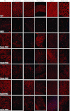
Immunohistochemical staining for dystrophin in C57BL10 control, mdx untreated, the 5-aa hydrophobic core Pip6-PMO-treated and Pip5e-PMO-treated mice. A single 12.5 mg/kg systemic injection of peptide-PMO was administered to mdx mice. Tissues were harvested 2 weeks later. Dystrophin immunostaining in TA, quadriceps, gastrocnemius, diaphragm, heart, and abdomen muscle groups for C57BL10, mdx-treated and mdx-untreated mice are shown. PMO, phosphorodiamidate morpholino oligonucleotide.
Figure 4
Figure 4
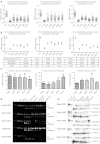
Dystrophin splicing and protein restoration in C57BL10 control, mdx untreated, the 5-aa hydrophobic core Pip6-PMO-treated and Pip5e-PMO-treated mice following a single 12.5 mg/kg, intravenous (i.v.) injection. (a) Quantification of dystrophin immunohistochemical staining relative to control laminin counter-stain in quadriceps, diaphragm and heart muscles of C57BL10, mdx-untreated and mdx-treated mice. Relative intensity values for each region of interest (120 regions) are plotted and the model estimate average calculated (presented in b) from the repeated measures, multilevel statistical model. For statistical significance tables see Tables 1 and 2. Percentage recovery score is represented below. (c) Percentage Δ23 exon skipping as determined by quantitative real time (q-RT)-PCR in quadriceps, diaphragm, and heart muscles. (d) Representative real time (RT)-PCR images demonstrating exon skipping (skipped) in TA, quadriceps, gastrocnemius, diaphragm, heart, and abdomen muscles. The top band indicates full-length (FL) or unskipped transcript. (e) Representative western blot images for each treatment. Ten micrograms of total protein was loaded (TA, quadriceps, gastrocnemius, diaphragm, heart, and abdomen muscles) relative to 50% (5 µg protein) and 10% (1 µg) C57BL10 controls, and normalized to α-actinin loading control (for quantification see Supplementary Figure S2a). PMO, phosphorodiamidate morpholino oligonucleotide.
Figure 5
Figure 5
Immunohistochemical staining for dystrophin in C57BL10 control, mdx untreated and the shortened hydrophobic core Pip6-PMO-treated mice (Pip6c and Pip6d). A single 12.5mg/kg systemic injection of PPMO was administered to mdx mice. Tissues were harvested 2 weeks later. Dystrophin immunostaining in TA, quadriceps, gastrocnemius, diaphragm, heart and abdomen muscle groups for C57BL10, mdx-treated and mdx-untreated mice are shown. PMO, phosphorodiamidate morpholino oligonucleotide.
Figure 6
Figure 6
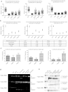
Dystrophin splicing and protein restoration in C57BL10 control, mdx untreated and the shortened hydrophobic core Pip6-PMO-treated mice (Pip6c and Pip6d) compared to Pip5e-PMO following a single 12.5 mg/kg, intravenous (i.v.) injection. (a) Quantification of dystrophin immunohistochemical staining relative to control laminin counter-stain in quadriceps, diaphragm and heart muscles of C57BL10, mdx-untreated and mdx-treated mice. Relative intensity values for each region of interest (120 regions) are plotted and the model estimate average calculated (presented in b) from the repeated measures, multilevel statistical model. For statistical significance tables, see Tables 1 and 2. Percentage recovery score is represented below. (c) Percentage Δ23 exon skipping as determined by quantitative real time (q-RT)-PCR in quadriceps, diaphragm, and heart muscles. (d) Representative real time (RT)-PCR images demonstrating exon skipping (skipped) in TA, quadriceps, gastrocnemius, diaphragm, heart, and abdomen muscles. The top band indicates full-length (FL) or unskipped transcript. (e) Representative western blot images for each treatment. Ten micrograms of total protein was loaded (TA, quadriceps, gastrocnemius, diaphragm, heart, and abdomen muscles) relative to 50% (5 µg protein) and 10% (1 µg) C57BL10 controls, and normalized to α-actinin loading control (for quantification see Supplementary Figure S2a). PMO, phosphorodiamidate morpholino oligonucleotide.
Figure 7
Figure 7
Dystrophin splicing and protein restoration in C57BL10 control, mdx untreated and the Pip6e-PMO derivatives, Pip6g and Pip6h, following a single 12.5 mg/kg, intravenous (i.v.) injection. (a) Immunohistochemical staining for dystrophin in C57BL10 control, mdx untreated and Pip6g- and Pip6h-PMO-treated mice. Dystrophin immunostaining in TA, quadriceps, gastrocnemius, diaphragm, heart, and abdomen muscle groups for C57BL10, mdx-untreated and mdx-treated mice are shown. (b) Quantification of dystrophin immunohistochemical staining relative to laminin counter-stain in quadriceps, diaphragm, and heart muscles of C57BL10, mdx-untreated and mdx-treated mice. Relative intensity values for each region of interest (120 regions) are plotted and the model estimate averages calculated (presented in c) from the repeated measures, multilevel statistical model. For statistical significance tables see Supplementary Figure S3a,b. Percentage recovery score is represented below. (d) Percentage Δ23 exon skipping as determined by quantitative real time (q-RT)-PCR in quadriceps, diaphragm and heart muscles. (e) Representative real-time (RT)-PCR images demonstrating exon skipping (skipped) in TA, quadriceps, gastrocnemius, diaphragm, heart, and abdomen muscles. The top band indicates full-length (FL) or unskipped transcript. (f) Representative western blot images for each treatment. Ten micrograms of total protein was loaded (TA, quadriceps, gastrocnemius, diaphragm, heart, and abdomen muscles) relative to 50% (5 µg protein) and 10% (1 µg) C57BL10 controls, and normalized to α-actinin loading protein (for quantification, see Supplementary Figure S3c). PMO, phosphorodiamidate morpholino oligonucleotide.

Similar articles

Cited by

References

    1. Emery AE. Population frequencies of inherited neuromuscular diseases–a world survey. Neuromuscul Disord. 1991;1:19–29. - PubMed
    1. Wilton SD, Lloyd F, Carville K, Fletcher S, Honeyman K, Agrawal S.et al. (1999Specific removal of the nonsense mutation from the mdx dystrophin mRNA using antisense oligonucleotides Neuromuscul Disord 9330–338. - PubMed
    1. Dunckley MG, Manoharan M, Villiet P, Eperon IC., and, Dickson G. Modification of splicing in the dystrophin gene in cultured Mdx muscle cells by antisense oligoribonucleotides. Hum Mol Genet. 1998;7:1083–1090. - PubMed
    1. Dickson G, Hill V., and, Graham IR. Screening for antisense modulation of dystrophin pre-mRNA splicing. Neuromuscul Disord. 2002;12 Suppl 1:S67–S70. - PubMed
    1. Alter J, Lou F, Rabinowitz A, Yin H, Rosenfeld J, Wilton SD.et al. (2006Systemic delivery of morpholino oligonucleotide restores dystrophin expression bodywide and improves dystrophic pathology Nat Med 12175–177. - PubMed

LinkOut - more resources