. 2013 Jun;70(12):2159-74.
doi: 10.1007/s00018-012-1248-2. Epub 2013 Jan 24.

Dystrophin deficiency leads to disturbance of LAMP1-vesicle-associated protein secretion

Affiliations

Dystrophin deficiency leads to disturbance of LAMP1-vesicle-associated protein secretion

Stephanie Duguez et al. Cell Mol Life Sci. 2013 Jun.

Abstract

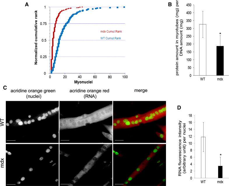
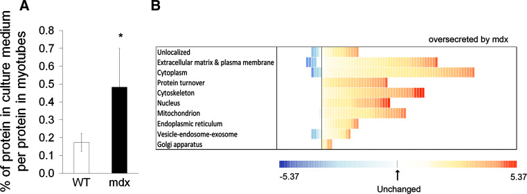
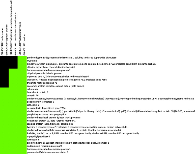
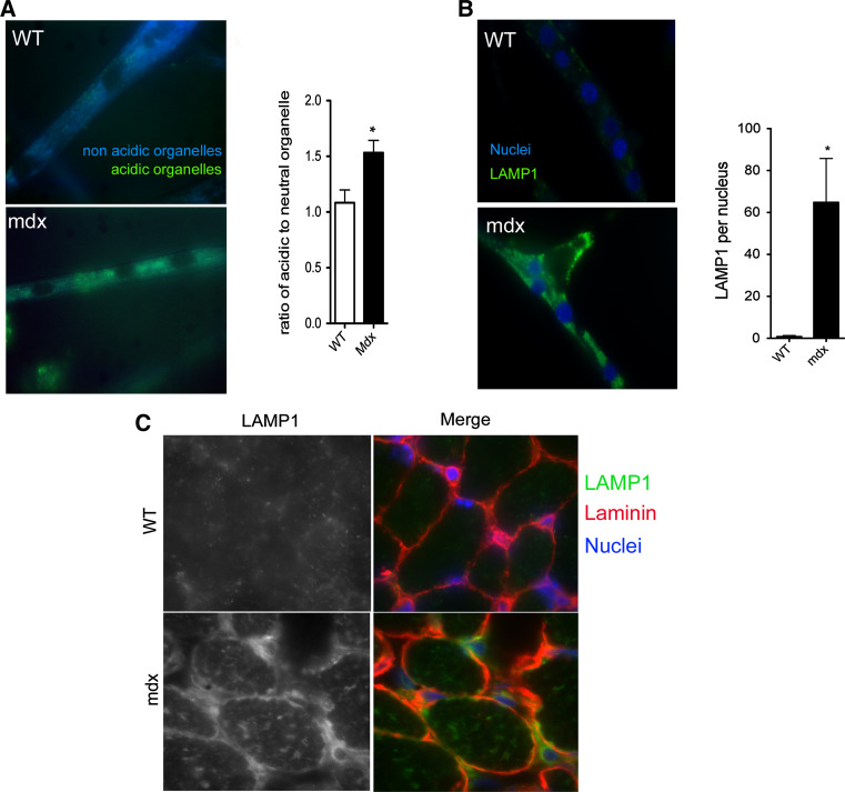
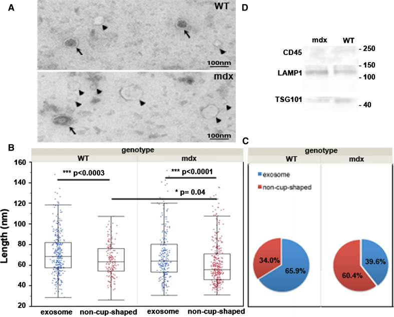
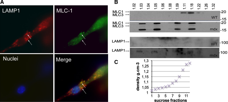
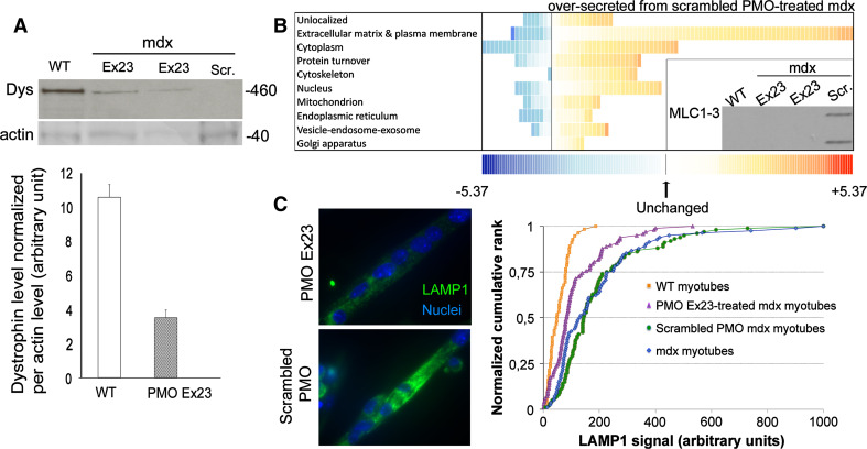
Duchenne muscular dystrophy results from loss of the protein dystrophin, which links the intracellular cytoskeletal network with the extracellular matrix, but deficiency in this function does not fully explain the onset or progression of the disease. While some intracellular events involved in the degeneration of dystrophin-deficient muscle fibers have been well characterized, changes in their secretory profile are undescribed. To analyze the secretome profile of mdx myotubes independently of myonecrosis, we labeled the proteins of mdx and wild-type myotubes with stable isotope-labeled amino acids (SILAC), finding marked enrichment of vesicular markers in the mdx secretome. These included the lysosomal-associated membrane protein, LAMP1, that co-localized in vesicles with an over-secreted cytoskeletal protein, myosin light chain 1. These LAMP1/MLC1-3-positive vesicles accumulated in the cytosol of mdx myotubes and were secreted into the culture medium in a range of abnormal densities. Restitution of dystrophin expression, by exon skipping, to some 30 % of the control value, partially normalized the secretome profile and the excess LAMP1 accumulation. Together, our results suggest that a lack of dystrophin leads to a general dysregulation of vesicle trafficking. We hypothesize that disturbance of the export of proteins through vesicles occurs before, and then concurrently with, the myonecrotic cascade and contributes chronically to the pathophysiology of DMD, thereby presenting us with a range of new potential therapeutic targets.


Conflict of interest statement

The authors declare that they have no conflicts of interest.

Figures

Fig. 1
Fig. 1
Mdx myotubes are atrophic and have less RNA. All samples were analyzed at day 6 of differentiation. a Distribution of myotube nuclear numbers, showing lower numbers in mdx than in wild-type myotubes (p < 0.00001). b Protein quantity per DNA in myotubes. n = 5/group. c Representative images of acridine orange staining in mdx and wild-type myotubes. RNA (red) was visualized using TRITC filter, and nuclei (green) using FITC filter (×20 objective). d RNA level per nucleus. n = 3/group. Values are mean ± SD. *p < 0.05, mdx vs. wild type. WT wild type. Bar: 50 μm
Fig. 2
Fig. 2
Mdx myotubes show greater protein release in vitro. a Protein quantity released into the culture medium. Values are expressed as a percentage of total cellular protein (n = 5/group). *p < 0.05, mdx vs. wild-type H-2K myotubes. b Distribution of proteins from mdx and wild-type myotubes differentially released into the culture medium. Proteins were sorted according to their known cellular localizations as attributed by the DAVID functional annotation software. Each square represents a protein. The value of the fold change is color-coded: red being over-secreted by mdx myotubes (maximum being at 5.37-fold) and blue under-secreted (minimum at −5.37-fold). See Suppl Table 1 for more details
Fig. 3
Fig. 3
Secretion by mdx myotubes of vesicle-related proteins. Functional annotation clustering in DAVID database allowed identification of a group of 31 proteins related to vesicles (see “Materials and methods” for the parameters used, see Suppl Table 3 for detailed results). Protein names are indicated on the y-axis with associated annotation terms on the x-axis. Green squares indicate that a particular protein belongs to that annotation category; black squares indicate that it does not
Fig. 4
Fig. 4
Accumulation of LAMP1 vesicles in mdx myotubes. a Representative images of acidic organelles in mdx and wild-type myotubes. Emission wavelength is dependent on organelle acidity, with more acidic organelles being detected using the FITC filter and more neutral organelles using the DAPI filter. The ratio of acidic vs. neutral was quantified. (×63 objective). n = 3/group. *p < 0.05. b Representative images of LAMP1 immunolabeling in mdx and wild-type myotubes. LAMP1 is in green and nuclei in blue. LAMP1 level was normalized per nucleus (×63 objective). n = 3/group. *p < 0.05. c Representative images of LAMP1 immunostaining of TA muscles of mdx and wild-type mice aged 16 days. LAMP1 is in green, laminin red and nuclei in blue (×100 objective). n = 3/group
Fig. 5
Fig. 5
Characterization of secreted vesicles: morphology and marker expression. a Representative electron microscopy images of vesicles secreted by WT and mdx myotubes. Arrows indicate classical “cup-shaped” exosomes. Arrowheads indicate smooth and clear “non-cup-shaped” vesicles delimited by a membrane. Bar = 100 nm. b Measurements of the sizes of exosomes and of smooth and clear vesicles from WT and mdx samples (a minimum of 500 particles per sample were analyzed). In both WT and mdx samples, the non-cup-shaped vesicles are significantly smaller than the exosomes. Average non-cup-shaped mdx vesicle size was slightly smaller than WT. Box-plots show means with upper and lower quartiles. *p < 0.05, ***p < 0.0001. c Mdx samples had a significantly higher proportion of non-cup-shaped vesicles (Chi-squared test p < 0.0001). d Vesicles express exosomal markers. Western blot showing WT and mdx vesicle expression of Tsg101 and LAMP1 exosomal markers. CD45, a shedding vesicle marker, was faintly detected in WT samples
Fig. 6
Fig. 6
Colocalization of LAMP1 and MLC1 in mdx myotubes. a BacMam system (Invitrogen) was used to deliver a cDNA encoding fluorescently tagged LAMP1, and MLC1 was immunolabeled. MLC1 is in green, LAMP1 in red, and nuclei in blue (×63 objective). Arrows colocalization of LAMP1 and MLC1. WT wild-type, MLC1 myosin light chain 1. b Immunoblots of MLC1-3 (upper blots) and of LAMP1 (lower blots) on vesicle fractions extracted from the culture media of wild-type and mdx myotubes. Vesicles were isolated using sucrose gradient suspension under ultracentrifugation. The sucrose density (g cm−3) of the 12 fractions is indicated above the Western blot. The lower blot shows an aberrant distribution of vesicle densities in mdx culture medium. The molecular weight (kDa) is indicated on the right of each blot. c Plot of fraction density against fraction number for sucrose gradient
Fig. 7
Fig. 7
PMO Ex23 treatment rescues the secretome profile and reduces the amount of LAMP1-positive vesicles. Restitution of dystrophin expression in PMO Ex23-treated mdx myotubes. a Top panel representative Western blot of dystrophin (460 kDa) and actin (40 kDa) on wild-type myotubes (lane 1), PMO-Ex23-treated (lanes 2, 3) and scrambled-PMO-treated mdx myotubes (lane 4). Lower panel quantification of dystrophin level, normalized per actin amount. Dys Dystrophin, Scr Scrambled. b Distribution of secreted proteins from Ex23 and scrambled-PMO-treated mdx myotubes into the culture medium. Proteins were sorted according to their known cellular localizations as attributed by the DAVID database. Each square represents a protein. The value of the fold change is color-coded: red being over-secreted by scrambled-PMO-treated mdx myotubes (maximum being at 5.37 fold) and blue under-secreted (minimum at −5.37). See Suppl. Table 4 for more details. Inset panel detection of MLC1-3 by Western blot in the culture medium of WT, scrambled-PMO-treated, and PMO-Ex23-treated mdx myotubes. c Quantification of LAMP1 level in scrambled-PMO-treated or PMO-Ex23-treated mdx myotubes. Left panel Representative images of LAMP1 immunolabeling in PMO-Ex23-treated and scrambled-PMO-treated mdx myotubes. LAMP1 is in green and nuclei in blue (×63 objective). Right panel normalized cumulative frequency plots of LAMP1 levels in each myotube of WT, mdx, scrambled-PMO-treated mdx and PMO-Ex23-treated mdx myotube cultures. n = 3/group. ***p < 0.00001: scrambled-PMO-treated vs. PMO-Ex23-treated mdx myotubes

Similar articles

Cited by

References

    1. Hoffman EP, Brown RH, Jr, Kunkel LM. Dystrophin: the protein product of the Duchenne muscular dystrophy locus. Cell. 1987;51(6):919–928. doi: 10.1016/0092-8674(87)90579-4. - DOI - PubMed
    1. Ohlendieck K, Ervasti JM, Snook JB, Campbell KP. Dystrophin-glycoprotein complex is highly enriched in isolated skeletal muscle sarcolemma. J Cell Biol. 1991;112(1):135–148. doi: 10.1083/jcb.112.1.135. - DOI - PMC - PubMed
    1. Williams MW, Bloch RJ. Extensive but coordinated reorganization of the membrane skeleton in myofibers of dystrophic (mdx) mice. J Cell Biol. 1999;144(6):1259–1270. doi: 10.1083/jcb.144.6.1259. - DOI - PMC - PubMed
    1. Engel A, Arahata K, Biesecker G. Mechanisms of muscle fiber destruction. In: Serratrice G, editor. Neuromuscular diseases. New York: Raven Press; 1984. pp. 137–141.
    1. Matsuda R, Nishikawa A, Tanaka H. Visualization of dystrophic muscle fibers in mdx mouse by vital staining with Evans Blue: evidence of apoptosis in dystrophin-deficient muscle. J Biochem. 1995;118(5):959–964. doi: 10.1093/jb/118.5.959. - DOI - PubMed

MeSH terms