

Molecular mechanisms of HIV immune evasion of the innate immune response in myeloid cells
- PMID: 23344558
- PMCID: PMC3564108
- DOI: 10.3390/v5010001
Molecular mechanisms of HIV immune evasion of the innate immune response in myeloid cells
Abstract
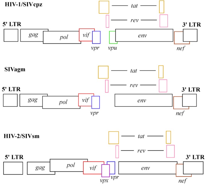
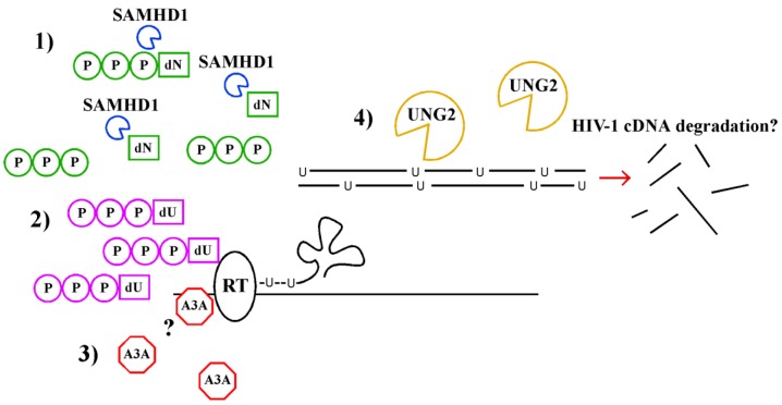
The expression of intrinsic antiviral factors by myeloid cells is a recently recognized mechanism of restricting lentiviral replication. Viruses that enter these cells must develop strategies to evade cellular antiviral factors to establish a productive infection. By studying the cellular targets of virally encoded proteins that are necessary to infect myeloid cells, a better understanding of cellular intrinsic antiviral strategies has now been achieved. Recent findings have provided insight into how the lentiviral accessory proteins, Vpx, Vpr and Vif counteract antiviral factors found in myeloid cells including SAMHD1, APOBEC3G, APOBEC3A, UNG2 and uracil. Here we review our current understanding of the molecular basis of how cellular antiviral factors function and the viral countermeasures that antagonize them to promote viral transmission and spread.
Figures
References
-
- Gupta P., Collins K.B., Ratner D., Watkins S., Naus G.J., Landers D.V., Patterson B.K. Memory cd4(+) t cells are the earliest detectable human immunodeficiency virus type 1 (hiv-1)-infected cells in the female genital mucosal tissue during hiv-1 transmission in an organ culture system. J. Virol. 2002;76:9868–9876. - PMC - PubMed
-
- Zhang Z., Schuler T., Zupancic M., Wietgrefe S., Staskus K.A., Reimann K.A., Reinhart T.A., Rogan M., Cavert W., Miller C.J., et al. Sexual transmission and propagation of siv and hiv in resting and activated cd4+ t cells. Science. 1999;286:1353–1357. doi: 10.1126/science.286.5443.1353. - DOI - PubMed
Publication types
MeSH terms
Grants and funding
LinkOut - more resources
Full Text Sources
Medical
Miscellaneous
