Review
. 2013 Jan 17;5(1):241-78.
doi: 10.3390/v5010241.

Arenavirus variations due to host-specific adaptation

Affiliations
Review

Arenavirus variations due to host-specific adaptation

Juan C Zapata et al. Viruses. .

Abstract

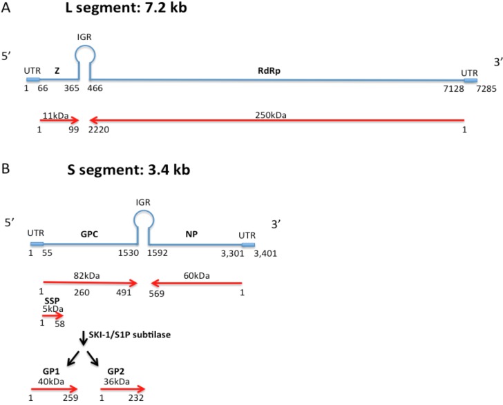
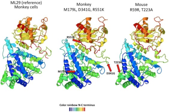
Arenavirus particles are enveloped and contain two single-strand RNA genomic segments with ambisense coding. Genetic plasticity of the arenaviruses comes from transcription errors, segment reassortment, and permissive genomic packaging, and results in their remarkable ability, as a group, to infect a wide variety of hosts. In this review, we discuss some in vitro studies of virus genetic and phenotypic variation after exposure to selective pressures such as high viral dose, mutagens and antivirals. Additionally, we discuss the variation in vivo of selected isolates of Old World arenaviruses, particularly after infection of different animal species. We also discuss the recent emergence of new arenaviruses in the context of our observations of sequence variations that appear to be host-specific.


Figures

Figure 1
Figure 1
Arenavirus genome structure from 5’ to 3’ end. A) The LASV L genome segment (7.2 kb) is represented in blue and is composed of an untranslated region (UTR) from nucleotide 1 to 66 and from 7129 to 7285, the gene encoding the zinc-binding protein (Z) from nucleotide 67 to 365, the intergenic region IGR from nucleotide 365 to 466, and the RNA-dependent RNA polymerase protein encoding gene (RdRp) from nucleotide 466 to 7128. B) The LASV S segment genome (blue lines) contains the untranslated region (UTR) from nucleotide 1 to 55 and from 3302 to 3401, the gene encoding the glycoprotein precursor protein (GPC) from nucleotide 57 to 1530, the intergenic region IGR from nucleotide 1531 to 1592, and the nucleocapsid protein encoding gene (NP) from nucleotide 1593 to 3301. The red arrows represent the 491 amino acid long GPC with its stable signal peptide (SSP) and glycoproteins 1 and 2 (GP1 and GP2) produced after maturation cleavage. The 569 amino acid long NP is shown encoded in the antisense orientation.
Figure 2
Figure 2
The LASV envelope glycoprotein precursor (GPC) structure is shown in A) from N- to C-terminus containing SSP, GP1 and GP2 proteins. The dark blue lines represent the cleavage points. The fusion domain (FD); transmembrane domain (TMD), and cytoplasmic domain (Cytop) are shown in brown letters (modified from [19]). B) The predicted Lassa-Josiah GPC structure obtained by open-source software (Phyre2). The structure goes from N-terminus (blue) to C-terminus (red). C) Schematic representation of the trimeric GPC subunit assembled in the cell membrane. GP1 is the most external protein bound to GP2 that is embedded in the lipid membrane. GP2 is thought to interact with SSP through an inter-subunit zinc finger (ball) (modified from [20]).
Figure 3
Figure 3
LCMV-ARM53b (A) and -Clone 13 GPC (B) predicted structure using the Phyre2 program. One amino acid change from F to L at position 260 alters the predicted GPC structure. The structure goes from N-terminus in blue color to the C-terminus in red color.
Figure 4
Figure 4
Predicted ML29 host-specific changes in GPC. The left-most structure shows the ML29 GPC predicted structure after passage in Vero cells. After inoculation into marmosets, the recovered viruses showed an isoleucine (I) to leucine (L) change at position 252 that affects the predicted GPC structure (middle structure). Another mutation at the same position, I to M, also changed GPC structure (figure on the right). The structure goes from N-terminus (blue) to C-terminus (red).
Figure 5
Figure 5
Predicted ML29 host-specific changes in NP. The left-most structure shows the ML29 NP predicted structure after passage in Vero cells. The middle structure represents the predicted NP changes (M179L, D341G, R551K) occurring in virus recovered from rhesus macaques only. The structure on the left shows two changes (R59R, and T223A) seen in mouse and marmosets. All host-specific mutations induced conformational changes in the NP predicted structure. The structure goes from N-terminus (blue) to C-terminus (red).
Figure 6
Figure 6
Arenavirus Evolution. The upper diagram shows the genomic structure and proteins encoded by filoviruses, arenaviruses, bunyaviruses, and the newly-discovered snake “arenavirus”. The blue arrows show the hypothetical changes that occurred in the arenaviral ancestors. The new snake virus has the L and NP genes of an arenavirus, but the GP of a filovirus and the Z gene similar to a cellular ubiquitin ligase (UL). The lower diagram shows how the reservoirs of arenaviruses, filoviruses, and bunyaviruses can interact with each other in the same ecological niche allowing co-infections and mixing of viral genomes to produce new viruses (Modify from [159]).

Similar articles

  • Arenavirus Quasispecies and Their Biological Implications.
    Grande-Pérez A, Martin V, Moreno H, de la Torre JC. Grande-Pérez A, et al. Curr Top Microbiol Immunol. 2016;392:231-76. doi: 10.1007/82_2015_468. Curr Top Microbiol Immunol. 2016. PMID: 26472215 Free PMC article. Review.
  • Arenavirus genetic diversity and its biological implications.
    Emonet SF, de la Torre JC, Domingo E, Sevilla N. Emonet SF, et al. Infect Genet Evol. 2009 Jul;9(4):417-29. doi: 10.1016/j.meegid.2009.03.005. Epub 2009 Mar 25. Infect Genet Evol. 2009. PMID: 19460307 Free PMC article. Review.
  • Widespread recombination, reassortment, and transmission of unbalanced compound viral genotypes in natural arenavirus infections.
    Stenglein MD, Jacobson ER, Chang LW, Sanders C, Hawkins MG, Guzman DS, Drazenovich T, Dunker F, Kamaka EK, Fisher D, Reavill DR, Meola LF, Levens G, DeRisi JL. Stenglein MD, et al. PLoS Pathog. 2015 May 20;11(5):e1004900. doi: 10.1371/journal.ppat.1004900. eCollection 2015 May. PLoS Pathog. 2015. PMID: 25993603 Free PMC article.
  • Novel arenavirus, Zambia.
    Ishii A, Thomas Y, Moonga L, Nakamura I, Ohnuma A, Hang'ombe B, Takada A, Mweene A, Sawa H. Ishii A, et al. Emerg Infect Dis. 2011 Oct;17(10):1921-4. doi: 10.3201/eid1710.10452. Emerg Infect Dis. 2011. PMID: 22000372 Free PMC article.
  • Past, present, and future of arenavirus taxonomy.
    Radoshitzky SR, Bào Y, Buchmeier MJ, Charrel RN, Clawson AN, Clegg CS, DeRisi JL, Emonet S, Gonzalez JP, Kuhn JH, Lukashevich IS, Peters CJ, Romanowski V, Salvato MS, Stenglein MD, de la Torre JC. Radoshitzky SR, et al. Arch Virol. 2015 Jul;160(7):1851-74. doi: 10.1007/s00705-015-2418-y. Arch Virol. 2015. PMID: 25935216

Cited by

References

    1. Auperin D.D., Romanowski V., Galinski M., Bishop D.H. Sequencing studies of pichinde arenavirus S RNA indicate a novel coding strategy, an ambisense viral S RNA. J. Virol. 1984;52:897–904. - PMC - PubMed
    1. Pinschewer D.D., Perez M., de la Torre J.C. Role of the virus nucleoprotein in the regulation of lymphocytic choriomeningitis virus transcription and RNA replication. J. Virol. 2003;77:3882–3887. doi: 10.1128/JVI.77.6.3882-3887.2003. - DOI - PMC - PubMed
    1. Eichler R., Strecker T., Kolesnikova L., ter Meulen J., Weissenhorn W., Becker S., Klenk H.D., Garten W., Lenz O. Characterization of the lassa virus matrix protein Z: Electron microscopic study of virus-like particles and interaction with the nucleoprotein (NP) Virus Res. 2004;100:249–255. doi: 10.1016/j.virusres.2003.11.017. - DOI - PubMed
    1. Casabona J.C., Levingston Macleod J.M., Loureiro M.E., Gomez G.A., Lopez N. The ring domain and the l79 residue of Z protein are involved in both the rescue of nucleocapsids and the incorporation of glycoproteins into infectious chimeric arenavirus-like particles. J. Virol. 2009;83:7029–7039. doi: 10.1128/JVI.00329-09. - DOI - PMC - PubMed
    1. Shtanko O., Imai M., Goto H., Lukashevich I.S., Neumann G., Watanabe T., Kawaoka Y. A role for the C terminus of mopeia virus nucleoprotein in its incorporation into Z protein-induced virus-like particles. J. Virol. 2010;84:5415–5422. doi: 10.1128/JVI.02417-09. - DOI - PMC - PubMed