. 2012 Jan 24;1(1):e4.
doi: 10.1038/mtna.2011.3.

Systemic RNAi-mediated Gene Silencing in Nonhuman Primate and Rodent Myeloid Cells

Affiliations

Systemic RNAi-mediated Gene Silencing in Nonhuman Primate and Rodent Myeloid Cells

Tatiana I Novobrantseva et al. Mol Ther Nucleic Acids. .

Abstract

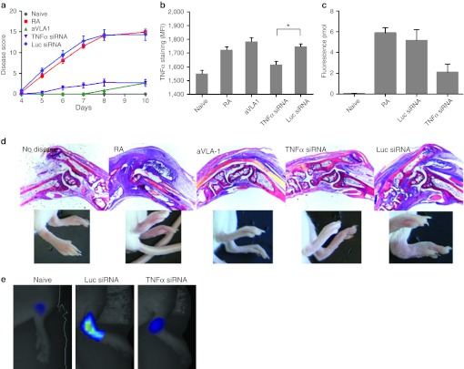
Leukocytes are central regulators of inflammation and the target cells of therapies for key diseases, including autoimmune, cardiovascular, and malignant disorders. Efficient in vivo delivery of small interfering RNA (siRNA) to immune cells could thus enable novel treatment strategies with broad applicability. In this report, we develop systemic delivery methods of siRNA encapsulated in lipid nanoparticles (LNP) for durable and potent in vivo RNA interference (RNAi)-mediated silencing in myeloid cells. This work provides the first demonstration of siRNA-mediated silencing in myeloid cell types of nonhuman primates (NHPs) and establishes the feasibility of targeting multiple gene targets in rodent myeloid cells. The therapeutic potential of these formulations was demonstrated using siRNA targeting tumor necrosis factor-α (TNFα) which induced substantial attenuation of disease progression comparable to a potent antibody treatment in a mouse model of rheumatoid arthritis (RA). In summary, we demonstrate a broadly applicable and therapeutically relevant platform for silencing disease genes in immune cells.


Figures

Figure 1
Figure 1
In vivo dynamic fluorescence-mediated tomography/X-ray computed tomography (FMT-CT) of lipid nanoparticles (LNP)-small interfering RNA (siRNA) delivery and siRNA localization within reservoir monocyte clusters in the spleen. (a) 3D reconstruction of FMT-CT data of KC2 (left) and C12-200 (right) is shown. The arrow points to the position of splenic signal. Signal in organ systems of interest was quantified, data are shown as mean and SE, n = 4–5 per group. (b). Ex vivo fluorescence reflectance imaging after injection of LNP siRNA side-by-side imaging of respective organs and highlights the signal intensity in the spleen. (c) Fluorescence images of spleens from mice injected or not injected with LNP-siRNA.
Figure 2
Figure 2
Efficacious and durable silencing in peritoneal cavity macrophages. (a) Cells were collected at 72 hours after a single intravenous (i.v.) bolus administration of the indicated doses of small interfering RNA (siRNA) in KC2 or C12-200 lipid nanoparticles (LNP) formulation. Leukocytes were stained for CD11b and CD45 and analyzed by flow cytometry. Representative profiles of CD45 staining in CD11b+ population are shown. Groups of three mice were analyzed, a representative experiment out of three independently conducted ones is shown. (b) Quantification of the percent silencing with C12-200 (gray bars) and KC2 (black bars) seen in panel (a). Bars represent average percent silencing within group with standard deviation. (c) Green fluorescent protein (GFP)-expressing peritoneal lavage cells from N5-RAGE GFP mice were transferred into C57BL/6 recipients by intraperitoneal (i.p.) injection. Thirty minutes after transfer mice were treated with 2 mg/kg KC2 formulation encapsulated CD45 or Luc siRNA i.v. At indicated days silencing of CD45 in GFP-expressing peritoneal cavity myeloid cells was monitored. Three mice per group were analyzed. (d) C57BL/6 mice were injected with 0.8 mg/kg of equivalent mixture of four siRNA directed against integrin β1, Rab5c, CD11b, and CD45 or with total amount of Luc siRNA. Twenty four hours post injection peritoneal cavity cells were collected and gene expression was analyzed by quantitative-PCR (Q-PCR). Each gene expression was measured as ratio to GAPDH expression. Knockdown results are expressed as % gene knock down with SEM, relative to the group injected with Luc siRNA. Three mice per group were analyzed.
Figure 3
Figure 3
Uptake of KC2 and C12-200 formulations in primary macrophages. (a) Quantitation of cellular uptake of Alexa Fluor-647 labeled small interfering RNA (siRNA) (AF647-si) in KC2 and C12-200 formulations in primary macrophages in vitro. RFU, relative fluorescence units. Data are average of 20 fields from three independent wells. (b) Colocalization of AF647-si in C12-200 or KC2 formulations with a marker of phagocytosis Alexa-488 labeled 1 µmol/l latex beads in primary macrophages, 1 hour time point C12-200, 4-hour time point KC2, arrows indicate signal overlap. (c) Quantitation of percent colocalization of AF488 beads with A647-si in KC2 or C12-200 formulations (4 hour time point). Data are the average of 20 fields from three different independent replicate wells for a total of 60 fields ± SEM.
Figure 4
Figure 4
Fast and efficient silencing in central monocytes/macrophages coupled with delayed but durable silencing in resident peritoneal cavity myeloid cells. (a) Cells were collected at 15 minutes, 1 hour, or 2 hours after a single intravenous (i.v.) bolus administration of 3 mg/kg of small interfering RNA (siRNA) in KC2 formulation. Leukocytes were seeded on plastic tissue culture plates and adherent cells were cultured for 3 days. Then harvested cells were stained for CD11b and CD45 and analyzed by flow cytometry. Mean fluorescent intensity shifts are shown when comparing CD45 siRNA to Luc siRNA in KC2 lipid nanoparticles (LNP). (b) Green fluorescent protein (GFP)-expressing peritoneal lavage cells from N5RAGE GFP mice were transferred into C57BL/6 recipients by i.p. injection. Thirty minutes after transfer mice were treated with 2 mg/kg KC2 formulation encapsulated CD45 or Luc-specific siRNA i.v. 72 hours later peritoneal cells were collected and the level of CD45 in GFP positive and negative peritoneal macrophage was quantified by flow cytometry. (c, d) C12-200 (c) or KC2 (d) encapsulated Alexa647 labeled siRNAs was injected at 1 mg/kg i.v. Blood, spleen, and peritoneal lavage were collected at indicated time points. Macrophages were identified by surface marker staining (see Materials and Methods) and the Alexa647 signal ±SEM is plotted.
Figure 5
Figure 5
Macrophage-specific CD11b silencing. mRNA ratio of CD11b to GAPDH was measured in peritoneal cavity or total perfused liver lobes 24 hours after bolus intravenous (i.v.) injection of small interfering RNA (siRNA) in lipid nanoparticles (LNP). Bars represent the mean level from three animals per group, three biological replicas per sample, with SEM in animals treated with CD11b siRNA relative to Luc siRNA-treated animals.
Figure 6
Figure 6
Inhibition of collagen mAb-induced arthritis by small interfering RNA (siRNA) against tumor necrosis factor-α (TNFα). (a) Preventative treatment of mice with siRNA against TNFα (purple curve) but not against Luc (blue curve) in C12-200 formulation decreases arthritis score. Anti-VLA1 mAb given several hours before arthitogenic antibody mix was used as a positive control (green curve) as previously shown. Mice were treated with anticollagen mAb's at day 0, followed by lipopolysaccharide (LPS) on day 3. Mice receiving only arthitogenic antibodies are graphed in red. 0.5 mg/kg siRNA in C12-200 formulation was administered on days −3, 0, 4, and 7. All mice were scored on days 4–8 and 10. Each limb was evaluated and scored on a 0–4 scale. Results are expressed as the mean arthritic score (±SEM) of all four limbs (maximum score of 16). Groups of 10 mice per condition were used; one out of two experiments is shown, both showed similar protection. (b) Intensity of intracellular TNFα staining in CD11b+ splenic macrophages from diseased mice at day 10. Groups of three mice were analyzed; bars represent average of the mean fluorescent intensity in the group with SEM, statistical analysis as described in the Results section. (c) Detection of inflammatory macrophage activity by fluorescence-mediated tomography (FMT) imaging. Arthritic mice treated with siRNAs targeted to TNFα or Luc were injected with Prosense-750, a pan-specific cathepsin substrate, and imaged by FMT on day 7. Fluorescence concentration reports on protease activity in the joint and was quantified as described in Supplementary Materials and Methods, n = 5, 2 paws per mouse were analyzed. Data are mean ± SEM. (d) Joint histology at 5x magnification in H&E staining correlated with images of the paws. (e) Representative images of macrophage activity measurement by FMT quantified in panel c. Naive and siRNA-treated animals are shown.
Figure 7
Figure 7
Silencing CD45 in vivo in peripheral blood monocytes/macrophages from nonhuman primate (NHP). Blood was drawn from each of the 12 experimental NHPs 1 hours prior and 1 hour after bolus administration of 3 mg/kg of small interfering RNA (siRNA) in KC2 and 1 mg/kg of C12-200. Leukocytes were seeded on plastic tissue culture plates and adherent cells were cultured for 3 days. Then harvested cells were stained for CD31, CD11b, and CD45 and analyzed by flow cytometry. (a) Representative profiles of CD45 staining in CD31/CD11b double-positive population are shown for both lipid nanoparticles (LNPs), blue profiles correspond to after CD45 siRNA dose, whereas black—for before dosing. (b) Average percent silencing in the total CD31/CD11b double-positive population as calculated from the shift in mean fluorescence intensity (MFI) in the surface of the cells before and after in vivo treatment with CD45 siRNA. (c) Summary of % CD45 knockdown in organ resident CD11b+ cells of monocyte/macrophage lineage in NHP dosed with 3 mg/kg of KC2 or 1 mg/kg of C12-200 3 days after dose. Bars represent average MFI value from three animals in the CD45 siRNA-treated group as it related to the average MFI of the Luc siRNA-treated group, plotted with SEM. Significant knockdown is seen in all organs analyzed except for peritoneal cavity. Respective histograms and representative dot plots from where the data is derived are shown in Supplementary Figure S6.

Similar articles

Cited by

References

    1. Bumcrot D, Manoharan M, Koteliansky V., and, Sah DW. RNAi therapeutics: a potential new class of pharmaceutical drugs. Nat Chem Biol. 2006;2:711–719. - PMC - PubMed
    1. Judge AD, Robbins M, Tavakoli I, Levi J, Hu L, Fronda A.et al. (2009Confirming the RNAi-mediated mechanism of action of siRNA-based cancer therapeutics in mice J Clin Invest 119661–673. - PMC - PubMed
    1. Whitehead KA, Langer R., and, Anderson DG. Knocking down barriers: advances in siRNA delivery. Nat Rev Drug Discov. 2009;8:129–138. - PMC - PubMed
    1. Semple SC, Akinc A, Chen J, Sandhu AP, Mui BL, Cho CK.et al. (2010Rational design of cationic lipids for siRNA delivery Nat Biotechnol 28172–176. - PubMed
    1. Love KT, Mahon KP, Levins CG, Whitehead KA, Querbes W, Dorkin JR.et al. (2010Lipid-like materials for low-dose, in vivo gene silencing Proc Natl Acad Sci USA 1071864–1869. - PMC - PubMed

LinkOut - more resources