. 2012 Oct 16;1(10):e48.
doi: 10.1038/mtna.2012.40.

Targeted exon skipping to address "leaky" mutations in the dystrophin gene

Affiliations

Targeted exon skipping to address "leaky" mutations in the dystrophin gene

Sue Fletcher et al. Mol Ther Nucleic Acids. .

Abstract

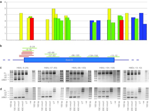
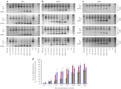
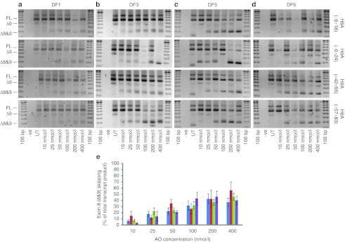
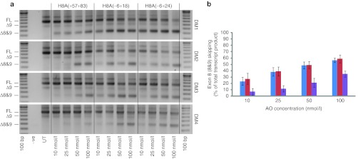
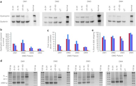
Protein-truncating mutations in the dystrophin gene lead to the progressive muscle wasting disorder Duchenne muscular dystrophy, whereas in-frame deletions typically manifest as the milder allelic condition, Becker muscular dystrophy. Antisense oligomer-induced exon skipping can modify dystrophin gene expression so that a disease-associated dystrophin pre-mRNA is processed into a Becker muscular dystrophy-like mature transcript. Despite genomic deletions that may encompass hundreds of kilobases of the gene, some dystrophin mutations appear "leaky", and low levels of high molecular weight, and presumably semi-functional, dystrophin are produced. A likely causative mechanism is endogenous exon skipping, and Duchenne individuals with higher baseline levels of dystrophin may respond more efficiently to the administration of splice-switching antisense oligomers. We optimized excision of exons 8 and 9 in normal human myoblasts, and evaluated several oligomers in cells from eight Duchenne muscular dystrophy patients with deletions in a known "leaky" region of the dystrophin gene. Inter-patient variation in response to antisense oligomer induced skipping in vitro appeared minimal. We describe oligomers targeting exon 8, that unequivocally increase dystrophin above baseline in vitro, and propose that patients with leaky mutations are ideally suited for participation in antisense oligomer mediated splice-switching clinical studies.Molecular Therapy - Nucleic Acids (2012) 1, e48; doi:10.1038/mtna.2012.40; published online 16 October 2012.


Figures

Figure 1
Figure 1
Location of exonic splicing enhancers (ESEs) and antisense oligomer (AO) targets in dystrophin exon 8. (a) Locations of ESEs in exon 8 and 50 bases of flanking intronic sequence, predicted by ESE finder 3.0.20,21 Predicted splice factor binding sites, and relative scores are indicated on the y-axis. Red: SF2/ASF; blue: SC35; green: SRp40; yellow: SRp55. (b) Annealing sites of AOs targeted to dystrophin exon 8, relative to positions of predicted splice motifs shown in (a). Evaluation of selected AOs targeted to exon 8 at transfection concentrations indicated, in (c) normal human myoblasts and (d) myogenic cells derived from a patient with Duchenne muscular dystrophy missing dystrophin exons 3–7. The red bars represent additional oligomers, designed to target the exon 8 acceptor site, that were tested and found to induce some exon skipping.
Figure 2
Figure 2
Evaluation of 2′-O-methyl (2OMe) antisense oligomers (AOs) targeting dystrophin exon 8 in Duchenne muscular dystrophy (DMD) myoblasts. (ac) RT-PCR analysis of dystrophin transcripts from three DMD myogenic cell strains transfected with the four lead 2OMe AOs as lipoplexes, targeted to exon 8 over the concentration range 2.5–400 nmol/l. Gel images show full-length (FL) and induced products (refer to Table 4) from (a) DM1 (Δ3–7), (b) DM3 (Δ3–7), and (c) DM4 (Δ5–7). (d) Densitometry data showing relative exons 8 and 9 skipping as in (ac) (n = 3), H8A(−6+18) shown in blue, H8A(−6+24) shown in red, H8A(+42+66) shown in green, and H8A(+57+83) shown in purple (mean + SEM).
Figure 3
Figure 3
Evaluation of 2′-O-methyl (2OMe) antisense oligomers (AOs) targeting dystrophin exon 8 in MyoD transformed Duchenne muscular dystrophy (DMD) fibroblasts. (ad) RT-PCR analysis from unrelated DMD patient fibroblast strains, all carrying a deletion of dystrophin exons 3–7, transfected with the four 2OMe AO lipoplexes, targeted to exons 8 at 10–400 nmol/l. Full-length (FL) and oligomer induced amplicons are indicated. (e) Densitometry was used to estimate relative exons 8 and 9 skipping induced by H8A(−6+18), shown in blue, H8A(−6+24), shown in red, H8A(+42+66), shown in green, and H8A(+57+83), shown in purple (n = 4) (mean + SEM).
Figure 4
Figure 4
Evaluation of phosphorodiamidate morpholino oligomers (PMOs), targeting dystrophin exon 8, in Duchenne muscular dystrophy (DMD) myoblasts. (a) RT-PCR products from four DMD myoblast strains (DM1 (Δ3–7), DM3 (Δ3–7), and DM4 (Δ5–7) myoblasts) transfected with three PMOs using nucleofection. Full-length and oligomer-induced products are shown. (b) Densitometry analysis (n = 4) showing relative exons 8 and 9 skipping induced by H8A(−6+18) (blue), H8A(−6+24) (red), H8A(+42+66) (green), and H8A(+57+83) (purple) (mean + SEM).
Figure 5
Figure 5
Dystrophin protein expression in PPMOk treated Duchenne muscular dystrophy (DMD) patient myoblasts. (a) Western blot showing dystrophin and dysferlin expression in PPMOk transfected (2 µmol/l), untreated (UT) DMD myogenic cells, and untreated normal human primary myogenic cells. Full-length dystrophin is 427 kDa, Δ5–9 dystrophin is ~402 kDa and Δ3–9 dystrophin is ~395 kDa. Dystrophin and dysferlin (230 kDa) were revealed by NCL-DYS1 and NCL-Hamlet1, respectively using the Western Breeze detection system. (b) Densitometry was used to estimate dystrophin expression relative to that in normal human myogenic cells, normalized to dysferlin. (c) Densitometry analysis showing relative dystrophin expression, determined by western blotting, as compared with untreated DMD myoblasts (fold increase) and normalized to dysferlin expression on the same blot. (d) RT-PCR analysis of exons 8 and 9 skipping levels. Full-length and oligomer-induced products are shown. (e) Densitometry analysis showing relative exons 8 and 9 skipping induced by H8A(−6+18) (blue), H8A(−6+24) (red), and H8A(+57+83) (purple). Data from untreated cells is shown in orange.

Similar articles

Cited by

References

    1. Prior TW., and, Bridgeman SJ. Experience and strategy for the molecular testing of Duchenne muscular dystrophy. J Mol Diagn. 2005;7:317–326. - PMC - PubMed
    1. Emery AE. The muscular dystrophies. Lancet. 2002;359:687–695. - PubMed
    1. Nicholson LV, Johnson MA, Bushby KM., and, Gardner-Medwin D. Functional significance of dystrophin positive fibres in Duchenne muscular dystrophy. Arch Dis Child. 1993;68:632–636. - PMC - PubMed
    1. Den Dunnen JT, Grootscholten PM, Bakker E, Blonden LA, Ginjaar HB, Wapenaar MC.et al. (1989Topography of the Duchenne muscular dystrophy (DMD) gene: FIGE and cDNA analysis of 194 cases reveals 115 deletions and 13 duplications Am J Hum Genet 45835–847. - PMC - PubMed
    1. Monaco AP, Bertelson CJ, Liechti-Gallati S, Moser H., and, Kunkel LM. An explanation for the phenotypic differences between patients bearing partial deletions of the DMD locus. Genomics. 1988;2:90–95. - PubMed