. 2013 Mar;140(5):1137-46.
doi: 10.1242/dev.087734. Epub 2013 Jan 23.

Pseudotyped retroviruses for infecting axolotl in vivo and in vitro

Affiliations

Pseudotyped retroviruses for infecting axolotl in vivo and in vitro

Jessica L Whited et al. Development. 2013 Mar.

Abstract

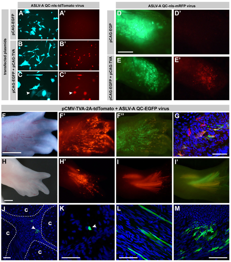
Axolotls are poised to become the premiere model system for studying vertebrate appendage regeneration. However, very few molecular tools exist for studying crucial cell lineage relationships over regeneration or for robust and sustained misexpression of genetic elements to test their function. Furthermore, targeting specific cell types will be necessary to understand how regeneration of the diverse tissues within the limb is accomplished. We report that pseudotyped, replication-incompetent retroviruses can be used in axolotls to permanently express markers or genetic elements for functional study. These viruses, when modified by changing their coat protein, can infect axolotl cells only when they have been experimentally manipulated to express the receptor for that coat protein, thus allowing for the possibility of targeting specific cell types. Using viral vectors, we have found that progenitor populations for many different cell types within the blastema are present at all stages of limb regeneration, although their relative proportions change with time.


Figures

Fig. 1.
Fig. 1.
VSV-G-psuedotyped QC retroviruses can infect axolotl cells in vitro and in vivo. (A,B) Axolotl AL1 cells were infected with VSV-G-pseudotyped QCMV-nls-EGFP. (A) Bright-field DIC. (B) Green channel shows cells descended from infected cells. (C-F) Cranial neural crest and underlying mesenchymal cells in axolotl embryos (stage 16) were infected with VSV-G-psuedotyped QCMV-EGFP, and their descendants were tracked when the animals reached hatchling stage. (C) Schematic for infections. (D,D′) Bright-field and fluorescent images of the head of an infected animal. The eye (outlined in yellow) is autofluorescent. (E) Transverse section of specimen pictured in D,D′ shows EGFP-expressing cells in the head muscles mm. levator mandibulae anterior (ant mand) and levator mandibulae externus (ext mand). Sections are stained with anti-GFP (green) and DAPI (blue). (F) Transverse section of the same specimen more caudally documenting EGFP+ cells in the mesoderm-derived m. levator mandibulae externus (ext mand, top arrowhead) and in the neural crest-derived Meckel’s cartilage (M). (G) Regenerating hind limb of live animal expressing EGFP 5 days after infection with QCMV-EGFP. (H) The same limb as in G, 11 days post-infection. (I,J) Cryosections of limb shown in G and H, harvested 11 days post-infection, 27 days post-amputation, stained with anti-GFP (green) and DAPI (blue). (I) In the posterior mesenchyme just beginning to undergo condensation to form digits, the majority of the overlying cells (both undifferentiated mesenchymal cells and nascent muscle fibers) are EGFP+. The epidermis (epi) is negative. (J) Condensations forming the tarsals (tar, outlined) have ∼46% EGFP+ cells, whereas ∼62% of intervening joint cells are EGFP+. (K) After full regeneration (unfixed, freshly harvested at 7 weeks post-amputation), limbs whose blastemas were infected at 2 weeks post-amputation show very robust EGFP fluorescence in many tissues distal to the amputation plane (dotted line) and no fluorescence more proximally. (L) Higher magnification view of autopod from limb shown in K. (M) Chondrocytes in a live limb. (N) Labeled vasculature (arrowheads) in the digits of a limb whose blastema was infected 3 weeks post-amputation. (O-R) Cryosections from the limb shown in K,L show a high percentage of infected cells in various tissues. (O) Distal tibia. (P) Tarsal cartilages and joint tissue. (Q) Muscle. (R) Digit tip. Scale bars: 1 mm in G,H,K,L; 200 μm in I,J,O-R; 500 μm in M,N.
Fig. 2.
Fig. 2.
Infection of blastemas at various time points post-amputation reveals progenitors for nearly every cell type are present in the blastema and are mitotically active throughout regeneration. (A) Schematic of experiment. Limbs were amputated at mid-stylopod (mid-humerus for forelimbs, mid-femur for hindlimbs) (arrowheads). Blastemas were infected with a VSV-G pseudotyped-, replication-incompetent QCMV retrovirus encoding a fluorophore, and limbs were harvested after full regeneration. (B) Example of an infection as performed in the schematic. (C) Regenerated forelimb infected with QCMV-EGFP 12 days post-amputation, cryosectioned and stained with anti-GFP (green), AlexaFluor-594-conjugated phalloidin (red) and DAPI (blue). Scale bar: 250 μm. h, humerus; u, ulna. (D-J″) High-magnification views of cell types identified when blastemas were infected with QCMV-Venus and regenerated limbs stained as in C. Scale bar: 50 μm. (D) Hypertrophic chondrocytes (bc, bone collar). (E) Two pairs of mother/daughter chondrocyte pairs in a distal phalange. (F) Perichondrial cell (pc) (adjacent muscle marked ‘m’ and cartilage ‘c’). (G) A single marked joint cell between two carpals (c). (H) Muscle fibers. (I) Dermal fibroblasts. (J-J″) A string of labeled vasculature cells within a capillary bed. (J) All three channels. (J′) DAPI channel subtracted for better visualization of unlabeled vessels, apparent by their F-actin organization. (J″) Adjacent and intersecting vessels outlined. (K) Relative proportion of descendant cell types at various infection times. Total numbers of cells counted for each time point are given below y-axis labels.
Fig. 3.
Fig. 3.
Regenerating limbs infected with QCAG-SHH have defective skeletal structures. (A) Uninfected control forelimb showing normal skeletal morphology. (A′) Higher-magnification view of carpals. (A″) Outline of central carpal elements. (B-C″) Regenerating forelimbs infected with QCAG-SHH show abnormal skeletal patterning following regeneration. (B-B″) Ectopic central carpal (arrowhead). (C-C″) Fusion between top and bottom central carpals (arrowhead). (D-D″) Uninfected control hindlimb including outline of central tarsal elements. (E-E″) Hindlimb infected with QCAG-SHH during regeneration showing ectopic tarsals/tarsal fusion (arrowheads). Scale bars: 1 mm in A-E; 200 μm in A′-E′.
Fig. 4.
Fig. 4.
Axolotl cells ectopically expressing the TVA receptor can be infected by ASLV-A-pseudotyped retroviruses. (A-B′) AL1 cells transfected with pCAG-EGFP (control, A,A′) or pCAG-EGFP and pCAG-TVA (B,B′) and exposed ASLV-A-psuedotyped QCMV-nls-tdTomato virus 4 days post-transfection. Cells were imaged live. (A) Green channel shows many transfected control cells. (A′) Red channel shows none of these cells have been infected. (B) Green channel shows many transfected cells. (B′) Red channel with identical imaging conditions as in A′ showing nearly all of the transfected cells have been infected with the virus. (C,C′) Higher magnification from the same experiment as B showing that cells weakly expressing the co-electroporation marker (arrowheads) can strongly express the viral marker. (D-E′) Limbs were electroporated with either pCAG-tdTomato (D,D′) or pCMV-TVA-2A-tdTomato (E,E′) at 1 week post-amputation and injected with ASLV-A pseudotyped QCMV-EGFP 2 days later, and imaged under identical conditions at 4 days post-injection. (D,D′) Without TVA, limbs cannot be infected with the virus (n=8). (E,E′) All limbs (n=8) expressing TVA were infected with the virus. (F-G) Late-palette stage regenerating limbs were electroporated with pCMV-TVA-2A-tdTomato, infected with ASLV-A psuedotyped QCMV-EGFP 2 days later, and imaged 4 days post-infection. (G) The limb shown in F-F″ was fixed, sectioned, counterstained with DAPI and percentage of tdTomato+ cells that were also EGFP+ was calculated to be ∼80% (n=221/277). (H-I′) An electroporation/viral infection experiment was followed until limbs were completely regenerated (n=6). (J-M) Examples of EGFP-expressing cells in TVA-expressing/ASLV-A-psuedotyped QCMV-EGFP infected regenerated limbs. (J) ‘c’ denotes condensed wrist cartilage elements, outlines of which are indicated by dashed lines. Blue is DAPI. Joint cells (arrowhead) are sandwiched between cartilage elements and are more densely packed than surrounding cartilage. (K) Labeled chondrocyte (arrowhead). (L) Labeled muscle fibers. (M) Labeled fibroblasts. Scale bars: 500 μm in D-E′; 1 mm in F-F″,H-I′; 50 μm in G; 100 μm in J-M.
Fig. 5.
Fig. 5.
The TVA/ASLV-A system can be used to target specific cell types in regenerating limbs. (A-B′) The mammalian PECAM enhancer/promoter element drives expression of marker genes in vascular endothelial cells in F0 transgenic axolotls. Bright-field (A) and fluorescent (A′) images of gills and vasculature in a live animal 10 days post-fertilization. Bright-field (B) and fluorescent (B′) images of limb vasculature in a separate live animal 7 weeks post-fertilization. (C) EGFP+ vessels in newly regenerated limb (limb outlined). (D-G) Confocal images of F0 transgenic PECAM-EGFP adult limb 9 weeks post-amputation. (D) Section stained with anti-GFP (green), phalloidin (red) and DAPI (blue). (E-G) Sections stained with anti-EGFP expression (red) showing expression in vascular endothelial cells, and cells expressing smooth muscle actin (green). (E) Large vessel showing the endothelial cells are ensheathed by smooth muscle actin-expressing cells. (F) Vessels in cross-section. Red staining (arrowhead) is luminal to green (double arrowheads). (G) Capillary (arrowheads) in longitudinal section. Red staining is luminal to green. (H,H′) Blastemas were electroporated with pPECAM-TVA (titer 108) and infected with ASLV-A-psuedotyped QCMV-tdTomato 3 days post-electroporation. Fully regenerated limbs were fixed and cryosectioned. (H) Bright-field image showing cartilage elements (c) of two adjacent digits. (H′) Red channel reveals tdTomato-expressing cells could be detected among the vasculature between the epidermis and cartilage in the regenerated limbs. (I-K) Confocal images showing specific expression of tdTomato in the vasculature of animals expressing TVA from the PECAM promoter who have been infected with ASLV-A-pseudotyped QCMV-tdTomato. Regenerated limbs were fixed, cryosectioned and stained with DAPI. (I) Uninfected animal not expressing TVA shows no fluorescent vessels in the vessel-rich area of the digits between cartilage and epidermis. (J) Using the same imaging conditions, red fluorescent vessels are apparent in the same region of a regenerated limb whose blastema was electroporated with pPECAM-TVA and infected with ASLV-A-pseudotyped QCMV-tdTomato, red fluorescent vessels are apparent. (K) A similar result was obtained by infecting blastemas in PECAM-TVA F0 transgenic limbs. Scale bars: 500 μm in A-B′; 1 mm in C; 200 μm in D; 50 μm in E,F,I-K; 32 μm in G; 100 μm in H,H′.
Fig. 6.
Fig. 6.
Expression of Cre recombinase from QC infections in axolotl cells. (A,A′) Cre-encoding elements delivered to AL1 cells in culture via the VSV-pseudotyped QCAG-Cre retrovirus can catalyze recombination between LoxP sites. AL1 cells were transfected with pCLESLT (transfected cells are green in E) and subsequently infected with QCAG-Cre. (A) Transfected cells express EGFP. (A′) A subset of pCLESLT-expressing cells underwent recombination at the LoxP sites enabling expression of nuclear tdTomato (arrowheads). (B,B′) Regenerating limbs electroporated with pCLESLT and subsequently infected with QCAG-Cre also show a subset of electroporated cells express tdTomato. Scale bar: 100 μm.

Similar articles

Cited by

References

    1. Atkinson D. L., Stevenson T. J., Park E. J., Riedy M. D., Milash B., Odelberg S. J. (2006). Cellular electroporation induces dedifferentiation in intact newt limbs. Dev. Biol. 299, 257–271 - PMC - PubMed
    1. Beier K. T., Samson M. E., Matsuda T., Cepko C. L. (2011). Conditional expression of the TVA receptor allows clonal analysis of descendents from Cre-expressing progenitor cells. Dev. Biol. 353, 309–320 - PMC - PubMed
    1. Berg D. A., Kirkham M., Beljajeva A., Knapp D., Habermann B., Ryge J., Tanaka E. M., Simon A. (2010). Efficient regeneration by activation of neurogenesis in homeostatically quiescent regions of the adult vertebrate brain. Development 137, 4127–4134 - PubMed
    1. Brockes J. P., Kumar A. (2005). Appendage regeneration in adult vertebrates and implications for regenerative medicine. Science 310, 1919–1923 - PubMed
    1. Burns J. C., Matsubara T., Lozinski G., Yee J. K., Friedmann T., Washabaugh C. H., Tsonis P. A. (1994). Pantropic retroviral vector-mediated gene transfer, integration, and expression in cultured newt limb cells. Dev. Biol. 165, 285–289 - PubMed

Publication types

Substances