. 2012 Feb 14;1(2):e11.
doi: 10.1038/mtna.2012.1.

Optimized in vivo transfer of small interfering RNA targeting dermal tissue using in vivo surface electroporation

Affiliations

Optimized in vivo transfer of small interfering RNA targeting dermal tissue using in vivo surface electroporation

Kate E Broderick et al. Mol Ther Nucleic Acids. .

Abstract

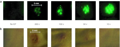
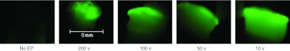
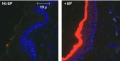
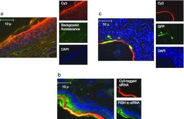
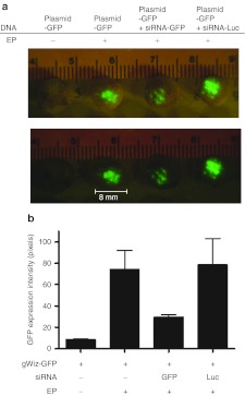
Electroporation (EP) of mammalian tissue is a technique that has been used successfully in the clinic for the delivery of genetic-based vaccines in the form of DNA plasmids. There is great interest in platforms which efficiently deliver RNA molecules such as messenger RNA and small interfering RNA (siRNA) to mammalian tissue. However, the in vivo delivery of RNA enhanced by EP has not been extensively characterized. This paper details the optimization of electrical parameters for a novel low-voltage EP method to deliver oligonucleotides (both DNA and RNA) to dermal tissue in vivo. Initially, the electrical parameters were optimized for dermal delivery of plasmid DNA encoding green fluorescent protein (GFP) using this novel surface dermal EP device. While all investigated parameters resulted in visible transfection, voltage parameters in the 10 V range elicited the most robust signal. The parameters optimized for DNA, were then assessed for translation of successful electrotransfer of siRNA into dermal tissue. Robust tagged-siRNA transfection in skin was detected. We then assessed whether these parameters translated to successful transfer of siRNA resulting in gene knockdown in vivo. Using a reporter gene construct encoding GFP and tagged siRNA targeting the GFP message, we show simultaneous transfection of the siRNA to the skin via EP and the concomitant knockdown of the reporter gene signal. The siRNA delivery was accomplished with no evidence of injection site inflammation or local tissue damage. The minimally invasive low-voltage EP method is thus capable of efficiently delivering both DNA and RNA molecules to dermal tissue in a tolerable manner.


Figures

Figure 1
Figure 1
A range of voltage parameters results in delivery of DNA plasmid expressing GFP to guinea pig skin using the SEP device. Guinea pig skin was injected intradermally with 100 µg of DNA plasmid expressing GFP (pgWIZ-GFP) and either left untreated or immediately pulsed with the SEP device at a voltage setting of 200, 100, 50 or 10 V for one pulse. Skin biopsies were harvested 48 hours later and observed by fluorescent microscopy (a) for determination of GFP positive signal or bright light (b) for determination of local treatment site reaction. Bar is included for approximation of site reaction size. GFP, green fluorescent protein; SEP, surface electroporation.
Figure 2
Figure 2
Lower voltage parameters result in higher antibody titers to NP protein in guinea pigs. Guinea pig skin was injected intradermally with 100 µg of DNA plasmid-expressing NP and either left untreated (ID only) or immediately pulsed with the SEP device at a voltage setting of 50 or 10 V. Animals were immunized at day 0 and day 21. Blood was harvested weekly and analyzed for the presence of binding antibodies against NP by ELISA. Data shown here is from day 49. ELISA, enzyme-linked immunosorbent assay; ID, intradermal; SEP, surface electroporation.
Figure 3
Figure 3
A range of voltage parameters results in successful delivery of tagged siRNA to guinea pig skin using the SEP device. Guinea pig skin was injected intradermally with 100 µg of siRNA-tagged with Alexa 488 and either left untreated or immediately pulsed with the SEP device at a voltage setting of 200, 100, 50 or 10 V for three pulses. Skin biopsies were harvested 48 hours later and observed by fluorescent microscopy for determination of positive signal. Bar is included for approximation of biopsy size. SEP, surface electroporation; siRNA, small interfering RNA.
Figure 4
Figure 4
Dermal electroporation results in robust siRNA-Cy3 signal present 48 hours post-treatment. Guinea pig skin was injected intradermally with 100 µg of siRNA-tagged with Cy3 and either left untreated or immediately pulsed with the SEP device at 10 V for three pulses. Skin biopsies were harvested 48 hours later, fixed in formalin, sectioned, and DAPI stained. Slide sections were observed by fluorescent microscopy for determination of positive signal. DAPI, 4′,6-diamidino-2-phenylindole; SEP, surface electroporation; siRNA, small interfering RNA.
Figure 5
Figure 5
Dermal electroporation results in siRNA-Cy3 signal with confirmation by siRNA-specific FISH, and GFP expression colocalized to the stratum corneum and epithelial cells. (a) Guinea pig skin was injected intradermally with 100 µg of siRNA-tagged with Cy3 and immediately pulsed with the dermal EP device at 10 V for three pulses. Skin biopsies were harvested 48 hours later, fixed in formalin, DAPI stained, and sectioned. Slide sections were observed by fluorescent microscopy for determination of positive signal. (b) The localization of siRNA was additionally detected by fluroescein-tagged fluorescent in situ hybridization (FISH) to the antisense strand of the Cy3-tagged siRNA in guinea pig skin sections serial to those displayed in part A of this figure. (c) Guinea pig skin was injected intradermally with a mix of 100 µg of siRNA-tagged with Cy3 and 100 µg DNA plasmid-expressing GFP and immediately pulsed with the dermal EP device at 10 V for three pulses. Skin biopsies were harvested 48 hours later, fixed in formalin, sectioned, and DAPI stained. Slide sections were observed by fluorescent microscopy for determination of positive signal. DAPI, 4′,6-diamidino-2-phenylindole; GFP, green fluorescent protein; siRNA, small interfering RNA.
Figure 6
Figure 6
Delivery of siRNA by the SEP device results in targeted gene knockdown of reporter gene in dermal tissue. Guinea pig skin was injected intradermally with 25 µg of DNA plasmid-expressing GFP (pgWIZ-GFP) left untreated, 25 µg of DNA plasmid-expressing GFP and immediately pulsed with the SEP device or plasmid mixed with either siRNA-GFP or siRNA-Luc (20 µg) and immediately pulsed with the SEP device. (a) Skin biopsies were harvested 24 hours later and observed under bright light for determination of treatment site location or fluorescent microscopy for determination of GFP positive signal. (b) GFP pixel density was determined using pixel counting software as described (see Materials and Methods). GFP, green fluorescent protein; luc, luciferase; SEP, surface electroporation; siRNA, small interfering RNA.

Similar articles

Cited by

References

    1. Bumcrot D, Manoharan M, Koteliansky V., and, Sah DW. RNAi therapeutics: a potential new class of pharmaceutical drugs. Nat Chem Biol. 2006;2:711–719. - PMC - PubMed
    1. de Fougerolles A, Vornlocher HP, Maraganore J., and, Lieberman J. Interfering with disease: a progress report on siRNA-based therapeutics. Nat Rev Drug Discov. 2007;6:443–453. - PMC - PubMed
    1. Akinc A, Querbes W, De S, Qin J, Frank-Kamenetsky M, Jayaprakash KN.et al. (2010Targeted delivery of RNAi therapeutics with endogenous and exogenous ligand-based mechanisms Mol Ther 181357–1364. - PMC - PubMed
    1. Higuchi Y, Kawakami S., and, Hashida M. Strategies for in vivo delivery of siRNAs: recent progress. BioDrugs. 2010;24:195–205. - PubMed
    1. Low L, Mander A, McCann K, Dearnaley D, Tjelle T, Mathiesen I.et al. (2009DNA vaccination with electroporation induces increased antibody responses in patients with prostate cancer Hum Gene Ther 201269–1278. - PubMed

LinkOut - more resources