. 2012 Feb 7;1(2):e9.
doi: 10.1038/mtna.2011.6.

Intracerebral Infusion of Antisense Oligonucleotides Into Prion-infected Mice

Affiliations

Intracerebral Infusion of Antisense Oligonucleotides Into Prion-infected Mice

Karah Nazor Friberg et al. Mol Ther Nucleic Acids. .

Abstract

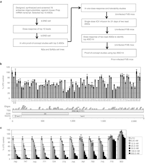
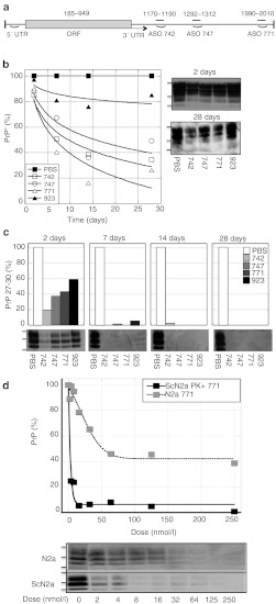
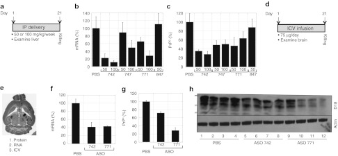
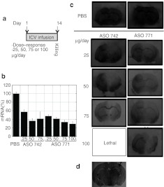
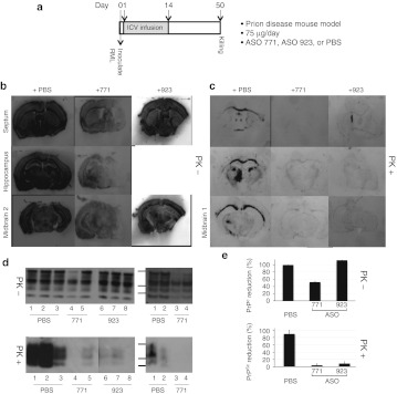
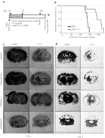
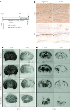
Mice deficient for the cellular prion protein (PrP(C)) do not develop prion disease; accordingly, gene-based strategies to diminish PrP(C) expression are of interest. We synthesized a series of chemically modified antisense oligonucleotides (ASOs) targeted against mouse Prnp messenger RNA (mRNA) and identified those that were most effective in decreasing PrP(C) expression. Those ASOs were also evaluated in scrapie-infected cultured cells (ScN2a) for their efficacy in diminishing the levels of the disease-causing prion protein (PrP(Sc)). When the optimal ASO was infused intracerebrally into FVB mice over a 14-day period beginning 1 day after infection with the Rocky Mountain Laboratory (RML) strain of mouse prions, a prolongation of the incubation period of almost 2 months was observed. Whether ASOs can be used to develop an effective therapy for patients dying of Creutzfeldt-Jakob disease remains to be established.


Figures

Figure 1
Figure 1
Identification of ASOs that diminish mouse Prnp mRNA expression measured by qRT-PCR. (a) Experimental design. (b) Prnp mRNA expression 24 hours post-transfection with 78 antisense oligonucleotides (120 nmol/l) targeting mouse Prnp mRNA in b.END cells. ASOs are displayed relative to their position on the 2,206-bp mouse Prnp mRNA (Accession NM_011170). (c) Dose-response of Prnp mRNA expression using the 10 most-effective ASOs in b.END cells. ASO concentrations ranged from 6.25–200 nmol/l, as indicated. Prnp mRNA is expressed as a percentage compared to untransfected control (UTC). ASO, antisense oligonucleotide; bp, base pair; ICV, intracerebral ventricular; mRNA, messenger RNA; qRT-PCR, quantitative real time-PCR.
Figure 2
Figure 2
ASOs targeting mouse Prnp mRNA reduced PrPC and PrPSc expression in N2a and ScN2a cells. (a) The three lead ASOs 742, 747, and 771 identified in b.END cells are displayed relative to their positions on the 2,206-bp mouse Prnp mRNA (Accession NM_011170). All fall within exon 4 outside of the coding sequence in the 3′ UTR. (b,c) Kinetics of PrPC suppression in N2a cells is shown in b and protease-resistant PrPSc in ScN2a cells is shown in c exposed to 500 nmol/l of each ASO for 2, 7, 14, and 28 days. ASO 923 and PBS were used as controls. (d) Dose-response curves of PrPC in N2a and PrPSc in ScN2a cells incubated with 0–250 nmol/l of ASO 771. In bd, PrP levels were quantified by densitometry of scanned western blot films using NIH Image J software and expressed as percentages relative to cells treated with PBS. Protein standards shown are 36, 30, and 22 kilodaltons. ASO, antisense oligonucleotide; mRNA, messenger RNA; ORF, open reading frame; PBS, phosphate-buffered saline; PrPC, cellular prion protein; PrPSc, disease-causing prion protein.
Figure 3
Figure 3
Administration of ASOs targeting mouse Prnp mRNA reduced PrPC expression in the livers (a–c) and brains (d–h) of FVB mice. ASOs were administered by intraperitoneal inoculations for liver studies (n = 4 per dose) and by ICV infusion (n = 4) for brain studies. (a,d) Experimental design. ASOs were administered for 21 days at the specified doses, after which time, mice were killed, tissues taken, and mRNA and protein levels were analyzed. (b,f) Prnp mRNA levels were measured by qRT-PCR; (c,g,h) PrPC levels were analyzed by a capture ELISA using antibody D18 (c) or by densitometry (g) of western blots probed with antibody D18 (h). (e) Brain map showing specific brain regions that were cannulated (region 3), harvested for protein (region 1), and RNA (region 2). (g,h) ASO 771 more effectively reduced PrPC levels in brain compared to ASO 742. Different lanes represent four different animals. Bottom panel of western blot was probed with actin. Protein standards shown are 36, 30, and 22 kilodaltons. For all panels, PrP expression levels are shown as percentages relative to those measured in mice treated with PBS. ELISA, enzyme-linked immunosorbent assay; ICV, intracerebral ventricular; ip, intraperitoneal; mRNA, messenger RNA; PBS, phosphate-buffered saline; PrPC, cellular prion protein; qRT-PCR, quantitative real time-PCR.
Figure 4
Figure 4
Dose-dependent reduction of mouse Prnp mRNA and PrPC by ASOs administered by ICV infusion. (a) Experimental design. Doses of 25, 50, 75, or 100 µg/day were administered by ICV delivery for 14 days, and Prnp mRNA and PrPC levels analyzed at day 14. (b) PrP mRNA levels were quantified by qRT-PCR and expressed as the relative percentage measured in PBS-treated controls. (c) Regional expression of PrPC in coronal cryosections (10 µm) at the level of the septum, shown by histoblotting with antibody D18. (d) Immunohistochemistry of a hippocampal coronal section stained with anti-PS antibody that detects the PS modification in the ASO. Arrow indicates the cannulation side. ASO, antisense oligonucleotide; ICV, intracerebral ventricular; mRNA, messenger RNA; PBS, phosphate-buffered saline; PrPC, cellular prion protein; PS, phosphorothioate; qRT-PCR, quantitative real-time PCR.
Figure 5
Figure 5
ASO 771 reduced PrPC expression and interfered with PrPSc deposition in prion-infected FVB mice. (a) Experimental design. On day 0, FVB mice were inoculated with RML prions on the right side of the brain. At day 1, ASO 771, ASO 923, or PBS was delivered ICV at 75 µg/day for 14 days on the left side of the brain. Mice were killed at day 50 dpi, brain sections taken and analyzed for PrPC and PrPSc levels. (b,c) Histoblot analysis was performed on 10-µm sections at the level of the septum, hippocampus, midbrain 1 and 2 regions, and stained with the D18 antibody. Sections were left undigested to visualize total PrP levels as shown in b or subjected to GdnHCl denaturation and PK digestion to visualize PrPSc levels as shown in c. (d) Western blots show total PrP (top) and PrPSc (bottom); each blot shows two different ASO-treated animals. (e) Densitometry of western blots using NIH Image J software, then expressed relative to PrP levels in the brains of mice treated with PBS. Protein standards shown are 36, 30, and 22 kDa. ASO, antisense oligonucleotide; ICV, intracerebral ventricular; PBS, phosphate-buffered saline; PrPC, cellular prion protein; PrPSc, disease-causing prion protein.
Figure 6
Figure 6
Treatment with ASO 771 reduced PrPSc in the brain and extended incubation periods by 42%. (a) Experimental design. On day 0, FVB mice were inoculated with RML prions on the right side of the brain. At day 1, either ASO 771 or PBS was delivered ICV at 75 µg/day for 14 days on the left side of the brain. At day 126, some mice were killed, and their brains analyzed for PrPC and PrPSc. (b) Survival curves of FVB mice treated with PBS (black) or ASO 771 (gray). (c,d) Mice were killed at 126 dpi, their brains analyzed by histoblotting for (c) total PrP and (d) PrPSc. Sections were left undigested as shown in c or subjected to GdnHCl denaturation and PK digestion as shown in d. Membranes were stained with the D18 antibody. ASO, antisense oligonucleotide; PBS, phosphate-buffered saline; PrPSc, disease-causing prion protein.
Figure 7
Figure 7
Treatment with ASO 771 at the midpoint of disease reversed PrPSc deposition but caused atypical illness. (a) Experimental design. On day 0, FVB mice were inoculated with RML prions. Beginning at 60 dpi, ASO 771 was delivered via ICV at 75 µg/day for 11 days, when they developed atypical illness. Mice were killed at 71 dpi and their brains analyzed for (c) PrPC and (b,d) PrPSc. (b) Immunohistochemistry for PrPSc was performed on formalin-fixed, paraffin-embedded tissue sections through the hippocampus by the hydrolytic autoclaving method and with recFab HuM-P. (c,d) Histoblots indicate that both PrPC and PrPSc were reduced following administration of ASO 771. Reduction of PrPC appeared more prominent on the cannulation side, whereas PrPSc depletion occurred bilaterally. Sections were left undigested to visualize total PrP levels as shown in c or subjected to GdnHCl denaturation and PK digestion to visualize PrPSc levels as shown in d, then stained with the D18 antibody. Bar in b represents 50 µm and applies to all the images in the panel. ASO, antisense oligonucleotide; cc, corpus collosum; hp, hippocampus; ICV, intracerebral ventricular; nc, neocortex; PBS, phosphate-buffered saline; PrPC, cellular prion protein; PrPSc, disease-causing prion protein.

References

    1. Tilly G, Chapuis J, Vilette D, Laude H., and, Vilotte JL. Efficient and specific down-regulation of prion protein expression by RNAi. Biochem Biophys Res Commun. 2003;305:548–551. - PubMed
    1. Daude N, Marella M., and, Chabry J. Specific inhibition of pathological prion protein accumulation by small interfering RNAs. J Cell Sci. 2003;116 Pt 13:2775–2779. - PubMed
    1. Pfeifer A, Eigenbrod S, Al-Khadra S, Hofmann A, Mitteregger G, Moser M.et al. (2006Lentivector-mediated RNAi efficiently suppresses prion protein and prolongs survival of scrapie-infected mice J Clin Invest 1163204–3210. - PMC - PubMed
    1. White MD, Farmer M, Mirabile I, Brandner S, Collinge J., and, Mallucci GR. Single treatment with RNAi against prion protein rescues early neuronal dysfunction and prolongs survival in mice with prion disease. Proc Natl Acad Sci USA. 2008;105:10238–10243. - PMC - PubMed
    1. Kocisko DA, Vaillant A, Lee KS, Arnold KM, Bertholet N, Race RE.et al. (2006Potent antiscrapie activities of degenerate phosphorothioate oligonucleotides Antimicrob Agents Chemother 501034–1044. - PMC - PubMed