Clinical Trial
. 2013 Mar 1;288(9):6248-58.
doi: 10.1074/jbc.M112.439356. Epub 2013 Jan 23.

Ca2+ signals generated by CatSper and Ca2+ stores regulate different behaviors in human sperm

Affiliations
Clinical Trial

Ca2+ signals generated by CatSper and Ca2+ stores regulate different behaviors in human sperm

Wardah Alasmari et al. J Biol Chem. .

Abstract

[Ca(2+)]i signaling regulates sperm motility, enabling switching between functionally different behaviors that the sperm must employ as it ascends the female tract and fertilizes the oocyte. We report that different behaviors in human sperm are recruited according to the Ca(2+) signaling pathway used. Activation of CatSper (by raising pHi or stimulating with progesterone) caused sustained [Ca(2+)]i elevation but did not induce hyperactivation, the whiplash-like behavior required for progression along the oviduct and penetration of the zona pellucida. In contrast, penetration into methylcellulose (mimicking penetration into cervical mucus or cumulus matrix) was enhanced by activation of CatSper. NNC55-0396, which abolishes CatSper currents in human sperm, inhibited this effect. Treatment with 5 μm thimerosal to mobilize stored Ca(2+) caused sustained [Ca(2+)]i elevation and induced strong, sustained hyperactivation that was completely insensitive to NNC55-0396. Thimerosal had no effect on penetration into methylcellulose. 4-Aminopyridine, a powerful modulator of sperm motility, both raised pHi and mobilized Ca(2+) stored in sperm (and from microsomal membrane preparations). 4-Aminopyridine-induced hyperactivation even in cells suspended in Ca(2+)-depleted medium and also potentiated penetration into methylcellulose. The latter effect was sensitive to NNC55-039, but induction of hyperactivation was not. We conclude that these two components of the [Ca(2+)]i signaling apparatus have strikingly different effects on sperm motility. Furthermore, since stored Ca(2+) at the sperm neck can be mobilized by Ca(2+)-induced Ca(2+) release, we propose that CatSper activation can elicit functionally different behaviors according to the sensitivity of the Ca(2+) store, which may be regulated by capacitation and NO from the cumulus.


Figures

FIGURE 1.
FIGURE 1.
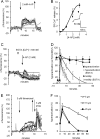
Alkalinization raises [Ca2+]i in human sperm. A, 2 mm 4-AP (green trace) and 25 mm NH4Cl (dark blue trace) cause similar changes in pHi of human sperm populations. The additions are marked by an arrow. Both aliquots were from the same ejaculate. B, amplitude of pHi increment imposed by 25 mm NH4Cl (dark blue), 10 and 20 mm TMA (light blue), 2 mm 4-AP (green), and 10 mm thimerosal (red). Each bar shows the mean of 4–10 experiments ± S.E. (error bars). C, 25 mm NH4Cl (added at the arrow) increases [Ca2+]o (OGB fluorescent intensity) in human sperm. Shown are the responses of eight cells in the same experiment. D, 20 mm TMA (first arrow) induces a large prolonged increment in pHi of human sperm. 2 mm 4-AP was added at the second arrow. E, 20 mm TMA (added at the first arrow) increases [Ca2+]o (OGB fluorescent intensity) in human sperm. The upward arrow shows TMA washout. Shown are the responses of eight cells in the same experiment. F, 3 μm progesterone (added at the arrow) causes a biphasic increase in [Ca2+]o (OGB fluorescent intensity) in human sperm. Shown are the responses of six cells in the same experiment, one of which generates [Ca2+]i oscillations after the initial transient.
FIGURE 2.
FIGURE 2.
Stored Ca2+ but not activation of CatSper induces hyperactivation. A and B, increment in percentage of hyperactivated cells induced by 25 mm NH4Cl (dark blue), 20 and 10 mm TMA (light blue), 2 mm 4-AP (green), 3 μm progesterone (yellow), and 5 μm thimerosal (red). A, cells prepared by swim-up into sEBSS; B, cells prepared by density gradient centrifugation into STF. Each bar shows mean ± S.E. (error bars) of 20–60 experiments except for thimerosal swim-up (n = 8), TMA (n = 4–5). *, p < 0.05; **, p < 0.01; ***, p < 0.001; ****, p < 0.0001 compared with control. C and D, frequency distributions of ALH (C) and linearity (D) under control conditions (black) and after stimulation with 25 mm NH4Cl (dark blue), 2 mm 4-AP (green), and 5 μm thimerosal (red). Each plot shows mean ± S.E. of 19 (control, 4-AP, progesterone), 9 (NH4Cl), and 10 (thimerosal) STF samples. E–H, example tracks of control (E) and cells exposed to 20 mm TMA (F), 2 mm 4-AP (G), and 5 μm thimerosal (H). All traces start at the origin (0, 0), and scales show distance (μm). Sample rate = 100 Hz.
FIGURE 3.
FIGURE 3.
4-AP causes hyperactivation by mobilizing stored Ca2+. A, sustained increase in [Ca2+]i (OGB fluorescent intensity) in human sperm exposed to 2 mm 4-AP (first arrow). 4-AP was washed off at the second arrow. Shown are responses of eight cells in same experiment. B, release of Ca2+ from sacroplasmic reticulum microsomes by 4-AP. The graph shows 4-AP-induced Ca2+ release (percentage of A23187-releasable Ca2+); each point represents the mean ± S.E. (error bars) of 4–7 experiments. Inset, example trace. 2 mm 4-AP was added at the first arrow, and remaining Ca2+ was released by 25 μm A23187 (second arrow). Calibration shows time and [Ca2+]. C, effect of 2 mm 4-AP in Ca2+-depleted medium. Sperm were superfused with EGTA-buffered medium (first arrow), which caused a rapid fall in [Ca2+]i. Subsequent application of 4-AP caused a [Ca2+]i transient in a subset of cells. Shown are responses of 11 cells in the same experiment. D, time course of hyperactivation (percentage of cells; squares) induced by 2 mm 4-AP in cells suspended in sEBSS (filled symbols) and in parallel experiments where cells were resuspended in EGTA-buffered sEBSS immediately before use (open symbols). Circles show the percentage of motile cells. Each point shows mean ± S.E. of four experiments. E, sustained increase in [Ca2+]i (OGB fluorescent intensity) in human sperm exposed to 5 μm thimerosal (first arrow). Thimerosal was washed off at the second arrow, and 3 μm progesterone was applied at the third arrow. Shown are the responses of eight cells in the same experiment. Temperature was 30 °C. F, time course and concentration dependence of thimerosal-induced hyperactivation (percentage of cells). Each line shows mean ± S.E. of four experiments.
FIGURE 4.
FIGURE 4.
CatSper activity enhances penetration into viscous medium and contributes to spontaneous hyperactivation. A and B, increment in cell density (percentage of control) 1 cm (A) and 2 cm (B) into methylcellulose. Cells were stimulated with 25 mm NH4Cl (dark blue), 10 mm TMA (light blue), 2 mm 4-AP (green), 3 μm progesterone (yellow), and 5 μm thimerosal (red). Bars show the mean ± S.E. (error bars) of 10–20 experiments except for TMA (4). C, increment in cell density (percentage of control) 2 cm into methylcellulose of cells stimulated with 2 mm 4-AP (green), 3 μm progesterone (yellow), and 25 mm NH4Cl (dark blue) and in parallel incubations pretreated with 10 μm NNC (black). Bars, mean ± S.E. of 8–20 experiments except for TMA (4). D, inhibition of spontaneous hyperactivation upon exposure of STF-capacitated cells to NNC (difference between control and NNC-treated cells; μm NNC) is dependent upon the level of spontaneous hyperactivation prior to application of the drug (r = 0.75, n = 19). E, increment in hyperactivation (percentage of cells) in response to 2 mm 4-AP (green), 3 μm progesterone (yellow), 25 mm NH4Cl (dark blue), and 5 μm thimerosal (red). Black bars, responses in parallel 10 μm NNC-pretreated experiments. Each bar shows mean ± S.E. of 9–20 experiments. *, p < 0.05; **, p < 0.01; ***, p < 0.001; ****, p < 0.0001 compared with control (A and B) or NNC alone (C and E). NS, not significant.
FIGURE 5.
FIGURE 5.
Tentative model for the interacting effects of Ca2+ influx mediated by CatSper (blue box) and release of Ca2+ from the store at the sperm neck (yellow box) based on evidence from this and previous studies. The acrosomal Ca2+ store is also shown (green box). CICR links these two parts of the Ca2+-signaling apparatus in a subset of cells where the Ca2+ store is sensitized (dashed arrow). Agents employed in this study are shown in red. STF is shown acting to enhance capacitation, including sensitization of the Ca2+ store at the sperm neck. Other (endogenous) agents and pathways are shown in black. Question marks indicate effects that are consistent with the model or may be predicted on the basis of studies on other cell types but that have not been established in sperm. Double question marks denote speculation.

Similar articles

Cited by

References

    1. Stauss C. R., Votta T. J., Suarez S. S. (1995) Sperm motility hyperactivation facilitates penetration of the hamster zona pellucida. Biol. Reprod. 53, 1280–1285 - PubMed
    1. Quill T. A., Sugden S. A., Rossi K. L., Doolittle L. K., Hammer R. E., Garbers D. L. (2003) Hyperactivated sperm motility driven by CatSper2 is required for fertilization. Proc. Natl. Acad. Sci. U.S.A. 100, 14869–14874 - PMC - PubMed
    1. Suarez S. S. (2008) Control of hyperactivation in sperm. Hum. Reprod. Update 14, 647–657 - PubMed
    1. Yanagimachi R. (1970) The movement of golden hamster spermatozoa before and after capacitation. J. Reprod. Fertil. 23, 193–196 - PubMed
    1. Drobnis E. Z., Yudin A. I., Cherr G. N., Katz D. F. (1988) Hamster sperm penetration of the zona pellucida. Kinematic analysis and mechanical implications. Dev. Biol. 130, 311–323 - PubMed

Publication types

MeSH terms

LinkOut - more resources