. 2013 Mar 15;73(6):1721-32.
doi: 10.1158/0008-5472.CAN-12-2233. Epub 2013 Jan 23.

LOX-mediated collagen crosslinking is responsible for fibrosis-enhanced metastasis

Affiliations

LOX-mediated collagen crosslinking is responsible for fibrosis-enhanced metastasis

Thomas R Cox et al. Cancer Res. .

Expression of concern in

Abstract

Tumor metastasis is a highly complex, dynamic, and inefficient process involving multiple steps, yet it accounts for more than 90% of cancer-related deaths. Although it has long been known that fibrotic signals enhance tumor progression and metastasis, the underlying molecular mechanisms are still unclear. Identifying events involved in creating environments that promote metastatic colonization and growth are critical for the development of effective cancer therapies. Here, we show a critical role for lysyl oxidase (LOX) in establishing a milieu within fibrosing tissues that is favorable to growth of metastastic tumor cells. We show that LOX-dependent collagen crosslinking is involved in creating a growth-permissive fibrotic microenvironment capable of supporting metastatic growth by enhancing tumor cell persistence and survival. We show that therapeutic targeting of LOX abrogates not only the extent to which fibrosis manifests, but also prevents fibrosis-enhanced metastatic colonization. Finally, we show that the LOX-mediated collagen crosslinking directly increases tumor cell proliferation, enhancing metastatic colonization and growth manifesting in vivo as increased metastasis. This is the first time that crosslinking of collagen I has been shown to enhance metastatic growth. These findings provide an important link between ECM homeostasis, fibrosis, and cancer with important clinical implications for both the treatment of fibrotic disease and cancer.


Figures

Figure 1
Figure 1
LOX is critical for the development of a fibrotic microenvironment following injury (A) Ashcroft scores representing the extent of fibrosis in lungs of control, bleomycin-treated, and irradiated mice. (B) Sirius Red staining (parallel light) of pulmonary tissue from control, bleomycin-treated and irradiated lung tissue. Fibrosis is highlighted by increased collagen deposition, significant disorganized thickening of alveolar septae, loss of normal alveolar architecture and considerable obstruction of alveoli due to collapse of alveolar space. (Scale bar = approx 50μm). (C) Increased collagen deposition in bleomycin-treated and irradiated pulmonary tissue as determined by SirCol quantification. (D) Immunofluorescence of control, bleomycin-treated and irradiated pulmonary tissue for LOX (alexafluor: green) shows increased levels of expression during fibrosis. (Scale bar = approx 50μm) (E) Quantitative analysis of LOX expression from immunofluorescence based on signal intensity measured using ImageJ. (F) Immunoblot analysis of LOX protein levels confirms elevated expression in both bleomycin-treated and irradiated pulmonary tissue following exposure compared to control. (G) Rising pulmonary collagen deposition in bleomycin-treated pulmonary tissue over time as determined by SirCol quantification (n = 3 mice per group).
Figure 2
Figure 2
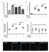
LOX inhibition decreases pulmonary fibrosis following injury (A) Collagen deposition in bleomycin-treated and irradiated pulmonary tissue with or without treatment with the function blocking LOX antibody as determined by SirCol quantification (Biocolor Ltd.). (B) Ashcroft scores representing the fibrotic changes in lungs of control, bleomycin-exposed, and irradiated mice receiving either vehicle or αLOX antibody. Blocking LOX activity decreases the degree of fibrosis observed (C) Quantitative analysis of fibrillar collagen in control, bleomycin-treated and irradiated lung treated with vehicle or αLOX antibody. Inhibition of LOX activity leads to a decrease in higher order organisation of collagen during fibrosis (D) Changes in body weight of mice are consistent between all treatment groups. (E) Sirius Red staining (orthogonal light) for cross-linked fibrillar collagen in lungs of control, bleomycin-treated and irradiated mice treated with vehicle or αLOX antibody. (Scale bar = approx 50μm).
Figure 3
Figure 3
Tissue fibrosis enhances primary breast cancer metastasis (A) Examples of H&E staining of pulmonary tissue from control, bleomycin-treated and irradiated mice showing presence of pulmonary metastases (arrowheads). An increased metastatic burden is observed in fibrotic lung compared to control (B) Quantitative analysis of total tumor burden (% of Total Lung), (C) frequency of lung metastases and (D) relative average size of metastases. (E) Examples of H&E staining of pulmonary tissue from control, bleomycin-treated and irradiated mice following tail vein injection of wt 4T1 tumor cells and foci formation (14d [arrowheads]) (F) Quantitative analysis of total tumor burden (% of Total Lung), (G) frequency of lung foci and (H) relative average size of foci showing an increase in foci formation in both bleomycin-induced and irradiation-induced fibrotic lung. (Scale bars = approx 200μm).
Figure 4
Figure 4
Therapeutic targeting of LOX prevents fibrosis-enhanced pulmonary metastases (A) Effects on 4T1 wt primary tumor volume by pre-induced fibrosis shows no significant changes in primary tumor growth (B) Examples of H&E staining of lung tissue showing decrease in the presence of pulmonary metastases in antibody treated fibrotic models (C) Relative quantitative analysis of total metastatic burden (% of Total Lung) shows decreases metastases in αLOX treated fibrotic lung compared to control. (Scale bars = approx 200μm).
Figure 5
Figure 5
Liver fibrosis enhances colonization of breast cancer cells in a LOX dependent manner (A) Sirius Red staining of control and DMN treated livers showing increased collagen deposition and cross-linking as a result of fibrosis (B) Quantitative analysis of collagen cross-linking as measured by signal intensity (C) METAVIR hepatic fibrosis scores for control, DMN treated and DMN treated with αLOX antibody or matched IgG control (D) Immunoblot analysis for LOX and alpha smooth muscle actin (αSMA) expression in liver samples showing increased levels during fibrosis (E) Effects on 4T1 wt primary tumor volume by pre-induced hepatic fibrosis shows no significant changes (F) Quantitative analysis of hepatic micrometastases in tumor bearing mice with or without DMN induced fibrosis and/or αLOX therapy (G) Examples of H&E staining of liver tissue showing micrometastatic lesions (Scale bars = approx 200μm).
Figure 6
Figure 6
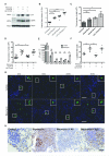
Inhibition of LOX driven fibrosis reduces pulmonary colonization and proliferation of breast cancer metastases (A) Immunoblot analysis of alpha smooth muscle actin (αSMA) and LOX expression in fibroblasts in response to bleomycin treatment at 48 hours. Inhibition of LOX activity does not affect activation of fibroblasts by bleomycin treatment nor LOX expression. (B) Relative stiffness of LOX- or ribose-modified collagen matrices as measured by shear rheology. Data is representative of two independent experiments. (C) Increasing stiffness of collagen I matrices leads to an increase in the rate of proliferation of 4T1 tumor cells. Data representative for 7 days growth on pre-modified collagen I matrix (D) Activated fibroblast remodelling of collagen I matrices leads to an increase in proliferation of seeded 4T1 mammary carcinoma cells at 7 days (E) Flow Cytometry analysis of GFP positive 4T1 mammary carcinoma cells present in disaggregated lung tissue following tail vein injection, n=3 mice per group per time point. (F) Quantitative scoring of Ki67 positive nuclei in 4T1 mammary carcinoma pulmonary metastases. Bleomycin induced fibrosis increases proliferation within the lung, which can be attenuated by inhibiting LOX mediated fibrosis. (G) Representative confocal images of lung tissue following tail vein injection of GFP expressing 4T1 mammary carcinoma cells. The lodging of tumor cells in the lung is clearly visible at all stages. (H) Representative immunohistochemistry for Ki67 in pulmonary metastases. (Scale bars = approx 200μm).

References

    1. Steeg PS. Tumor metastasis: mechanistic insights and clinical challenges. Nat Med. 2006;12:895–904. - PubMed
    1. Nguyen DX, Bos PD, Massague J. Metastasis: from dissemination to organ-specific colonization. Nat Rev Cancer. 2009;9:274–84. - PubMed
    1. Gupta GP, Massague J. Cancer metastasis: building a framework. Cell. 2006;127:679–95. - PubMed
    1. Fidler IJ. The pathogenesis of cancer metastasis: the ‘seed and soil’ hypothesis revisited. Nat Rev Cancer. 2003;3:453–8. - PubMed
    1. Coussens LM, Werb Z. Inflammation and cancer. Nature. 2002;420:860–7. - PMC - PubMed

Publication types