. 2013 Jan 23;33(4):1366-76.
doi: 10.1523/JNEUROSCI.3471-12.2012.

Serotonergic neurosecretory synapse targeting is controlled by netrin-releasing guidepost neurons in Caenorhabditis elegans

Affiliations

Serotonergic neurosecretory synapse targeting is controlled by netrin-releasing guidepost neurons in Caenorhabditis elegans

Jessica C Nelson et al. J Neurosci. .

Abstract

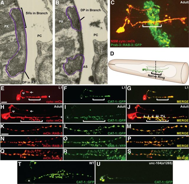
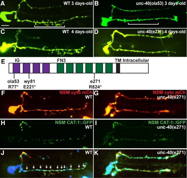
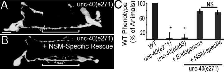
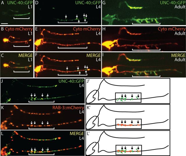
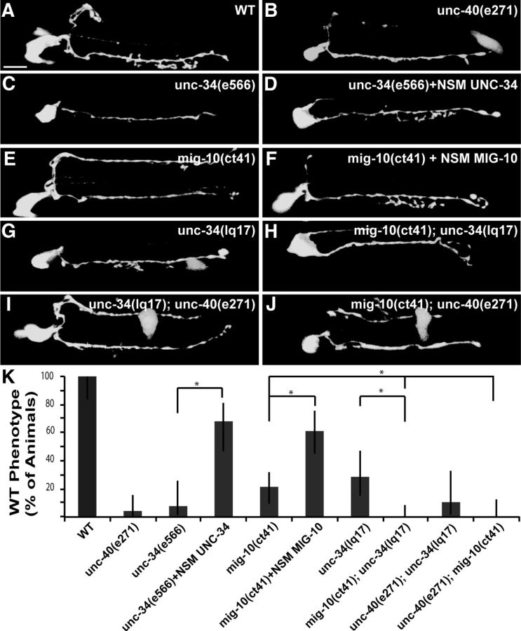
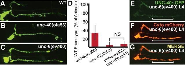
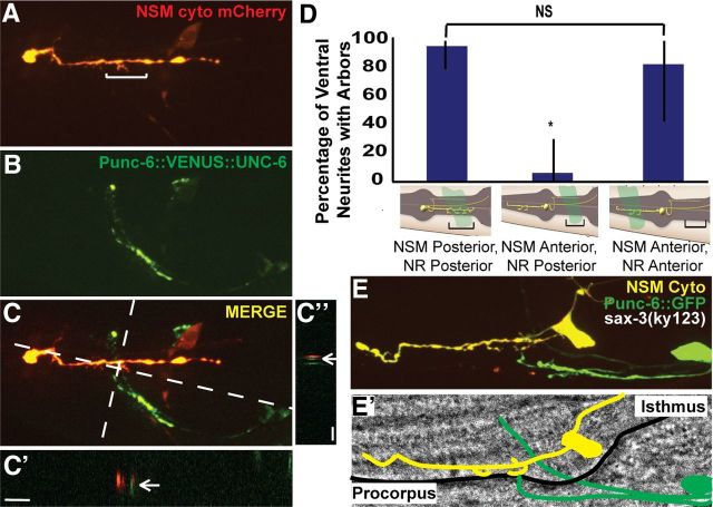
Neurosecretory release sites lack distinct postsynaptic partners, yet target to specific circuits. This targeting specificity regulates local release of neurotransmitters and modulation of adjacent circuits. How neurosecretory release sites target to specific regions is not understood. Here we identify a molecular mechanism that governs the spatial specificity of extrasynaptic neurosecretory terminal (ENT) formation in the serotonergic neurosecretory-motor (NSM) neurons of Caenorhabditis elegans. We show that postembryonic arborization and neurosecretory terminal targeting of the C. elegans NSM neuron is dependent on the Netrin receptor UNC-40/DCC. We observe that UNC-40 localizes to specific neurosecretory terminals at the time of axon arbor formation. This localization is dependent on UNC-6/Netrin, which is expressed by nerve ring neurons that act as guideposts to instruct local arbor and release site formation. We find that both UNC-34/Enabled and MIG-10/Lamellipodin are required downstream of UNC-40 to link the sites of ENT formation to nascent axon arbor extensions. Our findings provide a molecular link between release site development and axon arborization and introduce a novel mechanism that governs the spatial specificity of serotonergic ENTs in vivo.


Figures

Figure 1.
Figure 1.
The serotonergic NSM neuron forms ENT-containing axon arbors in a specific neuroanatomical coordinate. A, B, Electron micrograph sequence of cross-sections through the pharynx of worm N2W (www.wormimage.org). NSM (outlined in purple) forms an axon arbor (vertical projection) at the edge of the pharynx (PX) near the pseudocoelom (clear space annotated as PC). In A, synaptic vesicle clusters (SV) are visible at the base of the arbor (line) and in the tip of the arbor (bracket). A dense projection (DP) onto the pseudocoelom is visible in the base of the arbor (line). In B, the arbor has turned posteriorly and appears discontinuous from the main axon shaft (AS). Synaptic vesicle clusters are still visible in the main NSM axon shaft and in the tip of the arbor. A dense projection (DP) is also visible in the tip of the branch (line), facing the pseudocoelom (PC). C, Simultaneous visualization of NSM and the nematode nerve ring. NSM was imaged by expressing cytosolic mCherry using the tph-1 promoter (Sze et al., 2002), and the nerve ring was imaged by expressing transgene rab-3p::RAB-3::GFP. Note that arbors form in regions adjacent to the nerve ring (brackets). D, Schematic diagram of NSM neuron (modified image adapted from www.wormatlas.org with permission). As shown in the schematic, arbors form in the isthmus region of the pharynx, proximal to the nerve ring. Dashed line indicates the approximate location of the EM cross-sections shown in A and B. E–G, NSM in the L1 nematode. E, Cytosolic mCherry expressed cell specifically in NSM. Note the absence of arbors in the nerve ring region (bracket). Arrow marks dorsal neurite, open arrowhead marks proprioceptive dendrite, and filled arrowhead marks ventral neurite. F, Distribution of serotonergic vesicle clusters. G, Merge. H–J, NSM in the 3-d-old adult nematode. H, As in E for adult. I, As in F, for adult. Note the presence of vesicle clusters in the terminal arbors (brackets). J, Merge. Arrows indicate mature arbors with synaptic vesicle clusters at their bases. Open arrowhead indicates mature arbor lacking synaptic vesicle cluster at the base. A total of 78% of axon arbors are associated with synaptic vesicle clusters at their base (n = 70 with a 95% confidence interval extending from 66.4 to 85.7%). K–M, Localization of CAT-1::GFP (L) and mCherry::RAB-3 (K) in NSM, with merge in M. N–P, Localization of SNB-1::YFP (O) and mCherry::RAB-3 (N) in NSM, with merge in P. Q–S, Localization of GFP::SYD-2 (R) and mCherry::RAB-3 (Q) in NSM, with merge in S. T, U, CAT-1:GFP localization in WT (T) or unc-104(e1265) mutant animals (U). In all images, anterior is oriented to the left and dorsal is oriented up, as in the diagram (D). Bracket represents the expected position of the nerve ring as it circumvents the pharyngeal isthmus, and asterisk denotes position of the NSM cell body. Scale bars (in E): E–J, 5 μm; (in K) C, K–U, 5 μm.
Figure 2.
Figure 2.
UNC-40 is required for terminal arbor formation and synaptic vesicle clustering in the NSM neuron. A, B, NSM neuron in adult nematodes visualized by cell-specific expression of GFP. A, WT adult; note arbor length and position within nerve ring region (bracket). B, unc-40(ola53) mutant adult. Note the absence of terminal arbors. C, D, As in A and B for 4-d-old adults. E, Schematic diagram of the UNC-40 gene product with known alleles marked. unc-40(ola53) allele corresponds to a single C-to-T nucleotide substitution in exon 3 that results in a nonsense mutation R77*. The unc-40(ola53) stop codon is closer to the N terminus than other putative null alleles, unc-40(wy81) and unc-40(e271) (Colón-Ramos et al., 2007). F, G, NSM neuron in adult nematodes visualized by cell-specific expression of mCherry. Note that unc-40(e271) mutants phenocopy unc-40(ola53). H, I, NSM ENTs visualized with CAT-1::GFP. Note the diffuse appearance of synaptic vesicles in unc-40(e271) mutants (I). J, K, Ratiometric images of NSM in which CAT-1::GFP signal is compared with cytosolic mCherry signal. Regions of high ratio between CAT-1::GFP and mCherry appear red, and regions of low ratio appear blue. Note regions of high ratio punctuated by regions of low ratio in the WT NSM, representing regions of tight serotonergic vesicle clustering (arrows) separated by nonsynaptic regions (J). Note the relative absence of such individual puncta in unc-40(e271) and the presence of larger, diffuse accumulations of serotonergic vesicles (K).
Figure 3.
Figure 3.
UNC-40 acts cell autonomously in the NSM neurons to instruct axon arborization. A, B, NSM neuron in adult nematodes visualized with cytosolic GFP. unc-40(e271) phenotype (A) is rescued by NSM-specific expression of UNC-40 cDNA (B). C, Quantification of arborization phenotypes in the NSM neuron. Note that unc-40(e271) phenocopies ola53 and is similarly rescued by expression of UNC-40 under the endogenous regulatory elements or NSM-specific promoter. Error bars represent 95% confidence intervals. *p < 0.0001.
Figure 4.
Figure 4.
UNC-40 dynamically localizes to ENTs at the time of axon arborigenesis. A–I, Distribution of UNC-40::GFP and cytosolic mCherry in NSM in L1, L4, and adult animals. Note the enrichment of UNC-40::GFP at discrete puncta along the ventral neurite in L4 animals, as arbors are beginning to form (arrows). J–L, Simultaneous visualization of UNC-40::GFP and mCherry::RAB-3 in an L4 animal. Note that not all vesicle clusters are associated with UNC-40. However, UNC-40 clusters are associated with vesicle clusters (arrows) at the nerve ring region (brackets). J′–L′, Schematic diagram of UNC-40 (green) localization relative to RAB-3 (red).
Figure 5.
Figure 5.
UNC-34 and MIG-10 function cell autonomously in NSM to instruct axon arborization. A–J, tph-1p::gfp cell specifically expressed in NSM in WT (A), unc-40(e271) (B), unc-34(e566) (C), unc-34(e566) expressing UNC-34 cDNA cell specifically in NSM (D), mig-10(ct41) (E), mig-10(ct41) expressing MIG-10 cDNA cell specifically in NSM (F), unc-34(lq17) hypomorphic allele (G), mig-10(ct41);unc-34(lq17) (H), unc-34(lq17);unc-40(e271) (I), and mig-10(ct41); unc-40(e271) (J) animals. K, Quantitative analysis of UNC-40 pathway mutant phenotypes. All genotypes are significantly different from WT, p < 0.005. Cell-autonomous expression of UNC-34 significantly rescues the unc-34(e566) mutant phenotype (p < 0.0001). Cell-autonomous expression of MIG-10 significantly rescues the mig-10(ct41) mutant phenotype (p = 0.0002). unc-34(lq17); mig-10(ct41) mutants display a more severe mutant phenotype than unc-34(lq17) alone (p = 0.0001) or mig-10(ct41) alone (p = 0.0015). mig-10(ct41);unc-40(e271) mutants display a more severe mutant phenotype than mig-10(ct41) alone (p = 0.0091). Scale bar (in A): A–J, 5 μm. Error bars represent 95% confidence intervals, asterisk indicates significance, and significance was determined using Fisher's exact test.
Figure 6.
Figure 6.
UNC-6 instructs UNC-40 localization and axon arbor formation. A–C, NSM neuron in adult nematodes visualized with cytosolic GFP. Note that unc-6(ev400) mutants (C) phenocopy unc-40(ola53) mutants (B). D, Quantification of the percentage of animals displaying WT arborization patterns in unc-6(ev400), in unc-40(ola53) and in unc-6(ev400); unc-40(ola53) double mutants. E–G, UNC-40::GFP (E) fails to localize to puncta in unc-6(ev400) L4 mutant animals.
Figure 7.
Figure 7.
UNC-6 is expressed by guidepost neurons that provide a local signal to instruct NSM arborization at target regions. A–C″, Simultaneous visualization of NSM morphology (A) and endogenous UNC-6/Netrin subcellular localization in Netrin-expressing neurons (B) of L4 animals. C′, x–z projection of C. Note the adjacency between a nascent branch (arrow) and UNC-6/Netrin clusters at the nerve ring (green). C″, As in C′ but y–z projection. D, Quantification of NSM arborization in sax-3(ky123) mutants that disrupt the position of NSM with respect to UNC-6/Netrin-expressing neurons. NR, Nerve ring. Diagrams serve as histogram labels. In diagrams, pharynx is gray, NSM is yellow, and position of Netrin-expressing neurons is in green. Note that arbor formation in NSM depends on the adjacency of NSM to Netrin-expressing neurons. χ2 test, p < 0.0001. E, Representative image of a nematode in which the NSM ventral neurite (yellow) is misguided anteriorly into the pharyngeal procorpus, and the Netrin-expressing neurons (green) are misguided anteriorly as well. Note the appearance of ectopic terminal arbor structures on the misguided NSM neurite adjacent to the misguided Netrin-expressing neurons. E′, Diagram and corresponding differential interference contrast image of E.

Similar articles

Cited by

References

    1. Adler CE, Fetter RD, Bargmann CI. UNC-6/Netrin induces neuronal asymmetry and defines the site of axon formation. Nat Neurosci. 2006;9:511–518. - PMC - PubMed
    1. Albertson DG, Thomson JN. The pharynx of Caenorhabditis elegans. Philos Trans R Soc Lond B Biol Sci. 1976;275:299–325. - PubMed
    1. Alsina B, Vu T, Cohen-Cory S. Visualizing synapse formation in arborizing optic axons in vivo: dynamics and modulation by BDNF. Nat Neurosci. 2001;4:1093–1101. - PubMed
    1. Asakura T, Waga N, Ogura K, Goshima Y. Genes required for cellular UNC-6/netrin localization in Caenorhabditis elegans. Genetics. 2010;185:573–585. - PMC - PubMed
    1. Axäng C, Rauthan M, Hall DH, Pilon M. Developmental genetics of the C. elegans pharyngeal neurons NSML and NSMR. BMC Dev Biol. 2008;8:38. - PMC - PubMed

Publication types

MeSH terms