. 2013 Jan 23;33(4):1391-9.
doi: 10.1523/JNEUROSCI.2231-12.2013.

Ataxia with cerebellar lesions in mice expressing chimeric PrP-Dpl protein

Affiliations

Ataxia with cerebellar lesions in mice expressing chimeric PrP-Dpl protein

Catherine Lemaire-Vieille et al. J Neurosci. .

Abstract

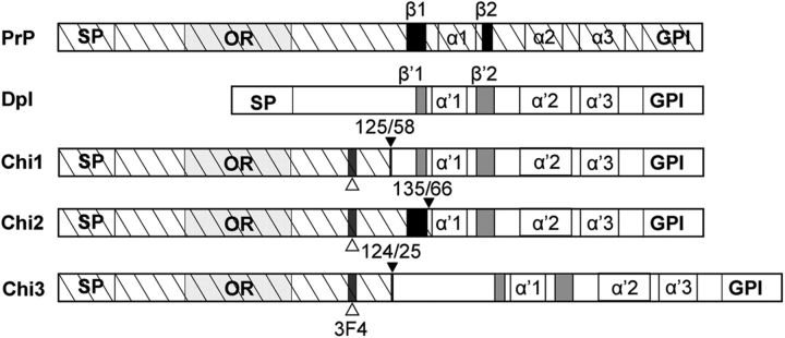
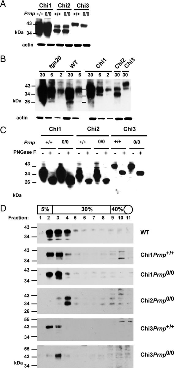
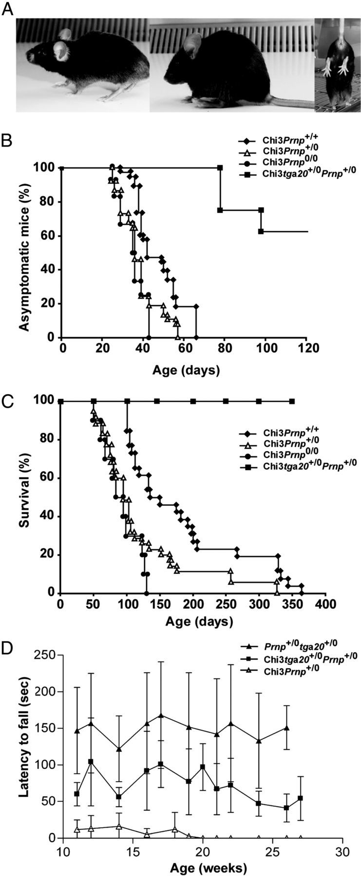
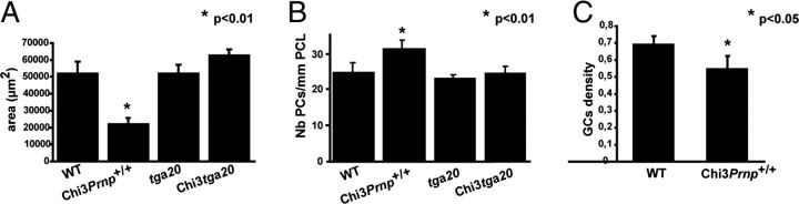
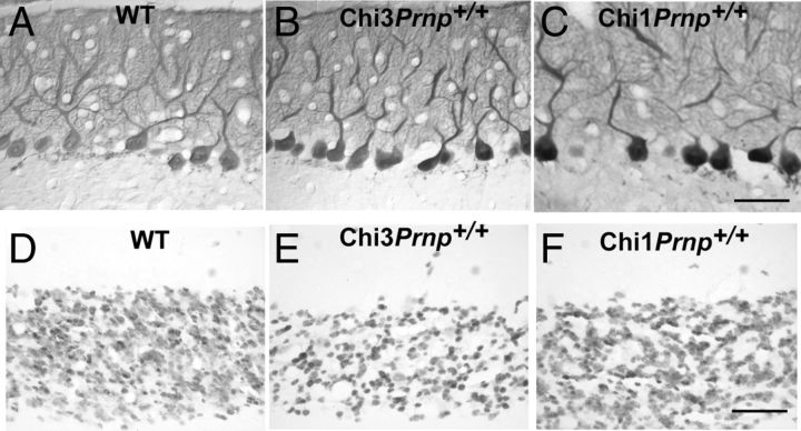
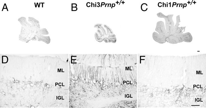
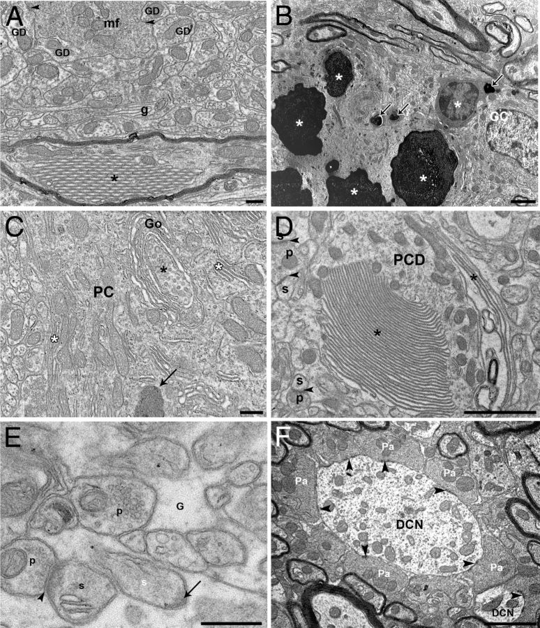
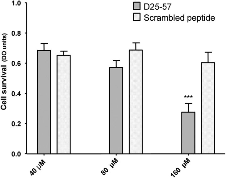
Mutations within the central region of prion protein (PrP) have been shown to be associated with severe neurotoxic activity similar to that observed with Dpl, a PrP-like protein. To further investigate this neurotoxic effect, we generated lines of transgenic (Tg) mice expressing three different chimeric PrP-Dpl proteins. Chi1 (amino acids 1-57 of Dpl replaced by amino acids 1-125 of PrP) and Chi2 (amino acids 1-66 of Dpl replaced by amino acids 1-134 of PrP) abrogated the pathogenicity of Dpl indicating that the presence of a N-terminal domain of PrP (23-134) reduced the toxicity of Dpl, as reported. However, when the amino acids 1-24 of Dpl were replaced by amino acids 1-124 of PrP, Chi3 Tg mice, which express the chimeric protein at a very low level, start developing ataxia at the age of 5-7 weeks. This phenotype was not counteracted by a single copy of full-length-PrP(c) but rather by its overexpression, indicating the strong toxicity of the chimeric protein Chi3. Chi3 Tg mice exhibit severe cerebellar atrophy with a significant loss of granule cells. We concluded that aa25 to aa57 of Dpl, which are not present in Chi1 and Chi2 constructs, confer toxicity to the protein. We tested this possibility by using the 25-57 Dpl peptide in primary culture of mouse embryo cortical neurons and found a significant neurotoxic effect. This finding identifies a protein domain that plays a role in mediating Dpl-related toxicity.


Figures

Figure 1.
Figure 1.
Schematic representation of WT PrP, Dpl, and chimeric PrP/Dpl mutants (Chi1, Chi2, and Chi3). Sequences are hatched for PrP and white for Dpl. SP, Secretory signal peptide, cleaved during synthesis; OR, octarepeats, five repeats of eight amino acids; α 1–3 and α′1–3, α-helices 1, 2, and 3 of the globular C-terminal domain of PrP and Dpl, respectively; β1–2 and β′1–2, β sheets 1 and 2 of PrP and Dpl, respectively; GPI, sequence cleaved after attachment of the glycosylphosphatidyl inositol anchor; 3F4, epitopes recognized by the mAb 3F4 (white arrowhead). For chimeric constructs, the junctions between PrP and Dpl are indicated by a black arrowhead.
Figure 2.
Figure 2.
Expression of Tg proteins. A, The expression levels of each chimeric protein are independent of the Prnp status (Prnp+/+ or Prnp0/0). Thirty micrograms of protein from crude brains homogenate were loaded. B, Expression of chimeric protein in brain compared with that of PrP in WT and tga20 mice. Chi3 expression is lower than that of WT PrP. Amounts of protein (in micrograms) loaded for each line are indicated on the top of the gel. C, The glycosylation pattern of full-length Chi proteins is similar in Chi Tg mice expressed on mouse Prnp+/+and Prnp0/0 background. Thirty micrograms of protein from total brain homogenates are subjected (+) or not (−) to PNGase F treatment. D, Analyses of density gradient of detergent-resistant membranes from brains of WT, Chi1, Chi2, and Chi3 mice (on Prnp+/+ and Prnp0/0 background). Western blots were performed with mAb SAF32 (B, D; ChiPrnp0/0 Tg mice) or mAb 3F4 (A, C, D; ChiPrnp+/+ Tg mice). Molecular size markers (in kilodaltons) are indicated on the left.
Figure 3.
Figure 3.
Phenotypic analysis of Chi3 mice. A, Typical clinical phenotypes at the terminal stage: ataxia (left), kyphosis (middle), and clasping when held head down by the tail (right). B, Chi3 mice were maintained on the Prnp+/+ (n = 38), Prnp+/0 (n = 37), Prnp0/0 (n = 12), and Prnp+/0tga20+/− (n = 8) allelotypes and monitored for the onset of the ataxia. Chi3Prnp+/0 and Chi3Prnp0/0 mice start showing coarse tremor slightly more rapidly than Chi3Prnp+/+ (p ≤ 0.001, Mann–Whitney test), whereas most Chi3tga20+/−Prnp+/0 remain asymptomatic. C, The mice were killed when the following neurological symptoms were observed: feet clasping when lifted, paresis of the hind legs, and prostration. The survival of mice is dependent of Prnp status, with Chi3Prnp+/0 and Chi3Prnp+/+ mice surviving longer than Chi3Prnp0/0 (p ≤ 0.001, Mann–Whitney test), whereas Chi3tga20+/−Prnp+/0 mice have a longer life expectancy (≥400d). D, Rotarod testing in acceleration mode of Chi3Prnp+/0 (n = 5), Chi3tga20+/−Prnp+/0 (n = 5) and tga20+/−Prnp+/0 (n = 5) mice. Overexpression of WT PrPc improves rotarod performance in Chi3 mice (Chi3tga20+/−Prnp+/0, p < 0.0001). This improvement still differs from tga20+/−Prnp+/0 control mice. Chi3Prnp+/0 mice continue to deteriorate, not being able to stay on the rod from 19 weeks of age. Significant differences were tested using one-way ANOVA with Tukey's post hoc comparison.
Figure 4.
Figure 4.
Histological studies. Toluidine blue (A–F) and Mason's trichrome (G, H) histological staining of sagittal sections in the cerebellum of 3-month-old WT mice (A), Chi3Prnp+/+ mice (B), 2-month-old Chi3Prnp+/0 mice (C), 3-month-old Chi3Prnp0/0 mice (D), tga20+/− mice (E), Chi3tga20+/− mice (F), 8-month-old WT mice (G), and 11-month-old Chi1Prnp+/+ mice (H). Scale bar, 500 μm.
Figure 5.
Figure 5.
A, Mean area of the median cerebellar vermis in 3- to 8-month-old WT mice (n = 2), 3- to 13.5-month-old Chi3Prnp+/+ mice (n = 4), 3-month-old tga20+/− mice (n = 2), and Chi3tga20+/− mice (n = 2) (*p < 0.01). B, Mean number of PCs per millimeter of PCL in Toluidin blue-stained semithin sections of the median cerebellar vermis of 3- to 8 month-old WT mice (n = 2), 3- to 13.5-month-old Chi3Prnp+/+ mice (n = 4), 3-month-old tga20+/− mice (n = 2), and 3-month-old Chi3tga20+/− mice (n = 2) (*p < 0.01). C, Mean density of NeuN immunolabeled GCs in the IGL in sagittal paraffin sections of the median cerebellar vermis of 3- to 8-month-old WT (n = 2) and 3- to 13.5-month-old Chi3Prnp+/+ (n = 4) mice (*p < 0.05).
Figure 6.
Figure 6.
Calcium-binding protein immunoperoxidase staining of the Purkinje cells in the cerebellum of 8-month-old WT (A) and Chi3Prnp+/+ (B) mice and 11-month-old Chi1Prnp+/+ mice (C). NeuN immunohistochemical staining of the GCs in the median cerebellar vermis of 8-month-old WT (D) and Chi3Prnp+/+ (E) mice and 11-month-old Chi1Prnp+/+ mice (F). Scale bars, 50 μm.
Figure 7.
Figure 7.
Immunohistochemical staining of GFAP in the cerebellum of WT, Chi3, and Chi1 mice. A, B, D, E, Vermal sections and cortex of the dorsal side of the lobule IXa of the cerebellum of 8-month-old WT (A, D), 8.5-month-old Chi3Prnp+/+ (B, E), and 11-month-old Chi1Prnp+/+ (C, F) mice. GFAP overexpression signals reactive astrogliosis in Chi3 mice compared with WT and Chi1Prnp+/+. Scale bars: A–C, 50 μm; D–F, 200 μm.
Figure 8.
Figure 8.
Ultrastructure analysis. A, B, Ultrastructure of the internal granular layer of Chi3 mice. A, A mossy fiber terminal (mf) makes asymmetric synapses (arrowheads) with GC dendrites (GD) and is surrounded by abnormal wraps of glial profiles (g). A myelinated PC-like axon contains an abnormal crystalloid stacking of tubular profiles (*). B, Steps of GC degeneration from shrunken cell body with cytoplasmic electron-opaque profiles (arrows) to overall electron-opacity of the neurons (*). See the normal-like GC on the right side. C–F, Ultrastructure of abnormal profiles in PC somata and dendrites of Chi3 mice. C, Autophagic-like phagophore (*) forming from the trans saccules of a Golgi (Go) apparatus dictyosome wrapping in a PC body. Note also the abnormal stacking of endoplasmic reticulum (white asterisks) and a dense body (arrow). D, A main PC dendrite (PCD) displays abnormal stacking of endoplasmic reticulum (*). Arrowheads indicate asymmetric synapses between presynaptic parallel fiber boutons (p) and postsynaptic PC dendritic spines (s). E, A PC dendritic spine (white s) is completely surrounded by glia (G) and displays a postsynaptic density (arrow) free of a presynaptic partner. Another PC spine (s) is innervated (arrowheads) by a presynaptic parallel fiber bouton (p). F, PC axon terminals (Pa) make symmetric synapses (arrowheads) on dendrites of a deep nuclear neuron (DCN). Scale bars: A, E, 500 nm; B, C, D, F, 2 μm.
Figure 9.
Figure 9.
Glycosylation pattern of Chi3 brain proteins compared with that of WT mice and Prnpngsk/ngsk mice. T, Testis extract from WT mice. Chi3 mice are expressed on mouse Prnp+/+, Prnp0/0, and on tga20+/− background with (+) or without (−) PNGase F pretreatment. Thirty micrograms of protein from total brain homogenates are subjected to PNGase F treatment as indicated and analyzed by Western blot revealed with rabbit anti-Dpl polyclonal antibody.
Figure 10.
Figure 10.
Neurotoxic effect of D25–57 doppel peptide on primary cortical neurons from embryonic 17.5 d WT mice. Neurons were chronically exposed to the indicated concentrations of D25–57 peptide for 48 h at 37°C. Neuronal viability was monitored by using the cell proliferation reagent CCK-8 assay. The neurotoxicity of 160 μm P25–57 peptide was statistically significant after a 48 h incubation time compared with 40 and 80 μm and with the control scrambled peptide. Significant differences (p < 0.01) were tested using a two-way ANOVA Bonferroni's post hoc test.

Similar articles

Cited by

References

    1. Aguzzi A, Sigurdson C, Heikenwaelder M. Molecular mechanisms of prion pathogenesis. Annu Rev Pathol. 2008;3:11–40. - PubMed
    1. Al Bersaoui R, Robert I, Lutz Y, Blanc F, Sommermeyer-Leroux G, Shibaguchi H, Aunis D, Fuchs JP. Purkinje-cell degeneration in prion protein-deficient mice is associated with a cerebellum-specific Doppel protein species signature. FEBS Lett. 2005;579:2715–2721. - PubMed
    1. Baumann F, Tolnay M, Brabeck C, Pahnke J, Kloz U, Niemann HH, Heikenwalder M, Rülicke T, Bürkle A, Aguzzi A. Lethal recessive myelin toxicity of prion protein lacking its central domain. EMBO J. 2007;26:538–547. - PMC - PubMed
    1. Baumann F, Pahnke J, Radovanovic I, Rülicke T, Bremer J, Tolnay M, Aguzzi A. Functionally relevant domains of the prion protein identified in vivo. PLoS One. 2009;4:e6707. - PMC - PubMed
    1. Büeler H, Fischer M, Lang Y, Bluethmann H, Lipp HP, DeArmond SJ, Prusiner SB, Aguet M, Weissmann C. Normal development and behaviour of mice lacking the neuronal cell- surface PrP protein [see comments] Nature. 1992;356:577–582. - PubMed