. 2013 Jan 23;33(4):1615-30.
doi: 10.1523/JNEUROSCI.2656-12.2013.

Peptide inhibitors disrupt the serotonin 5-HT2C receptor interaction with phosphatase and tensin homolog to allosterically modulate cellular signaling and behavior

Affiliations

Peptide inhibitors disrupt the serotonin 5-HT2C receptor interaction with phosphatase and tensin homolog to allosterically modulate cellular signaling and behavior

Noelle C Anastasio et al. J Neurosci. .

Abstract

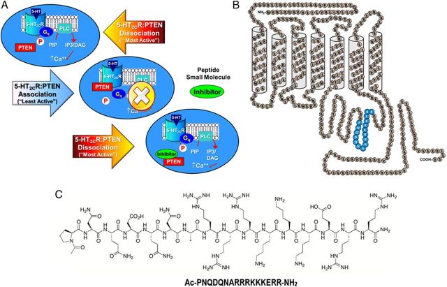
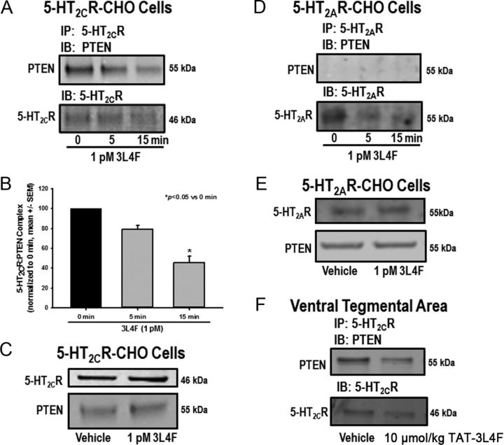
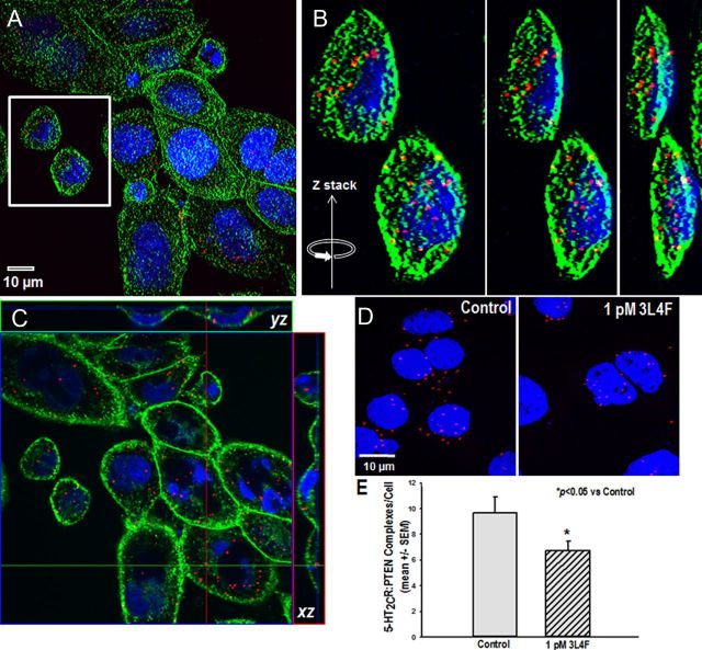
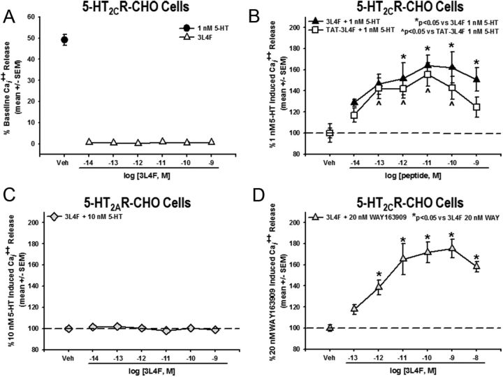
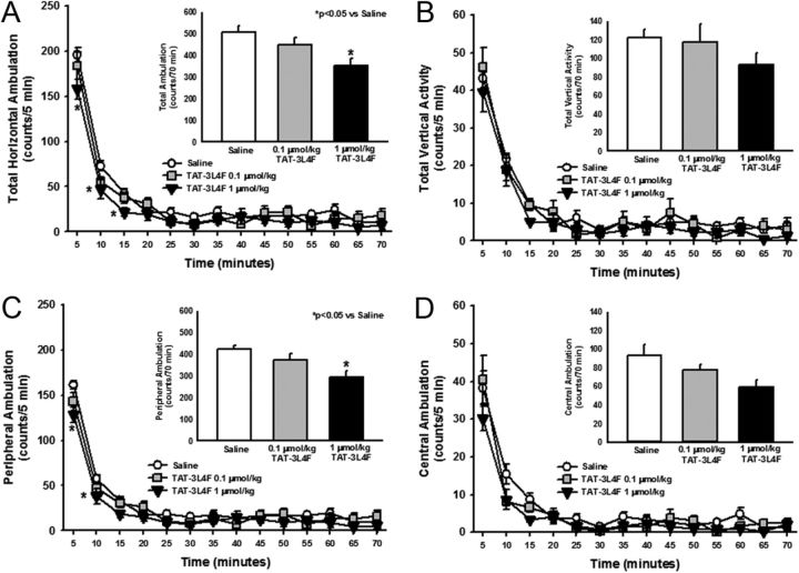
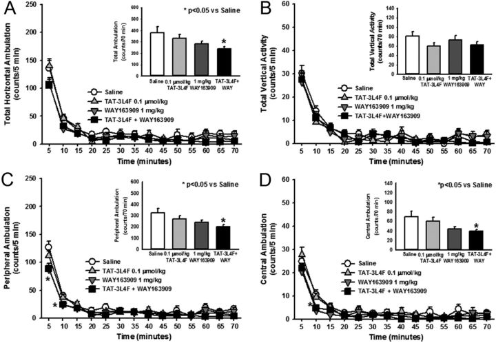
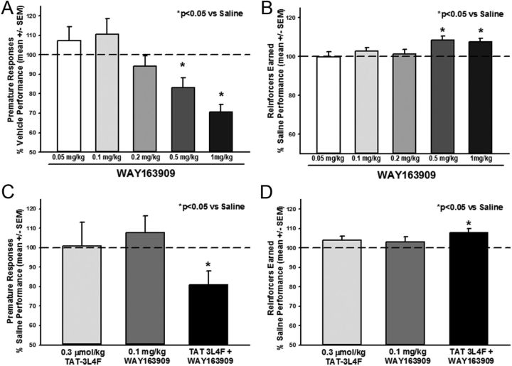
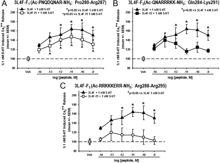
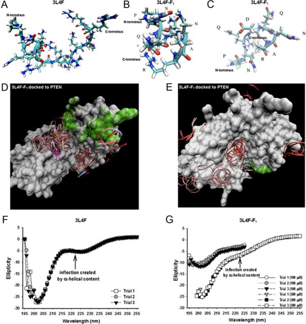
Serotonin (5-hydroxytryptamine; 5-HT) signaling through the 5-HT(2C) receptor (5-HT(2C)R) is essential in normal physiology, whereas aberrant 5-HT(2C)R function is thought to contribute to the pathogenesis of multiple neural disorders. The 5-HT(2C)R interacts with specific protein partners, but the impact of such interactions on 5-HT(2C)R function is poorly understood. Here, we report convergent cellular and behavioral data that the interaction between the 5-HT(2C)R and protein phosphatase and tensin homolog (PTEN) serves as a regulatory mechanism to control 5-HT(2C)R-mediated biology but not that of the closely homologous 5-HT(2A)R. A peptide derived from the third intracellular loop of the human 5-HT(2C)R [3L4F (third loop, fourth fragment)] disrupted the association, allosterically augmented 5-HT(2C)R-mediated signaling in live cells, and acted as a positive allosteric modulator in rats in vivo. We identified the critical residues within an 8 aa fragment of the 3L4F peptide that maintained efficacy (within the picomolar range) in live cells similar to that of the 3L4F peptide. Last, molecular modeling identified key structural features and potential interaction sites of the active 3L4F peptides against PTEN. These compelling data demonstrate the specificity and importance of this protein assembly in cellular events and behaviors mediated by 5-HT(2C)R signaling and provide a chemical guidepost to the future development of drug-like peptide or small-molecule inhibitors as neuroprobes to study 5-HT(2C)R allostery and therapeutics for 5-HT(2C)R-mediated disorders.


Figures

Figure 1.
Figure 1.
Disruption of the 5-HT2CR–PTEN complex is predicted to result in positive allosteric modulation of 5-HT2CR signaling. A, Signaling through 5-HT2CR is proposed to be “most active” under conditions of 5-HT2CR stimulation and dissociation of the third intracellular loop of the 5-HT2CR from PTEN (top). Association of 5-HT2CR with PTEN is predicted to limit 5-HT2CR signaling (middle). A peptide fragment of the 5-HT2CR (3L4F) (or a potential small molecule) is proposed to compete with 5-HT2CR for binding to PTEN (bottom) to enhance 5-HT2CR signaling (adapted from Ji et al., 2006). B, A schematic representation of the human 5-HT2CR protein (adapted from Julius et al., 1988, 1989) illustrates predicted amino acid residues (Pro280–Arg295) of 5-HT2CR that bind PTEN (blue). C, The chemical structure of PTEN and its amino acid content is presented.
Figure 2.
Figure 2.
The 3L4F peptide disrupts the complex between 5-HT2CR and PTEN in 5-HT2CR–CHO cells and rat brain. 5-HT2CR and PTEN are part of a protein complex in 5-HT2CR–CHO cells and rat brain as demonstrated on co-IP. The 5-HT2CR–PTEN complex is effectively disrupted by the small peptide fragment 3L4F, which competes with PTEN for binding to 5-HT2CR. The 5-HT2CR–CHO cells were incubated with 3L4F (1 pm) for 0, 5, or 15 min. A, B, Qualitative (A) and quantitative (B) data demonstrate that preincubation with 3L4F reduced the intensity of the PTEN band in a time-dependent manner after IP for 5-HT2CR. C, Preincubation with 1 pm 3L4F (15 min) did not alter protein expression levels of 5-HT2CR or PTEN in 5-HT2CR–CHO cells as assessed by Western blot. D, To assess specificity for 5-HT2CR over the highly homologous 5-HT2AR, IP for 5-HT2AR followed by IB for PTEN was conducted in 5-HT2AR–CHO cells. The PTEN immunoreactive band was not observed, indicating that 5-HT2AR did not associate with PTEN in 5-HT2AR–CHO cells. E, The 3L4F peptide did not alter 5-HT2AR or PTEN protein expression levels in 5-HT2AR–CHO cells as assessed by Western blot. F, IP of protein extracts from the VTA using the 5-HT2CR antibody followed by IB with the PTEN antibody yields a band at the expected MW (55 kDa) for PTEN, indicating the presence of the 5-HT2CR–PTEN complex in the VTA (Ji et al., 2006). The rat TAT–3L4F peptide (10 μmol/kg, i.p., 5 min treatment) reduced the intensity of the PTEN immunoreactive band detected after IP for 5-HT2CR relative to vehicle (saline) treatment. *p < 0.05 versus 0 min.
Figure 3.
Figure 3.
The 5-HT2CR–PTEN complex is observed and 3L4F disrupts its assembly in live cells in situ. A–D, Representative photomicrographs of 5-HT2CR–PTEN complexes as measured by the Duolink PLA in the 5-HT2CR–CHO cells. A, Confocal reconstruction of the 5-HT2CR–PTEN complex (red spots) in 5-HT2CR–CHO cells; the cellular membrane is labeled with wheat germ agglutinin (green), and the nucleus is labeled with DAPI (blue). B, Representative images from the reconstruction of confocal images were selected (A, white box), and progressive rotation of the Z-stack in the y direction illustrates the distribution of the 5-HT2CR–PTEN complex throughout the cell. C, Photomicrograph prepared from a confocal series of 5-HT2CR–CHO cells sectioned tangentially. Orthogonal views indicate that 5-HT2CR–PTEN complexes are clearly evident in the x–z and y–z directions relative to the image plane. The 5-HT2CR–PTEN complex appears to form prominently in plasma membrane and/or cytoplasmic compartments. D, E, Qualitative (D) and quantitative (E) representation demonstrating significant disruption of the number of 5-HT2CR–PTEN complexes by the 3L4F peptide (1 pm; 15 min) relative to control. *p < 0.05 versus control.
Figure 4.
Figure 4.
The 3L4F peptide enhances 5-HT-induced Cai2+ release in 5-HT2CR–CHO cells. A, Preincubation with the 3L4F peptide alone (15 min; ▵) did not induce Cai2+ release in the 5-HT2CR–CHO cells. The EC50 for 5-HT-mediated Cai2+ release is 1 nm in 5-HT2CR–CHO cells (●). B, Preincubation with 3L4F (15 min) dose dependently (10−12 to 10−9 m) increased Cai2+ release versus 1 nm 5-HT-evoked Cai2+ release in the 5-HT2CR–CHO cells (*p < 0.05 vs 1 nm 5-HT; ▴). Preincubation with rat TAT–3L4F (15 min) dose dependently (10−13 to 10−10 m) increased Cai2+ release versus 1 nm 5-HT-evoked Cai2+ release in the 5-HT2CR–CHO cells (p < 0.05 vs 1 nm 5-HT; □). C, Preincubation with the 3L4F peptide alone (15 min) did not induce Cai2+ release in the 5-HT2AR–CHO cells. D, Preincubation with 3L4F (15 min) dose dependently (10−12 to 10−8 m) increased the 5-HT2CR-selective agonist WAY163909-evoked (20 nm) Cai2+ release in the 5-HT2CR–CHO cells (*p < 0.05 vs 20 nm WAY163909; ▵). Veh, Vehicle.
Figure 5.
Figure 5.
The TAT–3L4F peptide suppresses ambulation but not vertical activity. We assessed the effects of saline (1 ml/kg, i.p.) or rat TAT–3L4F peptide (0.1 or 1 μmol/kg, i.p.) on spontaneous locomotor activity. A, The time course of horizontal ambulation is divided into 5 min time bins across the 70 min session; TAT–3L4F at 1 μmol/kg significantly reduced horizontal ambulation versus saline during the first 15 min. A, Inset, The mean ± SEM total horizontal ambulation is represented after administration of saline or TAT–3L4F; TAT–3L4F (1 μmol/kg) significantly reduced total horizontal ambulation versus saline. B, The time course of vertical activity is divided into 5 min time bins across the 70 min session. B, Inset, the mean ± SEM total vertical activity is represented after administration of saline or TAT–3L4F. C, The time course of peripheral ambulation is divided into 5 min time bins across the 70 min session; TAT–3L4F (1 μmol/kg) significantly reduced peripheral ambulation versus saline during the first 10 min. C, Inset, The mean ± SEM total peripheral ambulation is represented after administration of saline or TAT–3L4F; TAT–3L4F (1 μmol/kg) significantly reduced peripheral ambulation versus saline. D, The time course of central ambulation is divided into 5 min time bins across the 70 min session. D, Inset, The mean ± SEM total central ambulation is represented after administration of saline or TAT–3L4F. *p < 0.05 versus saline.
Figure 6.
Figure 6.
The combination of TAT–3L4F plus the selective 5-HT2CR agonist WAY163909 suppresses ambulation but not vertical activity. We assessed the effects of saline (1 ml/kg, i.p.), rat TAT–3L4F peptide (0.1 μmol/kg, i.p.), or the combination of TAT–3L4F plus WAY163909 (1 mg/kg, i.p.) on spontaneous locomotor activity. A, The time course of horizontal ambulation is divided into 5 min time bins across the 70 min session. A, Inset, The mean ± SEM total horizontal ambulation is represented after administration of saline, TAT–3L4F, or TAT–3L4F plus WAY163909; TAT–3L4F plus WAY163909 significantly reduced total horizontal ambulation versus saline. B, The time course of vertical activity is divided into 5 min time bins across the 70 min session. B, Inset, The mean ± SEM total vertical activity is represented after administration of saline, TAT–3L4F, or TAT–3L4F plus WAY163909. C, The time course of peripheral ambulation is divided into 5 min time bins across the 70 min session; WAY163909 alone and TAT–3L4F plus WAY163909 significantly reduced peripheral ambulation versus saline during the first 10 min. C, Inset, The mean ± SEM total peripheral ambulation is represented after administration of saline, TAT–3L4F, or TAT–3L4F plus WAY163909; TAT–3L4F plus WAY163909 significantly reduced peripheral ambulation versus saline. D, The time course of central ambulation is divided into 5 min time bins across the 70 min session; WAY163909 and TAT–3L4F plus WAY163909 significantly reduced central ambulation versus saline at the 10 min time point. D, Inset, The mean ± SEM total central ambulation is represented after administration of saline, TAT–3L4F, or TAT–3L4F plus WAY163909; TAT–3L4F plus WAY163909 significantly reduced central ambulation versus saline. *p < 0.05 versus saline.
Figure 7.
Figure 7.
The selective 5-HT2CR agonist WAY163909 alone or in combination with TAT–3L4F suppresses impulsive action in the 1-CSRT task. WAY163909 (0.05, 0.1, 0.2, 0.5, and 1 mg/kg) was administered 15 min before saline injections; 1 ml/kg saline was administered immediately before 1-CSRT task test sessions in a counterbalanced within-subjects design. Data are presented as a percentage of the number of responses made on the saline administration session that preceded each dose evaluated. A, B, WAY163909 (0.5 and 1 mg/kg, i.p.) decreased premature responses (A) and increased the number of reinforcers earned (B) versus saline. The combination effects of the rat TAT–3L4F peptide (0.03 μmol/kg) plus WAY163909 (0.1 mg/kg) on impulsive action indices in the 1-CSRT task were evaluated. The combination of TAT–3L4F plus WAY163909 administered 15 min before saline injections (1 ml/kg saline was administered immediately before 1-CSRT task test sessions in a between-subjects design) significantly altered impulsive action in the 1-CSRT task. C, D, TAT–3L4F plus WAY163909 decreased premature responses (C) and increased the number of reinforcers earned (D) versus saline. Neither WAY163909 (0.1 mg/kg) nor TAT–3L4F (0.3 μmol/kg) alone altered the number of premature responses (C) or reinforcers earned (D). *p < 0.05 versus saline (dashed line indicates saline levels).
Figure 8.
Figure 8.
The active fragment of 3L4F is a component of its first 8 aa span. A, Preincubation with human 3L4F-F1 (15 min) dose dependently (10−13 to 10−9 m) increased Cai2+ release versus 1 nm 5-HT-evoked Cai2+ release in the 5-HT2CR–CHO cells (*p < 0.05 vs 1 nm 5-HT; ▵) with similar efficacy as the lead peptide 3L4F (▴). B, The human 3L4F-F2 peptide did not alter Cai2+ release versus 1 nm 5-HT-evoked Cai2+ release in the 5-HT2CR–CHO cells (■). C, The human 3L4F-F3 peptide did not alter Cai2+ release versus 1 nm 5-HT-evoked Cai2+ release in the 5-HT2CR–CHO cells (□). Veh, Vehicle.
Figure 9.
Figure 9.
Molecular modeling of 3L4F and 3L4F-F1 peptides against PTEN predict structural elements important for PTEN recognition and inhibition. A, B, The predicted conformation as derived from REMD simulations of the 3L4F peptide (A) displays α-helical content at the N terminus, which is conserved in the 3L4F-F1 peptide (B) conformations. C, The 3L4F-F1 peptide REMD simulations also predicted β-turn structure. D, GRAMM dockings of the most populated α-helical conformation clustered in the central groove between the phosphatase active site and C2 membrane-targeting structural domains. The phosphatase active site of PTEN is denoted in purple, and the C2 membrane-targeting structural domain of PTEN is denoted in green. E, GRAMM dockings of a less-populated cluster site of α-helical conformations was found on the immediately opposite side of the central groove. CD detects the presence of some α-helical content (inflection point at 222 nm) in the 3L4F (300 μm) (F) and 3L4F-F1 peptide (100 and 300 μm) (G). Each peptide was subjected to three trial runs of CD.

References

    1. Aarts M, Liu Y, Liu L, Besshoh S, Arundine M, Gurd JW, Wang YT, Salter MW, Tymianski M. Treatment of ischemic brain damage by perturbing NMDA receptor- PSD-95 protein interactions. Science. 2002;298:846–850. - PubMed
    1. Abbas AI, Yadav PN, Yao WD, Arbuckle MI, Grant SG, Caron MG, Roth BL. PSD-95 is essential for hallucinogen and atypical antipsychotic drug actions at serotonin receptors. J Neurosci. 2009;29:7124–7136. - PMC - PubMed
    1. Agnati LF, Guidolin D, Leo G, Guescini M, Pizzi M, Stocchi V, Spano PF, Ghidoni R, Ciruela F, Genedani S, Fuxe K. Possible new targets for GPCR modulation: allosteric interactions, plasma membrane domains, intercellular transfer and epigenetic mechanisms. J Recept Signal Transduct Res. 2011;31:315–331. - PubMed
    1. Anastasio NC, Lanfranco MF, Bubar MJ, Seitz PK, Stutz SJ, McGinnis AG, Watson CS, Cunningham KA. Serotonin 5-HT2C receptor protein expression is enriched in synaptosomal and post-synaptic compartments of rat cortex. J Neurochem. 2010;113:1504–1515. - PMC - PubMed
    1. Anastasio NC, Stoffel EC, Fox RG, Bubar MJ, Rice KC, Moeller FG, Cunningham KA. Serotonin (5-hydroxytryptamine) 5-HT2A receptor: association with inherent and cocaine-evoked behavioral disinhibition in rats. Behav Pharmacol. 2011;22:248–261. - PMC - PubMed

Publication types

MeSH terms