. 2013 Jan 23;33(4):1651-9.
doi: 10.1523/JNEUROSCI.3191-12.2013.

Tau loss attenuates neuronal network hyperexcitability in mouse and Drosophila genetic models of epilepsy

Affiliations

Tau loss attenuates neuronal network hyperexcitability in mouse and Drosophila genetic models of epilepsy

Jerrah K Holth et al. J Neurosci. .

Abstract

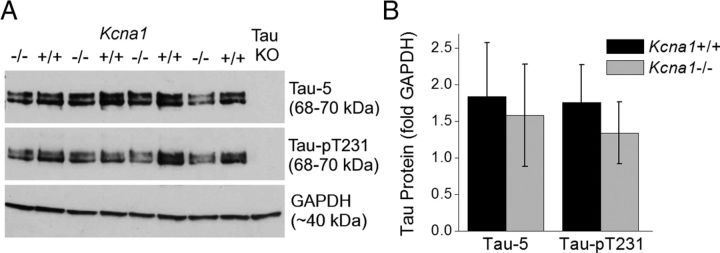
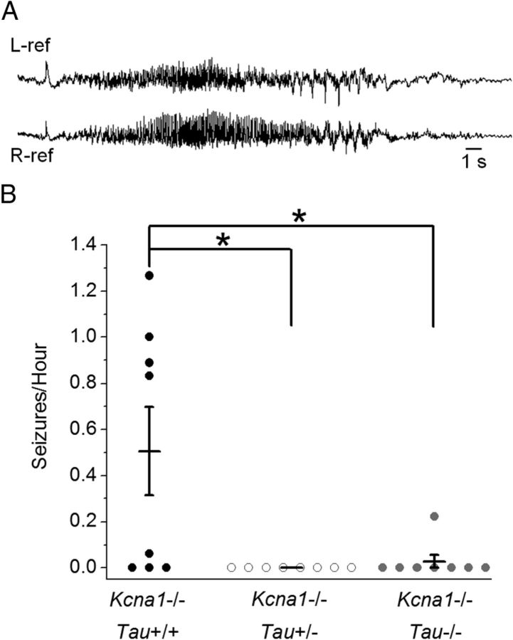
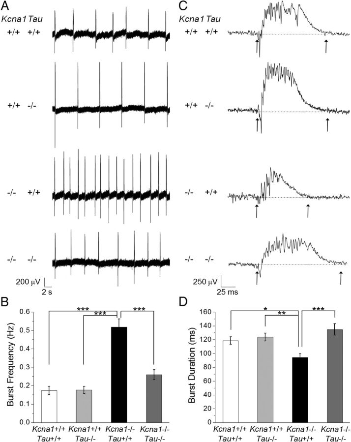
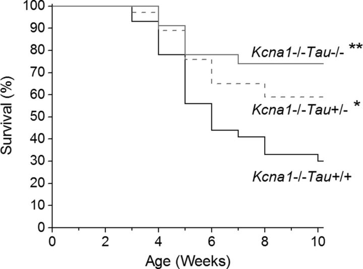
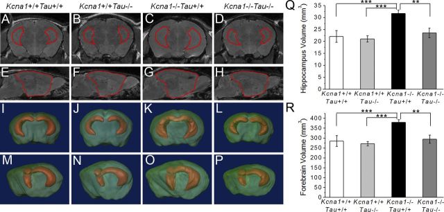
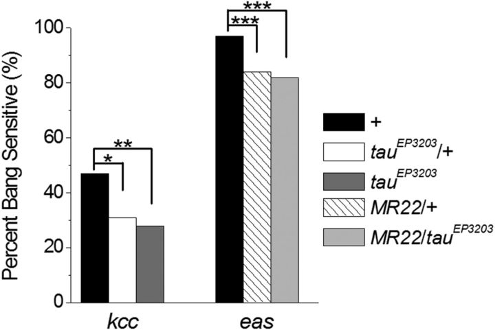
Neuronal network hyperexcitability underlies the pathogenesis of seizures and is a component of some degenerative neurological disorders such as Alzheimer's disease (AD). Recently, the microtubule-binding protein tau has been implicated in the regulation of network synchronization. Genetic removal of Mapt, the gene encoding tau, in AD models overexpressing amyloid-β (Aβ) decreases hyperexcitability and normalizes the excitation/inhibition imbalance. Whether this effect of tau removal is specific to Aβ mouse models remains to be determined. Here, we examined tau as an excitability modifier in the non-AD nervous system using genetic deletion of tau in mouse and Drosophila models of hyperexcitability. Kcna1(-/-) mice lack Kv1.1-delayed rectifier currents and exhibit severe spontaneous seizures, early lethality, and megencephaly. Young Kcna1(-/-) mice retained wild-type levels of Aβ, tau, and tau phospho-Thr(231). Decreasing tau in Kcna1(-/-) mice reduced hyperexcitability and alleviated seizure-related comorbidities. Tau reduction decreased Kcna1(-/-) video-EEG recorded seizure frequency and duration as well as normalized Kcna1(-/-) hippocampal network hyperexcitability in vitro. Additionally, tau reduction increased Kcna1(-/-) survival and prevented megencephaly and hippocampal hypertrophy, as determined by MRI. Bang-sensitive Drosophila mutants display paralysis and seizures in response to mechanical stimulation, providing a complementary excitability assay for epistatic interactions. We found that tau reduction significantly decreased seizure sensitivity in two independent bang-sensitive mutant models, kcc and eas. Our results indicate that tau plays a general role in regulating intrinsic neuronal network hyperexcitability independently of Aβ overexpression and suggest that reducing tau function could be a viable target for therapeutic intervention in seizure disorders and antiepileptogenesis.


Figures

Figure 1.
Figure 1.
Tau protein levels and phosphorylation at Thr231 in 4.5-week-old Kcna1−/− mouse forebrain do not significantly differ from Kcna1+/+ mice. A, Representative Western blots of total tau (Tau-5), tau phospo-Thr231 (Tau-pT231), and GAPDH-loading control for Kcna1−/− and Kcna1+/+ mice, as well as a tau knock-out, which showed no tau or tau phospho-Thr231 staining. B, Quantification of Tau-5 and Tau-pT231 normalized to GAPDH showed no significant difference in total tau or tau phospho-Thr231 levels between Kcna1−/− and Kcna1+/+ mice. p > 0.05; one-way ANOVA; n = 8; error bars represent SD.
Figure 2.
Figure 2.
Tau reduction significantly decreases seizure frequency in Kcna1−/− mice. A, Representative spontaneous cortical seizure recorded bilaterally in a Kcna1−/− mouse during chronic in vivo EEG monitoring. B, Analysis of seizures/h in double-mutant mice recorded for 9 or more hours. Both tau reduction and loss significantly decreased seizure frequency in Kcna1−/−Tau+/− (n = 8) and Kcna1−/−Tau−/− (n = 8) double mutants compared with Kcna1−/−Tau+/+ (n = 8). Seizures were observed in 5/8 Kcna1−/−Tau+/+ mice, 0/8 Kcna1−/−Tau+/− mice, and only 1/8 Kcna1−/−Tau−/− mice. Total deletion of tau reduced the average seizure frequency by >94%. *p < 0.05; Kruskal–Wallis; error bars represent SEM.
Figure 3.
Figure 3.
Tau loss decreases network hyperexcitability in Kcna1−/− hippocampal slices exposed to increased extracellular K+ levels. A, Spontaneous discharges in CA3 pyramidal cells were observed in 6–11 week-old mouse brain slices when K+ was raised from 2.5 to 7.5 mm. Representative 30 s traces illustrate differences in burst frequency between genotypes. B, Analysis of burst frequency during 5 min periods of spontaneous bursting per slice. Kcna1−/−Tau+/+ hippocampus (n = 22) had significantly increased burst frequency compared with Kcna1+/+Tau+/+ wild-type controls (n = 17), while Kcna1−/−Tau−/− slices (n = 19) were not significantly different from Kcna1+/+Tau+/+. Kcna1+/+Tau−/− burst frequency (n = 15) was also indistinguishable from wild type or Kcna1−/−Tau−/− double mutants. C, Representative 200 ms traces illustrate burst duration differences between genotypes, as defined by baseline crossings (arrows). D, Quantification of burst duration in 10 spontaneous bursts per brain slice. Kcna1−/−Tau+/+ hippocampus (n = 22) generated significantly shorter bursts than Kcna1+/+Tau+/+ (n = 17), Kcna1+/+Tau−/− (n = 15), and Kcna1−/−Tau−/− slices (n = 19), which were not significantly different. Loss of tau normalized Kcna1−/− spontaneous burst frequency and duration to wild-type levels. *p < 0.05, **p < 0.01, ***p < 0.0001; one-way ANOVA; error bars represent SEM.
Figure 4.
Figure 4.
Decreasing tau dosage significantly increases survival of Kcna1−/− mice. Kaplan–Meier survival curves show that Kcna1−/−Tau+/+ mice (n = 27) exhibited severe early lethality beginning in the third week of life, with only 30% surviving to 10 weeks of age. Decreasing tau dosage in Kcna1−/− mice by heterozygous (n = 37) and homozygous (n = 23) tau knock-out significantly increased survival, with 59 and 74% survival, respectively, to 10 weeks of age. *p = 0.013, **p = 0.003, all significant with respect to Kcna1−/−Tau+/+; Kaplan–Meier log rank test.
Figure 5.
Figure 5.
A–H, Tau loss prevents megencephaly in Kcna1−/− mouse brain. Left, MRI images of Kcna1+/+Tau+/+, Kcna1+/+Tau−/−, Kcna1−/−Tau+/+, and Kcna1−/−Tau−/− mouse brain were manually segmented and hippocampus (A–D) and forebrain (E–H) borders drawn to allow 3D reconstruction. I–P, 3D reconstructions shown in both coronal (I–L) and partial sagittal (M–P) views demonstrate the enlargement of the hippocampus and forebrain in Kcna1−/−Tau+/+ (K, O) mice relative to other genotypes. Q, R, Quantification of hippocampus (Q) and forebrain (R) volume in double-mutant mice. Kcna1−/−Tau−/− mice had significantly decreased hippocampus and forebrain volume compared with the megencephalic Kcna1−/−Tau+/+. Loss of tau in Kcna1−/− mice decreased hippocampus and forebrain volume to wild-type levels. **p = 0.003, ***p = 0.001; one-way ANOVA; n = 3; error bars represent SD.
Figure 6.
Figure 6.
Tau reduction significantly decreases hyperexcitability in bang-sensitive Drosophila mutants kcc and eas. Quantification of the percentage of flies that were bang sensitive in response to a vortex stimulus. Decreasing tau dosage by the tauEP3203 allele significantly decreased bang sensitivity in kcc mutants by 34% in kcc;tauEP3203/+ and 40% in kcc;tauEP3203 compared with kcc flies with wild-type tau (n≥87). Decreasing tau by the deficiency allele MR22 significantly decreased bang sensitivity in eas mutants by 13% (eas;MR22/+) and 15% (eas;MR22/tauEP3203) compared with eas flies with wild-type tau (n≥98). *p < 0.05, **p < 0.01, ***p < 0.001; χ2; genotypes: kccDHS1, kccDHS1;tauEP3203/+, kccDHS1;tauEP3203, easPC80/Y, easPC80/Y;Df(3R)MR22/+, easPC80/Y;Df(3R)MR22/tauEP3203.

Similar articles

Cited by

References

    1. Adelman JP, Bond CT, Pessia M, Maylie J. Episodic ataxia results from voltage-dependent potassium channels with altered functions. Neuron. 1995;15:1449–1454. - PubMed
    1. Almgren M, Persson AS, Fenghua C, Witgen BM, Schalling M, Nyengaard JR, Lavebratt C. Lack of potassium channel induces proliferation and survival causing increased neurogenesis and twofold hippocampus enlargement. Hippocampus. 2007;17:292–304. - PubMed
    1. Andrews-Zwilling Y, Bien-Ly N, Xu Q, Li G, Bernardo A, Yoon SY, Zwilling D, Yan TX, Chen L, Huang Y. Apolipoprotein E4 causes age- and Tau-dependent impairment of GABAergic interneurons, leading to learning and memory deficits in mice. J Neurosci. 2010;30:13707–13717. - PMC - PubMed
    1. Augustinack JC, Schneider A, Mandelkow EM, Hyman BT. Specific tau phosphorylation sites correlate with severity of neuronal cytopathology in Alzheimer's disease. Acta Neuropathol. 2002;103:26–35. - PubMed
    1. Buerger K, Teipel SJ, Zinkowski R, Blennow K, Arai H, Engel R, Hofmann-Kiefer K, McCulloch C, Ptok U, Heun R, Andreasen N, DeBernardis J, Kerkman D, Moeller H, Davies P, Hampel H. CSF tau protein phosphorylated at threonine 231 correlates with cognitive decline in MCI subjects. Neurology. 2002;59:627–629. - PubMed

Publication types

MeSH terms