. 2013 Jan 23;33(4):1741-52.
doi: 10.1523/JNEUROSCI.4003-12.2013.

Calcium channel agonists protect against neuromuscular dysfunction in a genetic model of TDP-43 mutation in ALS

Affiliations

Calcium channel agonists protect against neuromuscular dysfunction in a genetic model of TDP-43 mutation in ALS

Gary A B Armstrong et al. J Neurosci. .

Abstract

TAR DNA binding protein (TDP-43, encoded by the TARDBP gene) has recently been shown to be associated with amyotrophic lateral sclerosis (ALS), but the early pathophysiological deficits causing impairment in motor function are unknown. Here we expressed the wild-type human gene (wtTARDBP) or the ALS mutation G348C (mutTARDBP) in zebrafish larvae and characterized their motor (swimming) activity and the structure and function of their neuromuscular junctions (NMJs). Of these groups only mutTARDBP larvae showed impaired swimming and increased motoneuron vulnerability with reduced synaptic fidelity, reduced quantal transmission, and more orphaned presynaptic and postsynaptic structures at the NMJ. Remarkably, all behavioral and cellular features were stabilized by chronic treatment with either of the L-type calcium channel agonists FPL 64176 or Bay K 8644. These results indicate that expression of mutTARDBP results in defective NMJs and that calcium channel agonists could be novel therapeutics for ALS.


Figures
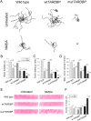
Figure 1.
Figure 1.
Zebrafish expressing mutTARDBP displayed impaired locomotion and NMDA application increased the number of dead neurons in the spinal cord. Locomotor behavior was reliably evoked by a light touch to the tail. A, Examples of 10 superimposed locomotor path traces from each treatment group starting at the center. Swim duration (B), swim distance (C), and maximum swim velocity (D) were calculated for individual fish. Animals expressing mutTARDBP displayed significant impairments in locomotor behavior when compared with wild-type larvae and larvae expressing wtTARDBP. NMDA application (200 μm) exacerbated locomotor performance in all groups. We observed an increase in the number of dead cells (revealed by AO staining) following NMDA application in mutTARDBP larvae only. E, Representative spinal cord images of AO-positive cells. F, Number of AO-positive cells per five somites. Numbers in parentheses represent sample sizes. Single asterisk represent statistical significant differences from wild-type zebrafish (*p < 0.05) and double asterisks represent (**p < 0.01).
Figure 2.
Figure 2.
Zebrafish expressing mutTARDBP displayed a decrease in fidelity of NMJ synaptic transmission and attenuated EPC amplitude. A, Top, Transmitted light. Middle, fluorescent image of Hb9 promoter driving GFP (cyan) expression in motoneurons. Bottom, Somites with ventral roots and in yellow a single sulforhodamine-filled muscle cell. Paired (primary motoneuron/muscle) recordings were performed. B, Example of whole-cell current-clamp trace of an action potential (AP) generated in the CaP motoneuron (top trace) and corresponding EPC measured in a fast-twitch muscle under whole-cell voltage-clamp (bottom trace). To assess synaptic transmission across the NMJ a 10 s train of depolarizing (AP generating) current steps was delivered at 10 Hz (C) or 30 Hz (D). Top traces show evoked APs and the bottom traces show corresponding fast-twitch muscle EPCs in a wild-type animal. Example traces of evoked APs and corresponding EPCs in a wtTARDBP at 10 (E) and 30 Hz (F) and in a mutTARDBP larva at 10 (G) and 30 Hz (H). Insets are corresponding enlarged example traces of EPCs denoted by the black dash above EPCs in C–G and H. Arrows in D and F indicate poststimulus asynchronous EPCs. The X in G and H indicates failure of release. I, mutTARDBP expressing larvae displayed a significant reduction in the fidelity of synaptic transmission at 30 Hz, qualified by the failure of an AP to produce a corresponding EPC. Furthermore, the mean amplitude of EPCs in mutTARDBP expressing larvae was found to be significantly reduced at both 10 and 30 Hz stimulation frequencies (J) and displayed a larger coefficient of variation at both stimulation frequencies (K). Numbers in parentheses represent sample sizes. Asterisks represent statistical significant differences from wild-type zebrafish.
Figure 3.
Figure 3.
Zebrafish expressing mutTARDBP displayed orphaned presynaptic endings and acetylcholine receptor clusters, attenuated mEPCs, and reduced quantal content. A, Representative images of one ventral root projection double labeled for SV2 (presynaptic marker, i) and αBTX (postsynaptic, ii). Wild-type and wtTARDBP larvae showed extensive colocalization of both SV2 and αBTX (iii, merged). However, we observed an increase in the number of orphaned αBTX labeling (arrowheads) and orphaned SV2 labeling (arrows) in mutTARDBP larvae. B, C, Recordings of mEPCs, which result from spontaneous release of a quantum, were recorded. D, Representative mEPCs. Animals expressing mutTARDBP displayed mEPCs with reduced frequency (E) and reduced amplitudes (F), indicating that the numbers of functional synapses are reduced or a proportion are nonfunctional. Kinetics of mEPC was not found to be significantly different. G, Rise time. H, Decay constant. I, Quantal content measured from paired recordings at 10 and 30 Hz were calculated by dividing the amplitude of individual EPC by the mean mEPC amplitude. Fish expressing mutTARDBP displayed significantly reduced quantal content from each AP. Numbers in parentheses represent sample sizes. Asterisks represent statistical significant differences from wild-type zebrafish.
Figure 4.
Figure 4.
Treatment with the calcium channel agonists FPL 64176 or Bay K 8644 recovers locomotor performance in intact zebrafish expressing mutTARDBP. A, Examples of 10 superimposed locomotor path traces from each treatment group. Swim duration (B), swim distance (C), and maximum swim velocity (D) were tabulated for individual animals. Chronic (12 h) treatment with 0.1 μm FPL 64176 significantly increased swim duration. Swim distance and maximum swim velocity in mutTARDBP larvae were also found to be restored following either 0.1 μm FPL 64176 or 1 μm Bay K 8644 application. Numbers in parentheses represent sample sizes. Asterisks represent statistical significant differences from fish treated with water within wild-type, wtTARDBP, or mutTARDBP treatment groups (p < 0.05).
Figure 5.
Figure 5.
Restoration of fidelity of NMJ synaptic transmission and EPC amplitude in zebrafish expressing mutTARDBP and treated with the calcium channel agonists FPL 64176 or Bay K 8644. Paired (primary motor neuron/muscle) recordings were performed in animals treated chronically (12 h) with either of these agents. A, B, Representative traces of 10 s trains of 10 and 30 Hz stimulation. Top traces show evoked action potentials (APs) and the bottom traces show corresponding fast-twitch muscle EPCs in a wild-type larva treated with 0.1 μm FPL 64176. Example traces of APs and corresponding EPCs in a wtTARDBP larva at 30 (C) and 10 Hz (D) and in a mutTARDBP larva at 10 (E) and 30 Hz (F) treated with 0.1 μm FPL 64176. Example traces of evoked EPCs in a mutTARDBP larva at 10 (G) and 30 Hz (H) treated with 1 μm Bay K 8644. Arrows in B, D, F, and H indicate poststimulus asynchronous events that were not present in untreated larvae expressing mutTARDBP (Fig. 2H). Insets are enlarged example traces of EPCs in A–H. Remarkably, treatment with either FPL 64176 or Bay K 8644 resulted in normal synaptic transmission success rates in fish expressing mutTARDBP (I), and evoked EPCs were large and not found to be significantly different from wild-type larvae or larvae expressing wtTARDBP (J). Finally, we observed no differences in the variability of EPC amplitude suggesting more stable synaptic transmission (K). White lines in I and J, and black lines in K represent the mean values obtained from fish expressing mutTARDBP and not treated with FPL 64176. Numbers in parentheses represent sample sizes.
Figure 6.
Figure 6.
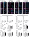
Chronic treatment with FPL 64176 or Bay K 8644 maintains NMJ morphology and function in zebrafish expressing mutTARDBP. A, Representative images of one ventral root projection double labeled for SV2 (presynaptic marker, i), αBTX (postsynaptic, ii), and merge (iii) in wild-type, wtTARDBP, and mutTARDBP larvae treated with 0.1 μm FPL 64176 for 12 h. ZNP-1 instead of SV2 was used as a presynaptic marker for larvae treated with 1.0 μm Bay K 8644. Arrowheads and arrows in insets represent orphaned SV2/ZNP-1 puncta and αBTX clusters, respectively. B–E, All animals treated either with FPL 64176 or Bay K 8644 displayed a low incidence of orphaned αBTX clusters and SV2/ZNP-1 puncta; however, zebrafish expressing mutTARDBP and treated with FPL 64176 did display a slightly high number of orphaned SV2 puncta compared with wild-type zebrafish (C). Representative mEPCs from animals treated with FPL 64176 (F) or Bay K 8644 (H). Importantly, no differences were found in the frequency (G, I) and amplitude (J, L) of mEPCs across treatment groups. K, M, Quantal content measured from paired recordings at 10 and 30 Hz was not found to be significantly different from wild-type, wtTARDBP, or mutTARDBP larvae, implicating that overall function of these NMJs was restored following treatment with FPL 64176 or Bay K 8644. White lines in G, I, J, K, L, M represent the mean values obtained from fish expressing mutTARDBP and not treated with FPL 64176 or Bay K 8644. Numbers in parentheses represent sample sizes. Asterisks represent statistical significant difference from wild-type zebrafish in C.

Similar articles

Cited by

References

    1. Arenson MS, Evans SC. Activation of protein kinase C increases acetylcholine release from frog motor nerves by a direct action on L-type Ca2+ channels and apparently not by depolarisation of the terminal. Neuroscience. 2001;104:1157–1164. - PubMed
    1. Bensimon G, Lacomblez L, Meininger V. A controlled trial of riluzole in amyotrophic lateral sclerosis. N Engl J Med. 1994;330:585–591. - PubMed
    1. Boon KL, Xiao S, McWhorter ML, Donn T, Wolf-Saxon E, Bohnsack MT, Moens CB, Beattie CE. Zebrafish survival motor neuron mutants exhibit presynaptic neuromuscular junction defects. Hum Mol Genet. 2009;18:3615–3625. - PMC - PubMed
    1. Bories C, Amendola J, Lamotte d'Incamps B, Durand J. Early electrophysiological abnormalities in lumbar motoneurons in a transgenic mouse model of amyotrophic lateral sclerosis. Eur J Neurosci. 2007;25:451–459. - PubMed
    1. Buss RR, Drapeau P. Synaptic drive to motoneurons during fictive swimming in the developing zebrafish. J Neurophysiol. 2001;86:197–210. - PubMed

Publication types

MeSH terms

Substances

LinkOut - more resources