. 2013 Apr;51(4):1110-7.
doi: 10.1128/JCM.02704-12. Epub 2013 Jan 23.

Development of a panel of recombinase polymerase amplification assays for detection of biothreat agents

Affiliations

Development of a panel of recombinase polymerase amplification assays for detection of biothreat agents

Milena Euler et al. J Clin Microbiol. 2013 Apr.

Abstract

Syndromic panels for infectious disease have been suggested to be of value in point-of-care diagnostics for developing countries and for biodefense. To test the performance of isothermal recombinase polymerase amplification (RPA) assays, we developed a panel of 10 RPAs for biothreat agents. The panel included RPAs for Francisella tularensis, Yersinia pestis, Bacillus anthracis, variola virus, and reverse transcriptase RPA (RT-RPA) assays for Rift Valley fever virus, Ebola virus, Sudan virus, and Marburg virus. Their analytical sensitivities ranged from 16 to 21 molecules detected (probit analysis) for the majority of RPA and RT-RPA assays. A magnetic bead-based total nucleic acid extraction method was combined with the RPAs and tested using inactivated whole organisms spiked into plasma. The RPA showed comparable sensitivities to real-time RCR assays in these extracts. The run times of the assays at 42°C ranged from 6 to 10 min, and they showed no cross-detection of any of the target genomes of the panel nor of the human genome. The RPAs therefore seem suitable for the implementation of syndromic panels onto microfluidic platforms.


Figures

Fig 1
Fig 1
Details of the RPA amplicon for VARV. All nucleotides in the alignment matching in the VARV sequence are presented as dots. Primer sequences are presented as full sequences. Gaps are presented as hyphens. VARV RPA FP is presented in sense, and VARV RPA P and RP are presented as reverse complement sequences. Gray fields: VARF RPA FP, degenerated IUB code positions; VARV RPA P, the TTT triplet used for the attachment of BTF (Table 2); VARF RPA RP, nucleotide at position 3 of the 3′ end mismatching all other orthopoxviruses. Sequences: cowpox virus, AY902252; camelpox virus, AF438165; monkeypox virus, AF380138; vaccinia virus, M35027; variola virus, X69198.
Fig 2
Fig 2
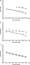
Real time RT-RPA assay performance. (A) Comparison of exo and fpg probe performance in RT-RPA. Standard regression lines (SRLs) for EBOV one-step-RT-RPA were generated from eight data sets (exo probe, black squares) and three data sets (fpg probe, white squares). (B) Influence of background DNA on EBOV one-step-RT-RPA. Black squares, SRL as described above; white squares, SRL of the same assay with 70 ng of human genome DNA background. (C) Influence of background DNA on RPA. Black squares, SRL derived from eight data sets of B. anthracis RPA; white squares, SRL of the same assay with 70 ng of human genome DNA background.
Fig 3
Fig 3
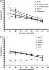
SRLs of all developed assays, including assays for F. tularensis and Rift Valley fever virus (described elsewhere [35b]). The SRLs were derived from eight data sets each. (A) DNA-RPA assays; (B) one-step-RT-RPA assays.
Fig 4
Fig 4
Extraction efficiency of the innuPREP MP basic kit. Plasma was spiked with whole organisms (range, 101 to 104 genomic copies/ml), and nucleic acids were extracted. Eluates were tested by respective quantitative real-time PCR assays in triplicate. The amount of measured molecules detected (md) per reaction is plotted against the 10-fold serial dilution of pathogens in plasma. The dotted line represents the calculated 100% efficiency of extraction. (A) B. anthracis (Gram-positive) extracts tested with pag-PCR; (B) Y. pestis (Gram negative) tested with pla-PCR; (C) VACV tested with LE-PCR; (D) MARV tested with NP-PCR.

Similar articles

Cited by

References

    1. Peeling RW, Mabey D. 2010. Point-of-care tests for diagnosing infections in the developing world. Clin. Microbiol. Infect. 16:1062–1069 - PubMed
    1. Cuchacovich R. 2006. Clinical implications of the polymerase chain reaction: an update. Dis. Clin. N. Am. 20:735–758 - PubMed
    1. Rothman R, Ramachandran P, Yang S, Hardick A, Won H, Kecojevic A, Quianzon C, Hsieh YH, Gaydos C. 2010. Use of quantitative broad-based polymerase chain reaction for detection and identification of common bacterial pathogens in cerebrospinal fluid. Acad. Emerg. Med. 17:741–747 - PMC - PubMed
    1. Yang S, Rothman RE. 2004. PCR-based diagnostics for infectious diseases: uses, limitations, and future applications in acute-care settings. Lancet Infect. Dis. 4:337–348 - PMC - PubMed
    1. Carman B. 2001. Molecular techniques should now replace cell culture in diagnostic virology laboratories. Rev. Med. Virol. 11:347–349 - PubMed

Publication types

LinkOut - more resources