. 2013 Apr;54(4):953-65.
doi: 10.1194/jlr.M032466. Epub 2013 Jan 23.

Cardiomyocyte-specific perilipin 5 overexpression leads to myocardial steatosis and modest cardiac dysfunction

Affiliations

Cardiomyocyte-specific perilipin 5 overexpression leads to myocardial steatosis and modest cardiac dysfunction

Hong Wang et al. J Lipid Res. 2013 Apr.

Abstract

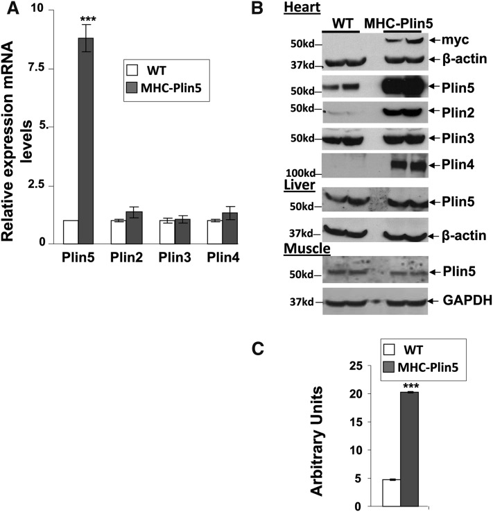
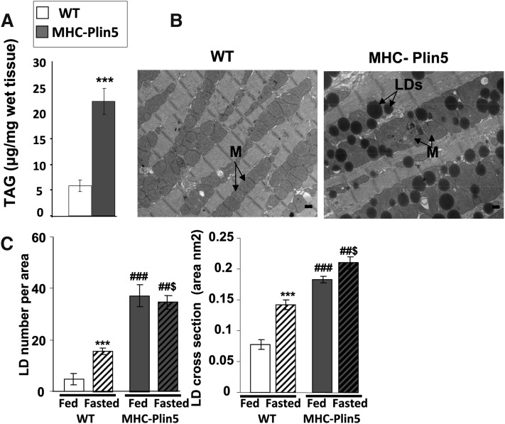
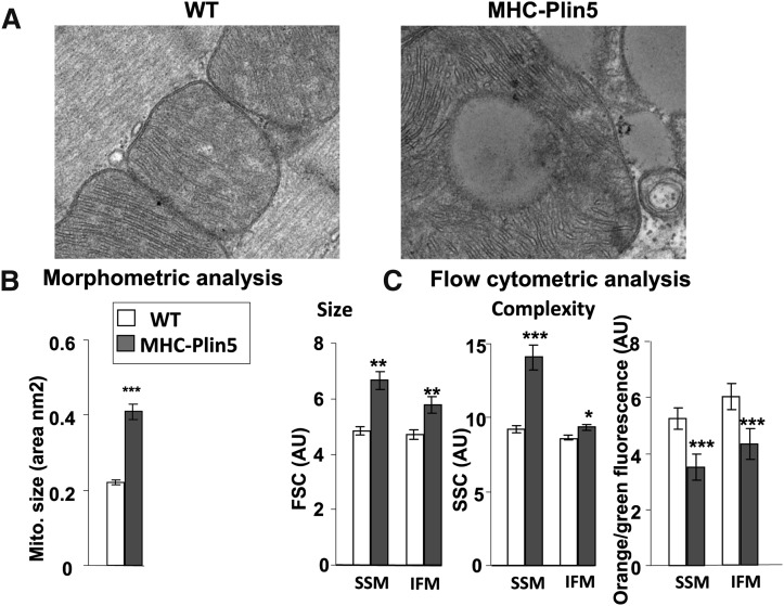
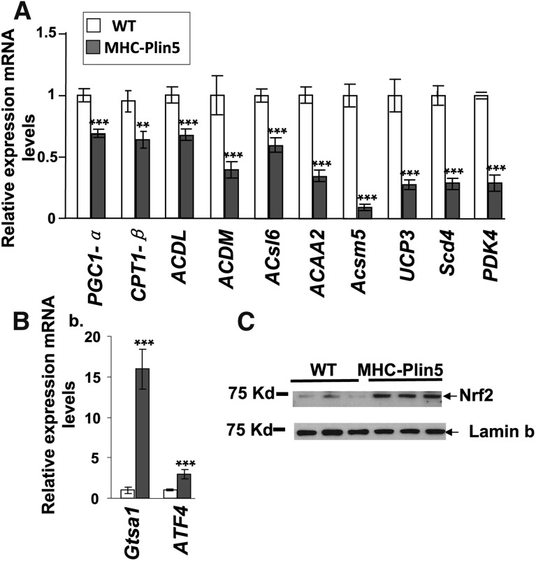
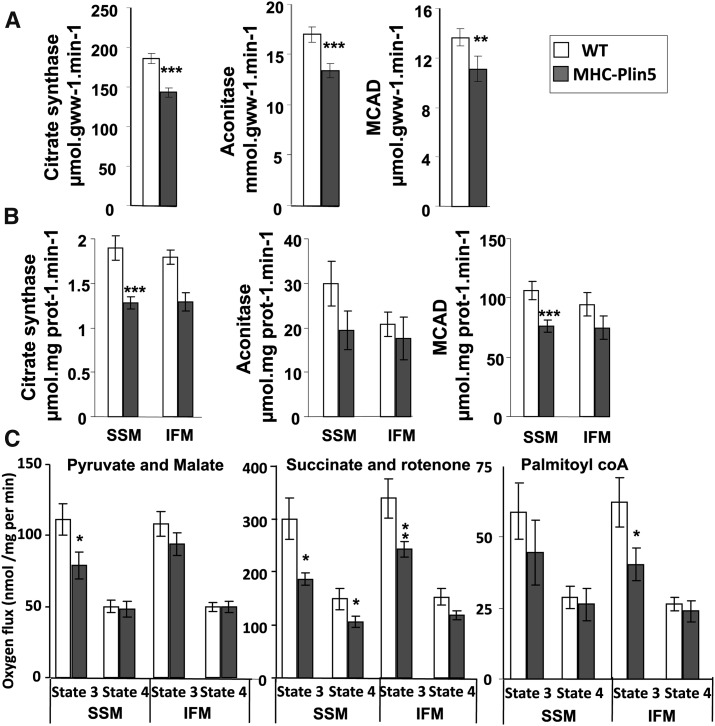
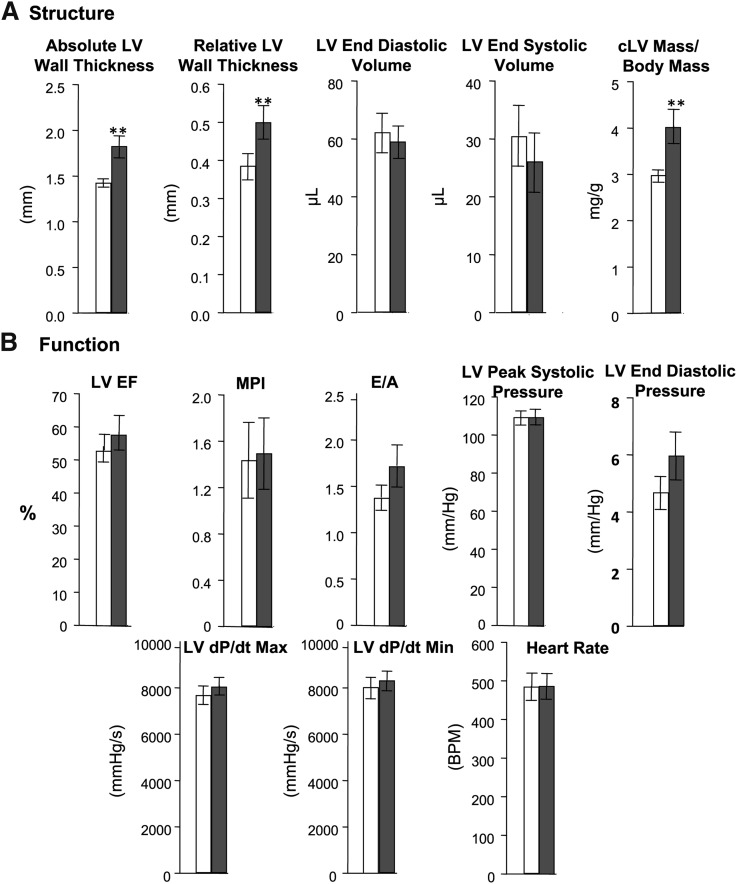
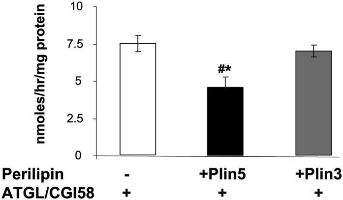
Presence of ectopic lipid droplets (LDs) in cardiac muscle is associated to lipotoxicity and tissue dysfunction. However, presence of LDs in heart is also observed in physiological conditions, such as when cellular energy needs and energy production from mitochondria fatty acid β-oxidation are high (fasting). This suggests that development of tissue lipotoxicity and dysfunction is not simply due to the presence of LDs in cardiac muscle but due at least in part to alterations in LD function. To examine the function of cardiac LDs, we obtained transgenic mice with heart-specific perilipin 5 (Plin5) overexpression (MHC-Plin5), a member of the perilipin protein family. Hearts from MHC-Plin5 mice expressed at least 4-fold higher levels of plin5 and exhibited a 3.5-fold increase in triglyceride content versus nontransgenic littermates. Chronic cardiac excess of LDs was found to result in mild heart dysfunction with decreased expression of peroxisome proliferator-activated receptor (PPAR)α target genes, decreased mitochondria function, and left ventricular concentric hypertrophia. Lack of more severe heart function complications may have been prevented by a strong increased expression of oxidative-induced genes via NF-E2-related factor 2 antioxidative pathway. Perilipin 5 regulates the formation and stabilization of cardiac LDs, and it promotes cardiac steatosis without major heart function impairment.


Figures

Fig. 1.
Fig. 1.
Heart-specific Plin5 overexpression results in posttranscriptional expression of other cardiac perilipin proteins. (A) mRNA expression of Plin5 is markedly increased in fed three-month-old male MHC-Plin5 compared with age-matched WT. Means are errors bars ± SEM; n = 9 per group; ***P < 0.001. (B) Western blot analysis of cardiac perilipin protein expression profile. Left ventricles were excised, and equal milligram equivalents of wet tissue were loaded in each lane. Experiments were performed twice. Liver and gastrocnemius muscle Plin5 content were determined to control for the specificity of Plin5 overexpression. A myc antibody was used to recognize ectopic expression of Plin5. β-actin and GAPDH were used as control loading. (C) Quantification of Plin5 protein expression in WT and MHC-Plin5 hearts by densitometry of Western blots. Means are errors bars ± SEM; n = 4 for each genotype; ***P < 0.001 for WT-fasted versus MHC-Plin5-fasted mice.
Fig. 2.
Fig. 2.
MHC-Plin5 mice show massive increase of LDs in their heart. (A) TAG content in cardiac muscle of male WT and MHC-Plin5 at three months old. Errors bars are means ± SEM; n = 6 for WT and n = 4 for MHC-Plin5; **P < 0.001 for WT versus MHC-Plin5. (B) Representative electron micrographs depict the histological appearance of left ventricles from fed WT and MHC-Plin5 mice at three months old (magnification 3,200, bar equals 500 nm). MHC-Plin5 hearts accumulated LDs (arrowheads) around clusters of mitochondria (M, arrowheads), whereas no lipid droplets were observed in similar regions of the left ventricle from WT mice. We used four mice per group and studied 35 positions per sample. (C) Morphometric analysis of LDs. Using six electron micrographs of left ventricles from three fed and overnight-fasted WT and MHC-Plin5 mice three months old, the LD number was manually counted and mitochondria area was determined using ImageJ software. Means are errors bars ± SEM; ***P < 0.001 for WT fed versus WT fasted, ###P < 0.001 for MHC-Plin5 fed versus MHC-Plin5 fasted, ##$P < 0.001 fasted MHC-Plin5 versus fasted WT.
Fig. 3.
Fig. 3.
Heart-specific Plin5 overexpression results in mitochondria morphology alteration. (A) Representative electron micrographs depict the histological appearance of mitochondria in left ventricles from fed WT and MHC-Plin5 mice at 3 months old (magnification 21,000, bar equals 100 nm). (B) Morphometric analysis of mitochondria. Using six electron micrographs of left ventricles from four overnight fed WT and MHC-Plin5 mice at 12 weeks old, mitochondria area were determined using ImageJ software. Means are errors bars ± SEM; ***P < 0.001. (C) Flow cytometry analysis of subpopulations of mitochondria and ΔΨm. The relative size and internal complexity of distinct mitochondrial subpopulations were determined using flow cytometric analyses. Mitochondrial subpopulations were stained with Mitotracker deep red 633, a membrane potential (ΔΨm)-dependent dye, and gated based on incorporation of the dye. Analysis of FSC and SSC were calculated per 20,000 gated events for all mitochondrial subpopulations. FSC and SSC are expressed in arbitrary units (AU). Means are errors bars ± SEM; n = 18 for WT and n = 12 for MHC-Plin5; *P < 0.05, **P < 0.01, ***P < 0.001 for WT versus MHC-Plin5. ΔΨm was assessed by staining mitochondrial subpopulations with JC-1 dye and assessing the shift from green to orange fluorescence with flow cytometry. Ψm was calculated based on orange-to-green fluorescence ratios in WT versus MHC-Plin5 cardiac mitochondrial subpopulations. Orange-to-green fluorescence ratios are expressed in AU. Means are errors bars ± SEM; n = 18 for WT and n = 12 for MHC-Plin5; ***P < 0.001 for WT versus MHC-Plin5.
Fig. 4.
Fig. 4.
Plin5 overexpression decreases selected PPARα target genes and increases selected Nrf2 target genes. (A) mRNA expression levels for selected PPARα target genes were determined by RT-qPCR analysis. mRNA expression of PPARα decreased in fed three-month-old MHC Plin5 hearts. Means are errors bars ± SEM; n = 9 for each group; **P < 0.01, ***P < 0.001 for WT versus MHC-Plin5. (B) mRNA levels of genes encoding Gtsa1 and ATF4 mRNA were increased in cardiac muscle. Means are errors bars ± SEM; n = 9 for each group; **P < 0.01, ***P < 0.001 for WT versus MHC-Plin5. (C) Nrf2 protein expression was found increased in MHC-Plin5 heart nuclear extracts. Lamin b was used as control loading.
Fig. 5.
Fig. 5.
α-MHC-Plin5 mice have impaired mitochondria function. (A) Myocardial activity of the mitochondrial oxidative enzymes citrate synthase, aconitase, and medium-chain acyl-CoA dehydrogenase in whole-heart extract from WT and MHC-Plin5. Means are errors bars ± SEM; n = 17 for WT and n = 12 for MHC-Plin5; **P < 0.01, ***P < 0.001 for WT versus MHC-Plin5. (B) SSM and IFM from WT and MHC-Plin5 hearts were isolated, and mitochondrial oxidative enzymes citrate synthase, aconitase, and medium-chain acyl-CoA dehydrogenase were measured. Means are errors bars ± SEM; n = 17 for WT and n = 11 for MHC-Plin5; *P < 0.05 for WT versus MHC-Plin5. (C) Respiration of isolated mitochondria from cardiac tissues of three-month-old MHC-Plin5 and WT mice. ADP-driven (state 3) and state 4 were measured with pyruvate (20 mM) and malate (10 μM) or succinate (10 mM) and rotenone (7.5 μM) or palmitoyl CoA (10 mM) in SSM and IFM preparations. Means are errors bars ± SEM; n = 17; *P < 0.05, **P < 0.05.
Fig. 6.
Fig. 6.
Echocardiography analysis of cardiac structure and function on four-month-old WT and MHC-Plin5 mice. (A) Cardiac structure data. Means are errors bars ± SEM; n =7 for WT and for MHC-Plin5; **P < 0.01. LV, left ventricular; cLV, concentric LV. (B) Contractile function measurements. For LV peak systolic pressure, LV end diastolic pressure, LV dP/dt max, LV dP/dt min, and heart rate, n =33 for WT and n = 22 for MHC-Plin5 mice. For measurements of LV, EF, MPI, and E/A, n = 7 for WT and for MHC-Plin5 mice.
Fig. 7.
Fig. 7.
Plin5 inhibits TAG hydrolase activity in an in vitro reconstituted system. CHO-K1 cells were transfected with ATGL-CFP and CGI-YFP plasmids, with Plin5 alone, or with Plin3 alone. Cellular extracts (20 μl) overexpressing ATGL and CGI-58 were incubated with 80 μl of cellular extracts overexpressing either Plin5 or Plin3 and 100 μl of a radioactive exogenous substrate. After 1 h of incubation, released radiolabeled NEFA were extracted and dpm was measured with a liquid scintillation counter. Means are errors bars ± SEM; n = 3 for empty GFP vector, n = 3 for Plin5-YFP, n = 3 for Plin3-GFP; *P < 0.05 for Plin5-YFP versus empty GFP vector or versus Plin3-GFP.

Comment in

References

    1. Wende A. R., Abel E. D. 2010. Lipotoxicity in the heart. Biochim. Biophys. Acta. 1801: 311–319. - PMC - PubMed
    1. Reingold J. S., McGavock J. M., Kaka S., Tillery T., Victor R. G., Szczepaniak L. S. 2005. Determination of triglyceride in the human myocardium by magnetic resonance spectroscopy: reproducibility and sensitivity of the method. Am. J. Physiol. Endocrinol. Metab. 289: E935–E939. - PubMed
    1. Khan R. S., Drosatos K., Goldberg I. J. 2010. Creating and curing fatty hearts. Curr. Opin. Clin. Nutr. Metab. Care. 13: 145–149. - PMC - PubMed
    1. Chokshi A., Drosatos K., Cheema F. H., Ji R., Khawaja T., Yu S., Kato T., Khan R., Takayama H., Knöll R., et al. 2012. Ventricular assist device implantation corrects myocardial lipotoxicity, reverses insulin resistance, and normalizes cardiac metabolism in patients with advanced heart failure. Circulation. 125: 2844–2853. - PMC - PubMed
    1. Listenberger L. L., Han X., Lewis S. E., Cases S., Jr.Farese R. V., Ory D. S., Schaffer J. E. 2003. Triglyceride accumulation protects against fatty acid-induced lipotoxicity. Proc. Natl. Acad. Sci. USA. 100: 3077–3082. - PMC - PubMed

Publication types

Associated data